

A screen for kinetochore-microtubule interaction inhibitors identifies novel antitubulin compounds
- PMID: 20657644
- PMCID: PMC2904697
- DOI: 10.1371/journal.pone.0011603
A screen for kinetochore-microtubule interaction inhibitors identifies novel antitubulin compounds
Abstract
Background: Protein assemblies named kinetochores bind sister chromatids to the mitotic spindle and orchestrate sister chromatid segregation. Interference with kinetochore activity triggers a spindle checkpoint mediated arrest in mitosis, which frequently ends in cell death. We set out to identify small compounds that inhibit kinetochore-microtubule binding for use in kinetochore-spindle interaction studies and to develop them into novel anticancer drugs.
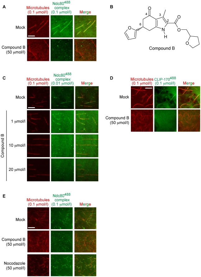
Methodology/principal findings: A fluorescence microscopy-based in vitro assay was developed to screen compound libraries for molecules that prevented the binding of a recombinant human Ndc80 kinetochore complex to taxol-stabilized microtubules. An active compound was identified that acted at the microtubule level. More specifically, by localizing to the colchicine-binding site in alphabeta-tubulin the hit compound prevented the Ndc80 complex from binding to the microtubule surface. Next, structure-activity analyses distinguished active regions in the compound and led to the identification of highly potent analogs that killed cancer cells with an efficacy equaling that of established spindle drugs.
Conclusions/significance: The compound identified in our screen and its subsequently identified analogs represent new antitubulin chemotypes that can be synthetically developed into a novel class of antimitotic spindle drugs. In addition, they are stereochemically unique as their R- and S-isomers mimic binding of colchicine and podophyllotoxin, respectively, two antitubulin drugs that interact differently with the tubulin interface. Model-driven manipulation of our compounds promises to advance insight into how antitubulin drugs act upon tubulin. These advances in turn may lead to tailor-made colchicine site agents which would be valuable new assets to fight a variety of tumors, including those that have become resistant to the (antispindle) drugs used today.
Conflict of interest statement
Figures


Similar articles
-
hNuf2 inhibition blocks stable kinetochore-microtubule attachment and induces mitotic cell death in HeLa cells.J Cell Biol. 2002 Nov 25;159(4):549-55. doi: 10.1083/jcb.200208159. Epub 2002 Nov 18. J Cell Biol. 2002. PMID: 12438418 Free PMC article.
-
Roles for the conserved spc105p/kre28p complex in kinetochore-microtubule binding and the spindle assembly checkpoint.PLoS One. 2009 Oct 28;4(10):e7640. doi: 10.1371/journal.pone.0007640. PLoS One. 2009. PMID: 19893618 Free PMC article.
-
The Hec1/Ndc80 tail domain is required for force generation at kinetochores, but is dispensable for kinetochore-microtubule attachment formation and Ska complex recruitment.Mol Biol Cell. 2020 Jul 1;31(14):1453-1473. doi: 10.1091/mbc.E20-05-0286. Epub 2020 May 13. Mol Biol Cell. 2020. PMID: 32401635 Free PMC article.
-
The Ndc80 complex: hub of kinetochore activity.FEBS Lett. 2007 Jun 19;581(15):2862-9. doi: 10.1016/j.febslet.2007.05.012. Epub 2007 May 11. FEBS Lett. 2007. PMID: 17521635 Review.
-
Kinetochore-microtubule coupling mechanisms mediated by the Ska1 complex and Cdt1.Essays Biochem. 2020 Sep 4;64(2):337-347. doi: 10.1042/EBC20190075. Essays Biochem. 2020. PMID: 32844209 Free PMC article. Review.
Cited by
-
The Effects of Podophyllotoxin Derivatives on Noncancerous Diseases: A Systematic Review.Int J Mol Sci. 2025 Jan 23;26(3):958. doi: 10.3390/ijms26030958. Int J Mol Sci. 2025. PMID: 39940726 Free PMC article.
-
Diverse small molecule inhibitors of human apurinic/apyrimidinic endonuclease APE1 identified from a screen of a large public collection.PLoS One. 2012;7(10):e47974. doi: 10.1371/journal.pone.0047974. Epub 2012 Oct 23. PLoS One. 2012. PMID: 23110144 Free PMC article.
-
Aurora-A inactivation causes mitotic spindle pole fragmentation by unbalancing microtubule-generated forces.Mol Cancer. 2011 Oct 19;10:131. doi: 10.1186/1476-4598-10-131. Mol Cancer. 2011. PMID: 22011530 Free PMC article.
-
A novel sulfonamide agent, MPSP-001, exhibits potent activity against human cancer cells in vitro through disruption of microtubule.Acta Pharmacol Sin. 2012 Feb;33(2):261-70. doi: 10.1038/aps.2011.156. Acta Pharmacol Sin. 2012. PMID: 22301862 Free PMC article.
-
Discovery and optimization of a series of 2-aryl-4-amino-5-(3',4',5'-trimethoxybenzoyl)thiazoles as novel anticancer agents.J Med Chem. 2012 Jun 14;55(11):5433-45. doi: 10.1021/jm300388h. Epub 2012 May 21. J Med Chem. 2012. PMID: 22578111 Free PMC article.
References
-
- Evan GI, Vousden KH. Proliferation, cell cycle and apoptosis in cancer. Nature. 2001;411:342–348. - PubMed
-
- Morgan DO. London: New Science Press Ltd.; 2007. The cell cycle: principles of control (Primers in biology).297
-
- Jordan MA, Wilson L. Microtubules as a target for anticancer drugs. Nature Rev Cancer. 2004;4:253–265. - PubMed
-
- Musacchio A, Salmon ED. The spindle-assembly checkpoint in space and time. Nat Rev Mol Cell Biol. 2007;8:379–393. - PubMed
-
- Gascoigne KE, Taylor SS. Cancer cells display profound intra- and interline variation following prolonged exposure to antimitotic drugs. Cancer Cell. 2009;14:111–122. - PubMed
Publication types
MeSH terms
Substances
Grants and funding
LinkOut - more resources
Full Text Sources
Other Literature Sources
Research Materials
