. 2010 Jul 15;5(7):e11583.
doi: 10.1371/journal.pone.0011583.

MMP-9, uPAR and cathepsin B silencing downregulate integrins in human glioma xenograft cells in vitro and in vivo in nude mice

Affiliations

MMP-9, uPAR and cathepsin B silencing downregulate integrins in human glioma xenograft cells in vitro and in vivo in nude mice

Krishna Kumar Veeravalli et al. PLoS One. .

Retraction in

Abstract

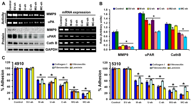
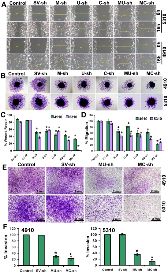
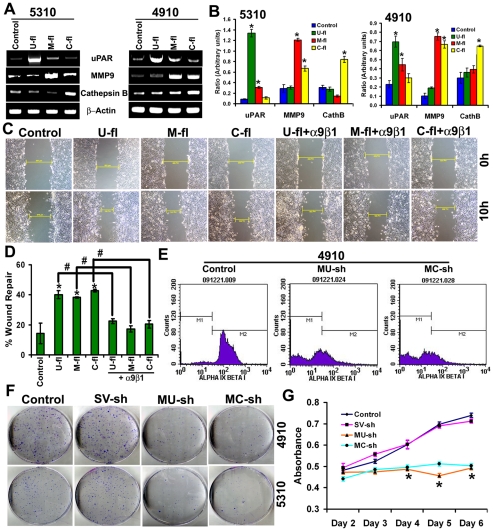
Background: Involvement of MMP-9, uPAR and cathepsin B in adhesion, migration, invasion, proliferation, metastasis and tumor growth has been well established. In the present study, MMP-9, uPAR and cathepsin B genes were downregulated in glioma xenograft cells using shRNA plasmid constructs and we evaluated the involvement of integrins and changes in their adhesion, migration and invasive potential.

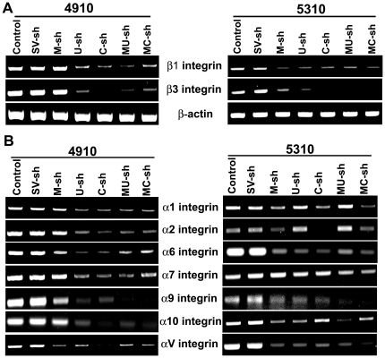
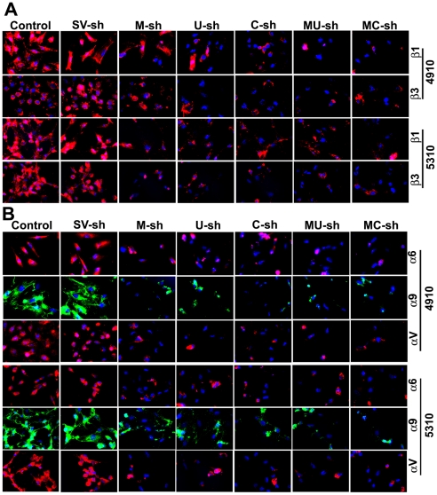
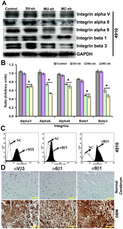
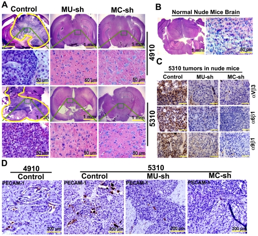
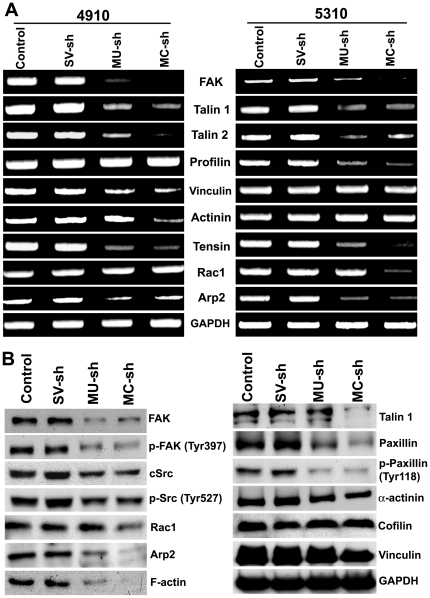
Methodology/principal findings: MMP-9, uPAR and cathepsin B single shRNA plasmid constructs were used to downregulate these molecules in xenograft cells. We also used MMP-9/uPAR and MMP-9/cathepsin B bicistronic constructs to evaluate the cumulative effects. MMP-9, uPAR and cathepsin B downregulation significantly inhibits xenograft cell adhesion to several extracellular matrix proteins. Treatment with MMP-9, uPAR and cathepsin B shRNA of xenografts led to the downregulation of several alpha and beta integrins. In all the assays, we noticed more prominent effects with the bicistronic plasmid constructs when compared to the single plasmid shRNA constructs. FACS analysis demonstrated the expression of alphaVbeta3, alpha6beta1 and alpha9beta1 integrins in xenograft cells. Treatment with bicistronic constructs reduced alphaVbeta3, alpha6beta1 and alpha9beta1 integrin expressions in xenograft injected nude mice. Migration and invasion were also inhibited by MMP-9, uPAR and cathepsin B shRNA treatments as assessed by spheroid migration, wound healing, and Matrigel invasion assays. As expected, bicistronic constructs further inhibited the adhesion, migration and invasive potential of the xenograft cells as compared to individual treatments.

Conclusions/significance: Downregulation of MMP-9, uPAR and cathespin B alone and in combination inhibits adhesion, migration and invasive potential of glioma xenografts by downregulating integrins and associated signaling molecules. Considering the existence of integrin inhibitor-resistant cancer cells, our study provides a novel and effective approach to inhibiting integrins by downregulating MMP-9, uPAR and cathepsin B in the treatment of glioma.


Conflict of interest statement

Competing Interests: The authors have declared that no competing interests exist.

Figures

Figure 1
Figure 1. Efficiency of shRNA plasmid constructs and their effect on adhesion of xenografts to various ECM proteins.
(A) Activity, protein and mRNA expressions of MMP-9, uPAR and cathepsin B in 4910 xenograft cells. MMP-9 and uPA activity was determined by gelatin and fibrin zymography after treatments with scrambled vector (SV-sh), MMP-9 (M-sh), uPAR (U-sh), cathepsin B (C-sh), MMP-9/uPAR (MU-sh) and MMP-9/cathepsin B (MC-sh) plasmid shRNAs. Western blot analysis showing MMP-9, uPAR and cathepsin B protein expression levels were reduced after M-sh, U-sh, C-sh, MU-sh and MC-sh treatments. n = 3. RT-PCR of 4910 cells transfected with SV-sh, M-sh, U-sh, C-sh, MU-sh and MC-sh was performed as per standard protocols. Further, quantification of the Western blots (B) revealed significant reductions in protein expressions after M-sh, U-sh, C-sh, MU-sh and MC-sh treatments. n = 3. Values shown are the mean (±SEM). *p<0.05 vs. control (C) Adhesion assay was performed to evaluate the effect of SV-sh, M-sh, U-sh, C-sh, MU-sh and MC-sh treatments in 4910 and 5310 glioma xenograft cells on their adhesive potential to collagen (type I), vitronectin, fibronectin and laminin coated plates. Percent adhesion was calculated from the mean obtained from 3 independent experiments and values shown are the mean (±SEM). *p<0.05 vs. control.
Figure 2
Figure 2. RT-PCR analysis of integrins.
(A) RT-PCR analysis of 4910 and 5310 cells transfected with scrambled vector (SV-sh), MMP-9 (M-sh), uPAR (U-sh), cathepsin B (C-sh), MMP-9/uPAR (MU-sh) and MMP-9/cathepsin B (MC-sh) plasmid shRNAs, showing the mRNA expressions of β integrins (β1 and β3) (A) and α integrins (α1, α2, α6, α7, α9, α10, and αV) (B).
Figure 3
Figure 3. Effect of shRNA plasmid constructs on the expression of integrins at protein level.
(A) Immunocytochemistry was performed to evaluate the effect of various shRNA treatments on the expressions of β1 and β3 integrins. Microscopic images demonstrate β1 and β3 integrin expressions (red fluorescence) in 4910 and 5310 cells transfected with scrambled vector (SV-sh), MMP-9 (M-sh), uPAR (U-sh), cathepsin B (C-sh), MMP-9/uPAR (MU-sh) and MMP-9/cathepsin B (MC-sh). All treatments reduced β1 and β3 integrins expression. (B) The effect of various shRNA treatments on the expressions of α6, α9 and αV integrins was also evaluated and the microscopic images demonstrate α6, α9 and αV integrins expression in 4910 and 5310 cells transfected with SV-sh, M-sh, U-sh, C-sh, MU-sh and MC-sh. All the treatments reduced α6, α9 and αV integrins expression in both the cell lines.
Figure 4
Figure 4. Effect of bicistronic constructs on integrins and the expression of αVβ3, α6β1 and α9β1 integrins on glioma xenografts and GBM clinical samples.
(A) Western blot analysis depicting the reduced protein expressions of αV, α6, α9, β1 and β3 integrins after MMP-9/uPAR (MU-sh) and MMP-9/cathepsin B (MC-sh) plasmid shRNA treatments in 4910 cells. n = 3. Further, quantification of the Western blots (B) revealed significant reductions in protein expressions after MU-sh and MC-sh treatments. n = 3. Values shown are the mean (±SEM). *p<0.05 vs. control. (C) FACS analysis showing the presence of αVβ3, α6β1 and α9β1 integrin heterodimers on 4910 glioma xenogaft cells. 4910 cells, in which iso match mouse IgG used as the primary antibody served as negative controls. NC-negative control (D) Immunohistochemistry of normal cerebrum and glioblastoma multiforme (GBM) for αVβ3, α6β1 and α9β1 integrin heterodimer expressions. GBM tissue micro arrays were processed for immunohistochemistry. Prominent brown staining indicative of αVβ3, α6β1 or α9β1 integrin expression was noticed in GBM samples. Scale bar = 100 µm.
Figure 5
Figure 5. Wound healing, spheroid migration and Matrigel invasion assays.
(A) Reduction in wound healing indicative of reduced migration potential was noticed after treatments with MMP-9 (M-sh), uPAR (U-sh), cathepsin B (C-sh), MMP-9/uPAR (MU-sh) and MMP-9/cathepsin B (MC-sh) plasmid shRNAs in both 4910 and 5310 cells. Untreated and SV-sh transfected cells served as controls. (B) Migration of the cells from 4910 and 5310 spheroids transfected with M-sh, U-sh, C-sh, MU-sh and MC-sh was prominently reduced. (C, D) Quantification of wound healing and spheroid migration assays. Percent wound repair was calculated from the mean of the average width of the wound obtained from 3 independent experiments. Similarly, percent migration was calculated from the mean of the average migration obtained from 3 independent experiments. Error bars indicate SEM. *p<0.05 vs. control. (E) Matrigel invasion assay of 4910 and 5310 cells transfected with SV-sh, MU-sh and MC-sh. MU-sh and MC-sh treatments prominently reduced the invasive potential of both 4910 and 5310 cells through Matrigel. (F) Invasion was quantified by counting the average number of invaded cells in five different fields with each treatment. Percent invasion was calculated from the mean of the average number of invaded cells obtained from 3 independent experiments. Error bars indicate SEM. *p<0.05 vs. control.
Figure 6
Figure 6. Involvement of α9β1 integrin on the migration potential of xenograft cells and the effect of bicistronic constructs on the glioma xenograft cell proliferation.
(A) RT-PCR of 5310 and 4910 cells transfected with uPAR (U-fl), MMP-9 (M-fl), and cathepsin B (C-fl) expressing plasmid DNAs was performed as per standard protocols. Prominent increase in respective mRNA expressions noticed after U-fl, M-fl, and C-fl treatments. Further, quantification of the RT-PCR data (B) revealed significant increases in uPAR mRNA and MMP-9 mRNA expression after treatments with M-fl and C-fl, respectively in both the xenograft cells. (C) Wound healing indicative of increased migration potential noticed after treatments with U-fl, M-fl, and C-fl in 5310 cells was reduced with the same treatments in presence of α9β1 antibody. Photographs are the representative images obtained from three independent experiments. (D) Quantification of wound healing assay. Percent wound repair was calculated from the mean of the average width of the wound obtained from 3 independent experiments. Error bars indicate SEM. *p<0.05 vs. control. # p<0.05. (E) FACS analysis showing reduced α9β1 integrin levels in 4910 cells after MMP-9/uPAR (MU-sh) and MMP-9/cathepsin B (MC-sh) treatments. (F) Clonogeneic assay depicting the reduced proliferation of 4910 and 5310 cells after MU-sh and MC-sh treatments. After 14 days of incubation, the colonies containing more than 50 cells were counted. Images shown are the representatives obtained from three independent experiments. (G) MTT assay showing significant reduction in the proliferation of 4910 cells from Day 4 to Day 6 after MU-sh and MC-sh treatments. n = 3. *p<0.05 vs. control.
Figure 7
Figure 7. Effect of treatment with bicistronic constructs on the brain tumors in nude mice pre-injected with xenograft cells.
(A) shRNA mediated regression of pre-established tumor growth. Hematoxylin and eosin staining performed on the brain sections obtained from various groups of animals revealed a prominent tumor reduction after MMP-9/uPAR (MU-sh) or MMP-9/cathepsin B (MC-sh) treatments. Each group consisted of 6 animals. Yellow curved line in 4910 and 5310 control brain sections indicate the tumor area. (B) Normal nude mice brain section stained with hematoxylin and eosin. (C) Immunohistochemical comparison of control, MU-sh and MC-sh-treated nude mice which are pre-injected with 5310 cells (and sacrificed 2–3 weeks prior to the end of the treatment), to analyze the expression of αVβ3, α6β1 and α9β1 integrin heterodimers. Brown staining indicative of αVβ3, α6β1 or α9β1 integrin expression was reduced in MU-sh and MC-sh treated sections compared to controls. n = 6. Scale bar = 50 µm. (D) Untreated 4910 and 5310 in vivo tumors were prominently stained for platelet endothelial cell adhesion molecule (PECAM-1) whereas the intensity of brown staining was reduced in 5310 in vivo tumors after MU-sh and MC-sh treatments.
Figure 8
Figure 8. RT PCR and Western blot analysis of the molecules associated with integrin-mediated migration.
(A) RT-PCR of 4910 and 5310 glioma xenograft cells transfected with scrambled vector (SV-sh), MMP-9/uPAR (MU-sh) and MMP-9/cathepsin B (MC-sh) plasmid shRNAs was performed as per standard protocol described in materials and methods. Several genes were downregulated with MU-sh and MC-sh treatments in both the xenograft cells. Results presented are the representative images of three independent experiments. (B) Western blot analysis showing the effect of bicistronic constructs (MU-sh and MC-sh) on the protein expressions of FAK, p-FAK (Tyr397), cSrc, p-Src (Tyr527), Rac1, Arp2, F-actin, talin1, paxillin, p-paxillin (Tyr118), α-actinin, cofilin and vinculin in 5310 human glioma xenograft cells. The expressions of majority of the proteins associated with integrin-mediated migration were reduced with MU-sh and MC-sh treatments. n = 3.

Similar articles

Cited by

References

    1. Albelda SM, Buck CA. Integrins and other cell adhesion molecules. FASEB J. 1990;4:2868–2880. - PubMed
    1. Hemler ME. VLA proteins in the integrin family: structures, functions, and their role on leukocytes. Annu Rev Immunol. 1990;8:365–400. - PubMed
    1. Hynes RO. Integrins: versatility, modulation, and signaling in cell adhesion. Cell. 1992;69:11–25. - PubMed
    1. Ruoslahti E. Fibronectin and its integrin receptors in cancer. Adv Cancer Res. 1999;76:1–20. - PubMed
    1. Schaller MD, Parsons JT. Focal adhesion kinase and associated proteins. Curr Opin Cell Biol. 1994;6:705–710. - PubMed

Publication types

MeSH terms