. 2010 Jul 21;5(7):e11689.
doi: 10.1371/journal.pone.0011689.

R5 clade C SHIV strains with tier 1 or 2 neutralization sensitivity: tools to dissect env evolution and to develop AIDS vaccines in primate models

Affiliations

R5 clade C SHIV strains with tier 1 or 2 neutralization sensitivity: tools to dissect env evolution and to develop AIDS vaccines in primate models

Nagadenahalli B Siddappa et al. PLoS One. .

Abstract

Background: HIV-1 clade C (HIV-C) predominates worldwide, and anti-HIV-C vaccines are urgently needed. Neutralizing antibody (nAb) responses are considered important but have proved difficult to elicit. Although some current immunogens elicit antibodies that neutralize highly neutralization-sensitive (tier 1) HIV strains, most circulating HIVs exhibiting a less sensitive (tier 2) phenotype are not neutralized. Thus, both tier 1 and 2 viruses are needed for vaccine discovery in nonhuman primate models.

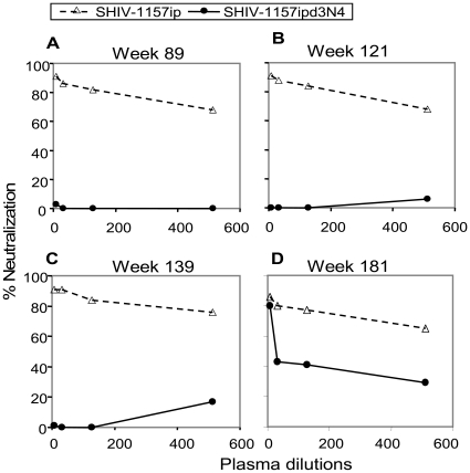
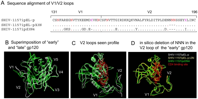
Methodology/principal findings: We constructed a tier 1 simian-human immunodeficiency virus, SHIV-1157ipEL, by inserting an "early," recently transmitted HIV-C env into the SHIV-1157ipd3N4 backbone [1] encoding a "late" form of the same env, which had evolved in a SHIV-infected rhesus monkey (RM) with AIDS. SHIV-1157ipEL was rapidly passaged to yield SHIV-1157ipEL-p, which remained exclusively R5-tropic and had a tier 1 phenotype, in contrast to "late" SHIV-1157ipd3N4 (tier 2). After 5 weekly low-dose intrarectal exposures, SHIV-1157ipEL-p systemically infected 16 out of 17 RM with high peak viral RNA loads and depleted gut CD4+ T cells. SHIV-1157ipEL-p and SHIV-1157ipd3N4 env genes diverge mostly in V1/V2. Molecular modeling revealed a possible mechanism for the increased neutralization resistance of SHIV-1157ipd3N4 Env: V2 loops hindering access to the CD4 binding site, shown experimentally with nAb b12. Similar mutations have been linked to decreased neutralization sensitivity in HIV-C strains isolated from humans over time, indicating parallel HIV-C Env evolution in humans and RM.

Conclusions/significance: SHIV-1157ipEL-p, the first tier 1 R5 clade C SHIV, and SHIV-1157ipd3N4, its tier 2 counterpart, represent biologically relevant tools for anti-HIV-C vaccine development in primates.


Conflict of interest statement

Competing Interests: The authors have declared that no competing interests exist.

Figures

Figure 1
Figure 1. Selection of a neutralization escape variant in rhesus monkey RPn-8.
This monkey was inoculated initially with an “early” form of the virus (the parental infectious molecular clone, SHIV-1157i; see Table 1), and 4 weeks after RPn-8 had progressed to AIDS (∼2.7 years), the “late” virus, SHIV-1157ipd3N4 , was generated by molecular cloning of the 3′ proviral half from RPn-8 PBMC DNA. Neutralization of SHIV-1157ip (“early” strain, open triangles) and SHIV-1157ipd3N4 (“late” strain, closed circles) with autologous plasma from animal RPn-8 at the time points post-inoculation indicated is shown (A–D), including contemporaneous plasma for the “late” virus collected at week 139 (panel C). Virus (grown in RM PBMC) was incubated with different dilutions of monkey plasma for 1 h before being added to TZM-bl cells in the presence of DEAE-dextran. Luciferase expression was measured on day 2 post-infection.
Figure 2
Figure 2. Construction and co-receptor usage of SHIV-1157ipEL.
A. SHIV-1157ipd3N4 was used as backbone to construct SHIV-1157ipEL. The 2.2 kb KpnI (K) - BamHI (B) fragment of the “early” SHIV-1157ip (spanning most of gp120, the entire gp41 extracellular domain, the transmembrane region (TM), and part of the cytoplasmic domain) (closed black bar) was used to replace the corresponding region of the proviral backbone (stippled bar). The modified 3′-half of was ligated with the 5′ half of SHIV-1157ipd3N4 DNA to form full-length SHIV-1157ipEL. NN: 2 NF-κB sites present in the 3′LTR. During viral replication, this duplication of NF-κB sites copied into the 5′LTR. B. Coreceptor usage of SHIV-1157ipEL and SHIV-1157ipEL-p. U87.CD4.CCR5 and U87.CD4.CXCR4 cells were exposed to parental SHIV-1157ipEL, passaged SHIV-1157ipEL-p, SHIV-1157ipd3N4 (clade C, R5 SHIV), and HIVNL4-3(HIV clade B env, X4). The levels of p27 Gag were measured in the supernatants as indicated.
Figure 3
Figure 3. Replication of SHIV-1157ipEL and SHIV-1157ipEL-p in rhesus macaque PBMC.
(A) PBMC from randomly selected naïve RM donors were stimulated with concanavalin A (Con-A) and exposed to SHIV-1157ipEL-containing supernatant (Methods). The PBMC of RM RFn-9 (thick red line) did not support the replication of parental SHIV-1157ipEL. (B) PBMC from random donors were stimulated with Con-A and exposed to passaged virus, SHIV-1157ipEL-p. Supernatants were harvested and p27 levels were measured at the time points indicated. PBMC cultures of RFn-9 (previously unable to support the replication of parental virus, see under A) now yielded high levels of p27 (thick red line), indicating SHIV-1157ipEL-p adaptation to RM. For RM listed in both A and B, aliquots of frozen PBMC from the same blood collection as in A were used for assays in B.
Figure 4
Figure 4. Serial passage of SHIV-1157ipEL in rhesus macaques.
(A) Parental SHIV-1157ipEL was passaged rapidly in four Indian-origin RM through serial blood transfer at peak viremia (week 2). (B) Viral loads were measured after serial passage at the time points indicated. †, these RM were euthanized at peak viremia.
Figure 5
Figure 5
(A) Sequence alignment of V1/V2 sites of and SHIV-1157ipEL-p, SHIV-1157ipEL-pΔ3N and SHIV-1157ipd3N4 (numbering according to HXB2). (B) Molecular modeling of SHIV-1157ipEL-p and SHIV-1157ipd3N4 sequences was performed using the X-ray structure of the CD4-bound YU2 gp120 core ; PDB code 1RZK). The V1, V2, and V3 loops were modeled onto the core. Mutations inducing structural heterogeneity in the V1 and V2 loops were identified. (C and D) Illustration of three-dimensional (3D) gp120 of SHIV-1157ipEL-p (white), SHIV-1157ipd3N4 (green), and (D) SHIV-1157ipEL-pΔ3N (yellow) showing that access to the CD4 binding site (red) is more restricted for SHIV-1157ipEL-pΔ3N and SHIV-1157ipd3N4 than for SHIV-1157ipEL-p.
Figure 6
Figure 6. Neutralization of SHIV-1157ipEL-p, SHIV-1157ipEL-pΔ3N and SHIV-1157ipd3N4 by sCD4 and b12.
The assays were performed in TZM-bl cells using virus stocks grown in RM PBMC. (A) Dotted lines, neutralization of SHIV-1157ipEL-p (circles), SHIV-1157ipEL-pΔ3N (red color with triangles) and SHIV-1157ipd3N4 (squares) using sCD4; solid lines, neutralization of the above SHIVs using the human nmAb b12. (B) Values represent the concentration (µg/ml) for sCD4 and human b12 at which relative luciferase units (RLU) were reduced 50% or 90% compared to virus control wells, respectively.
Figure 7
Figure 7. Intrarectal inoculation of SHIV-1157ipEL-p.
Six monkeys were used in a repeated low-dose i.r. titration; the aim was to find a virus dose that resulted in systemic infection (defined as viral RNA ≥104 copies/ml) after a maximum of five weekly i.r. inoculations. RM remaining uninfected at week 2 after the 5th weekly low-dose virus challenge were given a single high-dose of SHIV-1157ipEL-p (1.5×10⁵ TCID50). The green half-diamonds represent mean ± SD of 15 additional RM used in unpublished vaccine efficacy studies as unvaccinated controls; these animals were given maximally five weekly i.r. inoculations at 8,000 TCID50. The horizontal dotted line indicates lower limit of detection (<50 viral RNA copies/ml).
Figure 8
Figure 8. T-cell depletion during acute SHIV-1157ipEL-p viremia.
The percent CD4+ T cells in blood, lymph node and gut was estimated compared with cells from naïve monkeys (closed symbols). Biopsy specimens were collected during the acute phase of infection. Two of the 2-week time points were obtained from last two i.v.-inoculated RM used for viral adaptation, the remainder were collected from RM undergoing the 5x weekly i.r. challenge-dose titration. We assigned peak viremia as being week 2 after the pen-ultimate i.r. inoculation, the one that most likely was successful in achieving systemic infection. Individual time points are shown with different symbols (open squares, week 2; open triangles, week 5; open circles, week 12).

References

    1. Song RJ, Chenine AL, Rasmussen RA, Ruprecht CR, Mirshahidi S, et al. Molecularly cloned SHIV-1157ipd3N4: a highly replication- competent, mucosally transmissible R5 simian-human immunodeficiency virus encoding HIV clade C env. J Virol. 2006;80:8729–8738. - PMC - PubMed
    1. Rerks-Ngarm S, Pitisuttithum P, Nitayaphan S, Kaewkungwal J, Chiu J, et al. Vaccination with ALVAC and AIDSVAX to prevent HIV-1 infection in Thailand. N Engl J Med. 2009;361:2209–2220. - PubMed
    1. Walker BD, Burton DR. Toward an AIDS vaccine. Science. 2008;320:760–764. - PubMed
    1. Burke B, Barnett SW. Broadening our view of protective antibody responses against HIV. Curr HIV Res. 2007;5:625–641. - PubMed
    1. Pope M, Haase AT. Transmission, acute HIV-1 infection and the quest for strategies to prevent infection. Nat Med. 2003;9:847–852. - PubMed

Publication types

Associated data