. 2010 May;16(3):545-554.
doi: 10.1109/JSTQE.2009.2033609.

Optical Microangiography: A Label Free 3D Imaging Technology to Visualize and Quantify Blood Circulations within Tissue Beds in vivo

Affiliations

Optical Microangiography: A Label Free 3D Imaging Technology to Visualize and Quantify Blood Circulations within Tissue Beds in vivo

Ruikang K Wang. IEEE J Sel Top Quantum Electron. 2010 May.

Abstract

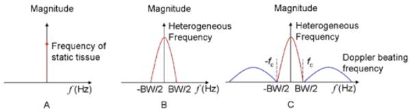
Optical microangiography (OMAG) is a recently developed volumetric imaging technique that is capable of producing 3D images of dynamic blood perfusion within microcirculatory tissue beds in vivo. The imaging contrast of OMAG image is based on the intrinsic optical scattering signals backscattered by the moving blood cells in patent blood vessels, thus it is a label free imaging technique. In this paper, I will first discuss its recent developments that use a constant modulation frequency introduced in the spectral interferograms to achieve the blood perfusion imaging. I will then introduce its latest development that utilizes the inherent blood flow to modulate the spectral interferograms to realize the blood perfusion imaging. Finally, examples of using OMAG to delineate the dynamic blood perfusion, down to capillary level resolution, within living tissues are given, including cortical blood perfusion in the brain of small animals and blood flow within human retina and choroids.


Figures

Fig.1
Fig.1
Diagram of frequency components for different tissue sample: (A) an ideal tissue sample (optically homogeneous sample) with no moving particles; (B) a real tissue sample (optically heterogeneous sample) with no moving particles; (C) a real tissue sample (optically heterogeneous sample) with moving particles.
Fig.2
Fig.2
Schematic of the OMAG system used in this study to image the velocities of blood flow, where PC represents the polarization controller and CCD the charged coupled device. The laser diode emitting the light at 633 nm was used for aiming purposes during imaging.
Fig.3
Fig.3
2D imaging of cerebral cortex in mice. (A) OMAG structural image, (B) corresponding OMAG blood flow image where abundant functional capillaries are evident, and (C) a plot of normalized OMAG flow signals across the dashed line in (B), corresponding to structural locations marked in (A). In (C), the flow signals (arrows) are ~15μm in size, close to the theoretical OMAG spatial resolution (x-y) of 16 μm.
Fig. 4
Fig. 4
A volume of 2.2×2.2×1.7 (x-y-z) mm3 of an adult mouse brain was imaged with the skull intact, using OMAG. The time to obtain this 3D OMAG image took ~25 s. (A) 3D volumetric rendering of the blood perfusion within the scanned tissue volume. (B), (C) and (D) are the sagittal, coronal, and transverse views of the blood flows fused with the depth-resolved OMAG microstructures within the scanned volume. Using suitable software, this 3-D image can be rotated, cut and examined from any angle to illustrate the spatial relationship between the blood flow and tissue microstructures. (E) and (D) are 2D x-y projection maps showing, respectively, the blood flow within the skull bone and mininges where the blood vessels were less abundant, and the cerebro-vascular flow that maps the detailed blood vessel network, including the capillaries over the cortex. White bar = 200 μm.
Fig. 5
Fig. 5
(A) OMAG, and (B) DOMAG images of CBF in adult mice under an intact cranium in vivo. The projection images were obtained from a tissue sample volume of 2.2×2.2×2.0 mm3. Images show superb ability of OMAG and DOMAG to quantify blood flows within scanned tissue.
Fig.6
Fig.6
The cerebral blood flow over the entire cortex of an adult mouse is imaged in vivo with the skull left intact. (A) and (B) are the projection views of blood perfusion with and without directional flow information, respectively. With the reference of middle sinus vein, the arteries (pointed by cyan arrows) and veins (white arrows) can be identified in (B). It took ~8 min to acquire the 3D data to obtain (A)/(B) using our current system. The projection image was obtained from OMAG scans of 8 different regions one by one, which were then mosaiced to form (A)/(B). (C) is a photograph of the skull with the skin folded aside where viewing the vasculatures through the skull is impossible. (D) is a photograph showing blood vessels over the cortex after the skull of the same mouse was removed. The superficial blood vessels show excellent correspondence with OMAG images in (A)/(B). The marked white box = 7.5×7.5mm^2
Fig.7
Fig.7
3D OMAG imaging of the cortex during temporary intraluminal filament-induced MCAO and thrombotic ischemic stroke in mice. Compared to baseline (A), progressive focal occlusion (white arrows) and reorganization of flow patterns (green arrow) develop during MCAO (B), with residual occlusions (white arrow) and increased proximal perfusion (blue arrow) 24 hr later (C). (B) was taken at 60 min into MCAO, and at this point the filament was withdrawn from the MCA.
Fig. 8
Fig. 8
In vivo volumetric imaging of posterior chamber of an eye from a volunteer. (A) OCT fundus image of the scanned volume as described in [33]. (B) OCT cross-sectional image at the position marked yellow in (A), in which four lines as shown are resulted from the segmentation method that are used to separate the blood flows in retina and choroids. (C) Corresponding 2D OMAG flow images within the scanned volume. (D) Volumetric rendering of the merged structural and flow images with a cut through in the center of structural image. (E) Volumetric rending of the blood flow image where flows in retina are coded with green and those in choroids with red. Scale bar = 500μm. [15]
Fig. 9
Fig. 9
x-y projection images from 3D OMAG blood flow images. (A) Projection image from the whole scanned volume with the blood vessels in retina are coded with green color, and those in choroids with red color. (B) x-y projection image from the blood vessels within retina only. (C) x-y projection image from the blood vessels within choroids only. Scale bar = 500μm. [15]

References

    1. Yamada E, Matsumura M, Kyo S, Omoto R. Usefulness of a prototype intravascular ultrasound imaging in evaluation of aortic dissection and comparison with angiographic study, transesophageal echocardiography, computed tomography and magnetic resonance imaging. Am J. Cardiol. 1995;75:161–171. - PubMed
    1. Misgeld T, Kerschensteiner M. In vivo imaging of the diseased nervous system. Nature Rev. Neurosci. 2006;7:449–463. - PubMed
    1. Molina CA, Saver JL. Extending reperfusion therapy for acute ischemic stroke: emerging pharmacological, mechanical, and imaging strategies. Stroke. 2005;36:2311–20. - PubMed
    1. Albers GW, Amarenco P, Easton JD, Sacco RL, Teal P. Antithrombotic and thrombolytic therapy for ischemic stroke. Chest. 2004;126S:483–512. the Seventh ACCP Conference on Antithrombotic and Thrombolytic Therapy. - PubMed
    1. Fisher M, Fernandez JA, Ameriso SF, Xie D, Gruber A, Paganini-Hill A, Griffin JH. Activated protein C resistance in ischemic stroke not due to factor V arginine506-->glutamine mutation. Stroke. 1996;27:1163–6. - PubMed

LinkOut - more resources