. 2010 Jul 21;5(7):e11682.
doi: 10.1371/journal.pone.0011682.

Mouse cytoplasmic dynein intermediate chains: identification of new isoforms, alternative splicing and tissue distribution of transcripts

Affiliations

Mouse cytoplasmic dynein intermediate chains: identification of new isoforms, alternative splicing and tissue distribution of transcripts

Anna Kuta et al. PLoS One. .

Erratum in

Abstract

Background: Intracellular transport of cargoes including organelles, vesicles, signalling molecules, protein complexes, and RNAs, is essential for normal function of eukaryotic cells. The cytoplasmic dynein complex is an important motor that moves cargos along microtubule tracks within the cell. In mammals this multiprotein complex includes dynein intermediate chains 1 and 2 which are encoded by two genes, Dync1i1 and Dync1i2. These proteins are involved in dynein cargo binding and dynein complexes with different intermediate chains bind to specific cargoes, although the mechanisms to achieve this are not known. The DYNC1I1 and DYNC1I2 proteins are translated from different splice isoforms, and specific forms of each protein are essential for the function of different dynein complexes in neurons.

Methodology/principal findings: Here we have undertaken a systematic survey of the dynein intermediate chain splice isoforms in mouse, basing our study on mRNA expression patterns in a range of tissues, and on bioinformatics analysis of mouse, rat and human genomic and cDNA sequences. We found a complex pattern of alternative splicing of both dynein intermediate chain genes, with maximum complexity in the embryonic and adult nervous system. We have found novel transcripts, including some with orthologues in human and rat, and a new promoter and alternative non-coding exon 1 for Dync1i2.

Conclusions/significance: These data, including the cloned isoforms will be essential for understanding the role of intermediate chains in the cytoplasmic dynein complex, particularly their role in cargo binding within individual tissues including different brain regions.


Conflict of interest statement

Competing Interests: The authors have declared that no competing interests exist.

Figures

Figure 1
Figure 1. Splicing pattern of mouse dynein intermediate chain 1 gene.
A. Summary of alternative splicing in Dync1i1, showing splice variants Dync1i1.A to Dync1i1.F, and the first 6 exons (numbered 1, 2, 3, 4, 5, 6); primer binding sites used for isoform specific RT-PCRs are as indicated ( Table 2 , S2), and AS1, 2 and 3 are the alternative splice sites in exon 4. Exons are drawn to scale. B. Alternative splice sites in Dync1i1 exon 4. The nucleotide in bold is the first base pair of the exon. Bases in lower case are intronic, bases in upper case are exonic.
Figure 2
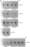
Figure 2. Amplification of different isoforms of Dync1i1 in mouse tissues using one primer pair.
Isoforms of Dync1i1 were amplified in mouse tissues using primer pair DIC1 Ex1 for and DIC1_R rev which produces six products: Dync1i1.A (531 bp), Dync1i1.B (480 bp), Dync1i1.E (471 bp), Dync1i1.D (447 bp), Dync1i1.C (420 bp), Dync1i1.F (387bp). Dync1i1.B (480 bp) and Dync1i1.E (471 bp) are not resolved due to similar length of PCR products, and were subsequently individually confirmed as present using isoform specific primer pairs ( Table 2 ), for example see amplification of Dync1i1.E using isoform specific primer pair DIC1_1.1 for and DIC1_5 rev (Figure S2). Note that in some tissues amplification products are faint, and so results were confirmed with other primer pairs as indicated in Table 2 ; for example, see amplification of Dync1i1.A in Figure 3 . In some tissues we also saw a high molecular weight fragment running at approximately 560bp (for example see spinal cord or cerebellum); this may represent another isoform but we were unable to clone or sequence this fragment. A. Adult tissues: brain, ovary, testis and liver. In brain 6 amplicons are detected as shown. In ovary and testis we detect isoform Dync1i1.C only (420bp). No product is amplified in liver. B. Adult neuronal tissues: in spinal cord, cortex, hippocampus, cerebellum, brainstem and olfactory bulb, between four and six amplicons (for example compare brainstem and olfactory bulb) could be detected as shown. C. Embryonic tissues: five amplicons are detected as shown. ‘+’ lanes are cDNA, ‘−’ lanes control for genomic DNA contamination and have no reverse transcriptase.
Figure 3
Figure 3. An isoform specific RT-PCR: screening for Dync1i1.A in mouse tissues.
Primers DIC1_1.1 for and DIC1_5 rev amplify a 206 bp product from isoform Dync1i1.A. ‘+’ lanes are cDNA, ‘−’ lanes control for genomic DNA contamination and have no reverse transcriptase. Dync1i1.A is found in all brain regions screened, but may not be highly expressed in the E17.5 whole embryo, although this is not a quantitative PCR (data not shown).
Figure 4
Figure 4. Splicing pattern of mouse dynein intermediate chain 2 gene.
A, B. Summary of alternative splicing in Dync1i2 with exon 1a or 1b respectively and the first seven exons (numbered 1a or 1b, 2, 3, 3b, 4, 5, 6, 7). Primer binding sites used for isoform specific RT-PCRs are as indicated ( Table 4 , S2). AS refers to alternative splice sites in exon 3b. Note that we could not detect the presence of the putative Dync1i2(1b).F in any tissue surveyed. Exons are drawn to scale. C. Alternative splice sites in Dync1i2 exon 3b. The nucleotide in bold is the first base pair of the exon. Bases in lower case are intronic, bases in upper case are exonic.
Figure 5
Figure 5. Amplification of different isoforms of Dync1i2 Exon 1b in mouse tissues using one primer pair.
Isoforms of Dync1i2 Exon 1b were amplified in mouse tissues using primer pair DIC2_Ex 1b for and DIC2_R rev which produce five products: Dync1i2.D (581 bp), Dync1i2.E (566 bp), Dync1i2.A (531 bp), Dync1i2.B (513 bp) and Dync1i2.C (453 bp). Dync1i2.D (581 bp) is not resolved from Dync1i2.E (566 bp) due to the similar length of PCR products; therefore we undertook nested PCRs with primers DIC2_Ex 1b for and DIC2_2.1 rev to determine the transcription pattern of these two isoforms (data not shown). We did not detect 2(1b).F in this assay. A. Adult tissues: brain, ovary, testis and liver. In brain five amplicons are detected as shown (and confirmed by nested PCRs). In ovary, testis and liver we detect isoform Dync1i2.C only. B. Adult neuronal tissues: In spinal cord, cortex, hippocampus, cerebellum, brainstem and olfactory bulb five amplicons are detected as shown. C. Embryonic tissues: Three or five amplicons are detected as shown.
Figure 6
Figure 6. Examples of Dync1i2 Exon 1a isoform specific PCR.
Four isoforms of Dync1i2 Exon1a are amplified in the mouse nervous system and E17.5 embryonic nervous tissues using primer pair DIC2_Ex1a for and DIC2_6 rev. Amplicons are as shown and in addition while Dync1i2(1a).D was not resolved in this amplification it was amplified as shown in Table 5 using primers DIC2_Ex 1a for and DIC2_2.1 rev (data not shown). We were unable to resolve, even by nested PCR, which of isoforms Dync1i2(1a).D and Dync1i2(1a).E were present in olfactory bulb. ‘+’ lanes are cDNA, ‘−’ lanes control for genomic DNA contamination and have no reverse transcriptase.
Figure 7
Figure 7. Differential expression of Dync1i2 with exon 1a and exon1b containing isoforms.
Brain cDNA was amplified by nested RT-PCR with exon 1a primers DIC2_Ex 1a for and DIC2_2.1 rev which amplify 2(1a).D (315bp) and 2(1a).E (300bp), and with exon 1b primers DIC2_Ex 1b for and DIC2_2.1 rev which amplify 2(1b).D (340bp) and 2(1b).E (325bp). We detect Dync1i2.E containing exons 1a or 1b; we detect Dync1i2.D containing exon 1b only and not exon 1a, showing differential expression of this isoform depending on which first non-coding exon is spliced in the adult brain. E17.5 whole embryo cDNA was amplified the same primer pairs (DIC2_Ex1a and DIC2_2.1 rev, and DIC2_Ex 1b and DIC2_2.1rev). We detect no Dync1i2.D isoforms. However, we detect 2(1b).E but not 2(1a).E, showing differential expression of Dync1i2.E depending on which first non-coding exon is spliced in.
Figure 8
Figure 8. Exon – protein domain relationship for dynein intermediate chain genes.
Protein domains marked as blocks, numbered exons marked in grey. The coiled coil region where protein interactions occur is shown as is DYNLT – binding region of Tctex dynein light chains, DYNLL – binding region of LC8 dynein light chains, DIM – intermediate chain dimerisation domain, DYNLRB – binding domain of Roadblock dynein light chains; seven WD40 repeats marked in green. The domain boundaries are as shown in Table S3 , , , , , , . A. Mouse DYNC1I1 isoform 1.A (645 amino acids). B. Mouse DYNC1I2 isoform 2.D (655 amino acids). Note that isoform 2.D includes exons 1 to 3 and new exon 3b; exon 4 to 18. C. Schematic alignment of DYNC1I1 and DYNC1I2 protein sequences coded by exons 3 to 6 of Dync1i1 and exons 3 to 7 of Dync1i2 genes; alternative splice sites in Dync1i1 exon 4 (AS1, AS2, AS3) and Dync1i2 exon 3b (AS1, AS2) are marked. The percent of identity (ID) and similarity (Sim) between segments of protein sequences coded by specific exons were compared using AliSubSimP. Protein sequences of DYNC1I1 and DYNC1I2 encoded by exons 3 are 51.2% identical and 73.2% similar; protein sequence of DYNC1I1 exon 4 was compared as two segments marked with asterix: 4* from AS1 to AS2 and 4** from AS2 to the end of exon 4. There is only 8.7% identity and 21.7% similarity between Dync1i1 exon 4* and DYNC1I2 exons 3b and 4 while DYNC1I1 exon 4** and DYNC1I2 exon 5 show 77.4% identity and 83.9% similarity. Furthermore, DYNC1I1 exon 5 is 85.0% identical and 90.0% similar to DYNC1I2 exon 6. Protein sequences of DYNC1I1 encoded by exon 6 and DYNC1I2 encoded by exon 7 are 57.9% identical and 76.3% similar. The whole fragments of the intermediate chains presented show 56.3% identity and 71.5% similarity. D. Alignment of DYNC1I1 and DYNC1I2 protein sequences encoded by exons 3 to 6 of Dync1i1 and exons 3 to 7 of Dync1i2 genes; alternative splice sites in Dync1i1 exon 4 (AS1, AS2, AS3) and in Dync1i2 exon 3b (AS1, AS2) marked, amino acids shaded overlap splice sites.

References

    1. Hook P, Vallee RB. The dynein family at a glance. J Cell Sci. 2006;119:4369–4371. - PubMed
    1. Pfister KK, Fisher EM, Gibbons IR, Hays TS, Holzbaur EL, et al. Cytoplasmic dynein nomenclature. J Cell Biol. 2005;171:411–413. - PMC - PubMed
    1. Pfister KK, Shah PR, Hummerich H, Russ A, Cotton J, et al. Genetic analysis of the cytoplasmic dynein subunit families. PLoS Genet. 2006;2:e1. - PMC - PubMed
    1. Levy JR, Holzbaur EL. Cytoplasmic dynein/dynactin function and dysfunction in motor neurons. Int J Dev Neurosci. 2006;24:103–111. - PubMed
    1. Gennerich A, Carter AP, Reck-Peterson SL, Vale RD. Force-induced bidirectional stepping of cytoplasmic dynein. Cell. 2007;131:952–965. - PMC - PubMed

Publication types