

Otx2 gene deletion in adult mouse retina induces rapid RPE dystrophy and slow photoreceptor degeneration
- PMID: 20657788
- PMCID: PMC2908139
- DOI: 10.1371/journal.pone.0011673
Otx2 gene deletion in adult mouse retina induces rapid RPE dystrophy and slow photoreceptor degeneration
Abstract
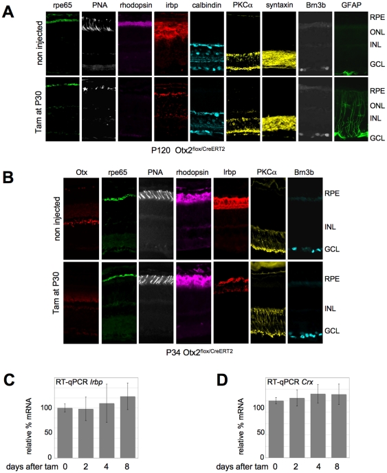
Background: Many developmental genes are still active in specific tissues after development is completed. This is the case for the homeobox gene Otx2, an essential actor of forebrain and head development. In adult mouse, Otx2 is strongly expressed in the retina. Mutations of this gene in humans have been linked to severe ocular malformation and retinal diseases. It is, therefore, important to explore its post-developmental functions. In the mature retina, Otx2 is expressed in three cell types: bipolar and photoreceptor cells that belong to the neural retina and retinal pigment epithelium (RPE), a neighbour structure that forms a tightly interdependent functional unit together with photoreceptor cells.
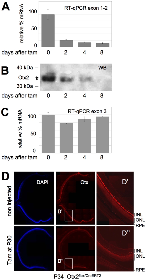
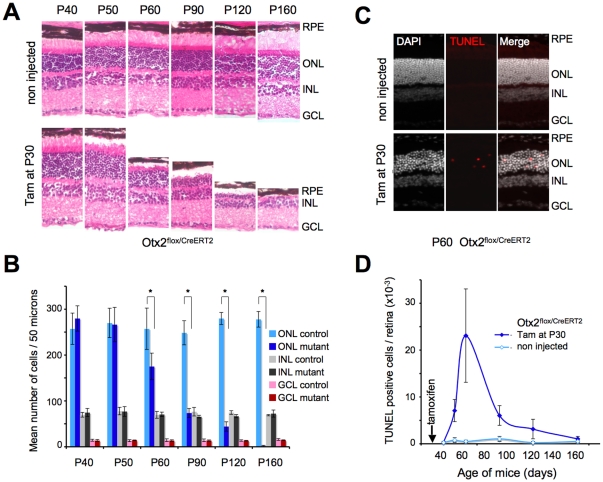
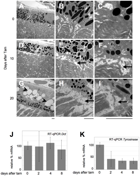
Methodology/principal findings: Conditional self-knockout was used to address the late functions of Otx2 gene in adult mice. This strategy is based on the combination of a knock-in CreERT2 allele and a floxed allele at the Otx2 locus. Time-controlled injection of tamoxifen activates the recombinase only in Otx2 expressing cells, resulting in selective ablation of the gene in its entire domain of expression. In the adult retina, loss of Otx2 protein causes slow degeneration of photoreceptor cells. By contrast, dramatic changes of RPE activity rapidly occur, which may represent a primary cause of photoreceptor disease.
Conclusions: Our novel mouse model uncovers new Otx2 functions in adult retina. We show that this transcription factor is necessary for long-term maintenance of photoreceptors, likely through the control of specific activities of the RPE.
Conflict of interest statement
Figures
Similar articles
-
Loss of Otx2 in the adult retina disrupts retinal pigment epithelium function, causing photoreceptor degeneration.J Neurosci. 2013 Jun 12;33(24):9890-904. doi: 10.1523/JNEUROSCI.1099-13.2013. J Neurosci. 2013. PMID: 23761884 Free PMC article.
-
Otx2 ChIP-seq reveals unique and redundant functions in the mature mouse retina.PLoS One. 2014 Feb 18;9(2):e89110. doi: 10.1371/journal.pone.0089110. eCollection 2014. PLoS One. 2014. PMID: 24558479 Free PMC article.
-
Photoreceptor cKO of OTX2 Enhances OTX2 Intercellular Transfer in the Retina and Causes Photophobia.eNeuro. 2021 Oct 6;8(5):ENEURO.0229-21.2021. doi: 10.1523/ENEURO.0229-21.2021. Print 2021 Sep-Oct. eNeuro. 2021. PMID: 34475267 Free PMC article.
-
The homeobox gene Otx2 in development and disease.Exp Eye Res. 2013 Jun;111:9-16. doi: 10.1016/j.exer.2013.03.007. Epub 2013 Mar 21. Exp Eye Res. 2013. PMID: 23523800 Review.
-
The many different cellular functions of MYO7A in the retina.Biochem Soc Trans. 2011 Oct;39(5):1207-10. doi: 10.1042/BST0391207. Biochem Soc Trans. 2011. PMID: 21936790 Free PMC article. Review.
Cited by
-
Retinal pigment epithelium development, plasticity, and tissue homeostasis.Exp Eye Res. 2014 Jun;123:141-50. doi: 10.1016/j.exer.2013.09.003. Epub 2013 Sep 21. Exp Eye Res. 2014. PMID: 24060344 Free PMC article. Review.
-
Maintenance of postmitotic neuronal cell identity.Nat Neurosci. 2014 Jul;17(7):899-907. doi: 10.1038/nn.3731. Epub 2014 Jun 15. Nat Neurosci. 2014. PMID: 24929660 Free PMC article. Review.
-
Deletion of OTX2 in neural ectoderm delays anterior pituitary development.Hum Mol Genet. 2015 Feb 15;24(4):939-53. doi: 10.1093/hmg/ddu506. Epub 2014 Oct 14. Hum Mol Genet. 2015. PMID: 25315894 Free PMC article.
-
Features of Retinal Neurogenesis as a Key Factor of Age-Related Neurodegeneration: Myth or Reality?Int J Mol Sci. 2021 Jul 9;22(14):7373. doi: 10.3390/ijms22147373. Int J Mol Sci. 2021. PMID: 34298993 Free PMC article. Review.
-
OTX Genes in Adult Tissues.Int J Mol Sci. 2023 Nov 30;24(23):16962. doi: 10.3390/ijms242316962. Int J Mol Sci. 2023. PMID: 38069286 Free PMC article. Review.
References
-
- Bok D. Contributions of genetics to our understanding of inherited monogenic retinal diseases and age-related macular degeneration. Arch Ophthalmol. 2007;125:160–164. - PubMed
-
- Lavado A, Montoliu L. New animal models to study the role of tyrosinase in normal retinal development. Front Biosci. 2006;11:743–752. - PubMed
-
- Acampora D, Gulisano M, Broccoli V, Simeone A. Otx genes in brain morphogenesis. Prog Neurobiol. 2001;64:69–95. - PubMed
-
- Puelles E, Acampora D, Lacroix E, Signore M, Annino A, et al. Otx dose-dependent integrated control of antero-posterior and dorso-ventral patterning of midbrain. Nat Neurosci. 2003;6:453–460. - PubMed
-
- Puelles E, Annino A, Tuorto F, Usiello A, Acampora D, et al. Otx2 regulates the extent, identity and fate of neuronal progenitor domains in the ventral midbrain. Development. 2004;131:2037–2048. - PubMed
Publication types
MeSH terms
Substances
LinkOut - more resources
Full Text Sources
Other Literature Sources
Molecular Biology Databases
Research Materials
