. 2010 Jul 15;6(7):e1000998.
doi: 10.1371/journal.ppat.1000998.

Extreme CD8 T cell requirements for anti-malarial liver-stage immunity following immunization with radiation attenuated sporozoites

Affiliations

Extreme CD8 T cell requirements for anti-malarial liver-stage immunity following immunization with radiation attenuated sporozoites

Nathan W Schmidt et al. PLoS Pathog. .

Abstract

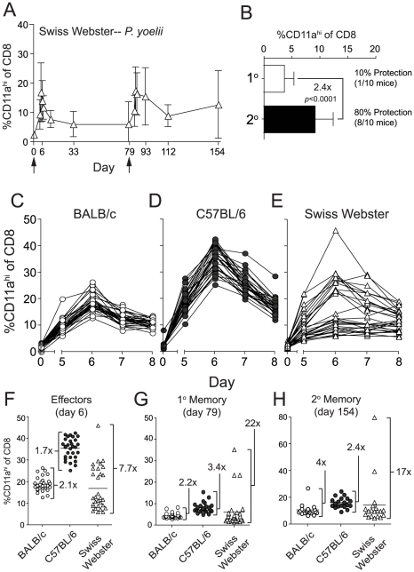
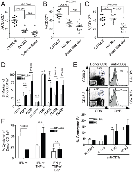
Radiation-attenuated Plasmodium sporozoites (RAS) are the only vaccine shown to induce sterilizing protection against malaria in both humans and rodents. Importantly, these "whole-parasite" vaccines are currently under evaluation in human clinical trials. Studies with inbred mice reveal that RAS-induced CD8 T cells targeting liver-stage parasites are critical for protection. However, the paucity of defined T cell epitopes for these parasites has precluded precise understanding of the specific characteristics of RAS-induced protective CD8 T cell responses. Thus, it is not known whether quantitative or qualitative differences in RAS-induced CD8 T cell responses underlie the relative resistance or susceptibility of immune inbred mice to sporozoite challenge. Moreover, whether extraordinarily large CD8 T cell responses are generated and required for protection following RAS immunization, as has been described for CD8 T cell responses following single-antigen subunit vaccination, remains unknown. Here, we used surrogate T cell activation markers to identify and track whole-parasite, RAS-vaccine-induced effector and memory CD8 T cell responses. Our data show that the differential susceptibility of RAS-immune inbred mouse strains to Plasmodium berghei or P. yoelii sporozoite challenge does not result from host- or parasite-specific decreases in the CD8 T cell response. Moreover, the surrogate activation marker approach allowed us for the first time to evaluate CD8 T cell responses and protective immunity following RAS-immunization in outbred hosts. Importantly, we show that compared to a protective subunit vaccine that elicits a CD8 T cell response to a single epitope, diversifying the targeted antigens through whole-parasite RAS immunization only minimally, if at all, reduced the numerical requirements for memory CD8 T cell-mediated protection. Thus, our studies reveal that extremely high frequencies of RAS-induced memory CD8 T cells are required, but may not suffice, for sterilizing anti-Plasmodial immunity. These data provide new insights into protective CD8 T cell responses elicited by RAS-immunization in genetically diverse hosts, information with relevance to developing attenuated whole-parasite vaccines.


Conflict of interest statement

The authors have declared that no competing interests exist.

Figures

Figure 1
Figure 1. Sensitivity and specificity of the CD8αloCD11ahi surrogate activation marker approach to identify RAS vaccination-induced CD8 T cell responses.
(A) Protection against P. berghei (Pb) or P. yoelii (Py) sporozoite challenge in BALB/c and B6 mice singly vaccinated with either 2×10⁴ Pb- or Py-RAS and challenged with 1000 Pb or Py sporozoites >80 days later. Numbers indicate % protected (no. protected/no. challenged×100). (B) Peripheral blood mononuclear cells from a single BALB/c mouse collected before (day 0) and after 2×10⁴ Pb-RAS vaccination (day 7, 61) were stained for CD8α and CD11a. Left column of dot plots shows the fraction of circulating CD8 T cells exhibiting an antigen-experienced phenotype (CD8αloCD11ahi) at each time point following Pb-RAS vaccination. Right columns of dot plots show the fraction of cells within the CD11ahi and CD11alo gates that stain with Kd/CS252–260 tetramer. (C) BALB/c mice were vaccinated with the indicated number of Pb-RAS and CD8αloCD11ahi responses were tracked in the peripheral blood of individual mice on day 0, 5, 6 and 7. N = 10 mice/dose, except for 1×10⁵ Pb-RAS group (N = 5). Data are the mean ± S.D. (D) Irradiated salivary gland homogenate from non-infected mosquitoes was injected into naïve BALB/c mice i.v. The CD8αloCD11ahi response was evaluated in the peripheral blood of individual mice before (day 0) and after (day 6) injection. ‘Equivalent’ refers to the final dilution of salivary gland homogenate injected into mice within each group. Dilutions were made based on an average recovery of ∼15,000 sporozoites per mosquito, calculated over >15 independent mosquito dissections. Data are mean ± S.D. for 3 mice per group. (E,F) similar to C,D except C57BL/6 mice were analyzed.
Figure 2
Figure 2. RAS vaccination of BALB/c and C57BL/6 mice increases the frequency and total number of CD8αloCD11ahi cells in spleen, liver and peripheral blood.
Naïve BALB/c and C57BL/6 mice were vaccinated with 2×10⁴ Py-RAS. Seven days later, mononuclear cells were isolated from the spleen, liver and blood of immune (N = 3/group) and naïve (N = 3) mice and stained for CD8α and CD11a. Data (mean ± S.D.) are expressed as the frequency (A) or total number (B) of CD8 T cells exhibiting the antigen-experienced phenotype (CD8αloCD11ahi) in each tissue. Statistics were determined by unpaired, two-tailed t-tests.
Figure 3
Figure 3. The magnitude and kinetics of RAS-vaccination-induced CD8 T cell responses in BALB/c and C57BL/6 inbred mice.
(A and B) Kinetics and magnitude of the total CD8 T cell response in the blood of BALB/c and C57BL/6 mice following 2×10⁴ Pb-RAS (A) or Py-RAS (B) vaccination. Data (mean±S.D.) are from 30 mice/strain. *P<0.0001, **P<0.0001. (C) BALB/c mice (N = 20) were vaccinated with 2×10⁴ Pb-RAS. Ninety-six days later, the frequency of CD8αloCD11ahi T cells in the peripheral blood was evaluated and individual mice were ranked according to the magnitude of the memory CD8 T cell response. Mice were treated with rat IgG, anti-CD4 (GK1.5) or anti-CD8 (2.43) 3 days and 1 day prior to challenge with 1000 Pb sporozoites. Numbers above refer to percent of T cell-depleted mice protected (no. protected/no. challenged ×100) following sporozoite challenge. (D) BALB/c mice were vaccinated with 2×10⁴ Pb-RAS. On days 5 and 6 post-vaccination, mice were injected i.p. with PBS or 60 mg/kg primaquine/PBS solution (arrows). Circulating CD8 T cell responses were evaluated in mice at the indicated time points. (E) Fifty-six days following Pb-RAS vaccination, CD8 T cell responses in the blood were evaluated. Mice were challenged 3 days later (day 59) with 1000 Pb sporozoites. Numbers refer to % protected (no. protected/no. challenged×100). Data in D,E are mean±S.D. from 8–10 mice/group. Statistics in A, B and E were determined by unpaired, two-tailed t-tests.
Figure 4
Figure 4. Homologous Pb- or Py-RAS boosting of mice elicits robust memory CD8 T cell responses but does not enhance protection of C57BL/6 mice against Py sporozoite challenge.
(A) The kinetics and magnitude of the circulating RAS vaccine-induced CD8 T cell response in C57BL/6 mice primed and boosted (arrows) with 2×10⁴ Pb-RAS. (B) Fold increase in secondary memory (d168) CD8 T cell responses in the blood of C57BL/6 mice following homologous Pb-RAS boost. Numbers to the right indicate the fraction of primary (1°) or secondary (2°) memory C57BL/6 mice that were protected following Pb sporozoite challenge (no. protected/no. challenged ×100). (C,D) Similar to A,B, except BALB/c mice were prime-boosted (arrows) with 2×10⁴ Py-RAS and challenged with P. yoelii sportozoites. Data in A–D are mean±S.D. from 20 mice. (E,F) Similar to C,D, except C57BL/6 mice were primed and boosted twice (arrows) with 2×10⁴ Py-RAS. Data in E,F are mean±S.D. from 30 mice. Statistics were determined by unpaired, two-tailed t-tests. For B,D and F, one hundred percent (10/10) of strain- and age-matched, naïve mice challenged in parallel were parasitized.
Figure 5
Figure 5. Py-RAS-specific CD8 T cell responses and protective immunity are markedly enhanced following prime-boost vaccination of an outbred mouse population.
(A) The kinetics and magnitude of CD8 T cell responses in the blood of individual Swiss Webster mice following priming and boosting (arrows) with 2×10⁴ Py-RAS. (B) Fold expansion of Py-RAS-induced primary (1°, d79) and secondary (2°, d154) memory CD8 T cell responses in the blood of Swiss Webster mice. Numbers to the right indicate the fraction of 1° or 2° memory Swiss Webster mice that were protected following Py sporozoite challenge (no. protected/no. challenged×100). One hundred percent (10/10) of age-matched, naïve Swiss Webster mice challenged in parallel were parasitized. Data in B are mean±S.D. from 20 mice. Statistics were determined by unpaired, two-tailed t-tests. BALB/c (C), C57BL/6 (D) and Swiss Webster mice (E) (N = 30 each) were vaccinated with 2×10⁴ Py-RAS. Peripheral blood was collected before (day 0) and on days 5–8 following vaccination. CD8αloCD11ahi T cell responses were measured in individual BALB/c, C57BL/6 and Swiss Webster mice at the initial effector stage (F), primary memory (G) or secondary memory (H) following priming and homologous boost with 2×10⁴ Py-RAS as in A. Each symbol represents an individual mouse. Numbers in F-H refer to the fold difference between the highest and lowest responders within each mouse strain at each time point.
Figure 6
Figure 6. Protection correlates with an effector memory (TEM) phenotype on circulating Py-RAS-induced secondary memory CD8 T cells in inbred and outbred mice.
(A–C) C57BL/6, BALB/c and Swiss Webster mice (N = 10/strain) were vaccinated with 2×10⁴ Py-RAS. Seventy-nine days later, mice received a homologous boost of 2×10⁴ Py-RAS. Seventy-five days after boost (day 154), circulating T cells were stained for CD8α, CD11a, CD62L, CD27 and CD127. (D–F) Py-RAS-specific, secondary memory BALB/c and C57BL/6 CD8 T cells were generated following adoptive transfer as described in Materials and Methods. (D) Donor-derived cells were analyzed directly ex vivo for surface expression of the indicated activation- or survival-associated marker. (E) Cells were stimulated ex vivo for 5 hrs using titrations of plate-bound anti-CD3ε and subsequently analyzed for the intracellular expression of granzyme B. Representative dot plots and histograms (top) and summary graph (bottom) are shown. (F) Cells were stimulated with 1µg/mL plate-bound anti-CD3ε for 5.5 hrs prior to staining for intracellular co-expression of IFN-γ, TNF-α and IL-2. In A–D, data represent the fraction of CD8αloCD11ahi T cells within each group expressing the indicated marker. Statistics were determined by unpaired, two-tailed t-tests. Data in D–F are mean±S.D. and represent analyses from 5 individual BALB/c and B6 recipient mice.
Figure 7
Figure 7. Sterilizing anti-P. berghei sporozoite immunity in BALB/c mice is associated with memory CD8 T cell responses of single antigenic-specificity that exceed 8% of all circulating CD8 T cells.
BALB/c mice were DC-CS252–260 prime, LM-CS252–260 boost immunized as previously described . Eighty-six days following immunization, peripheral blood from individual mice was assayed for the frequency of CS252–260-specific memory CD8 T cells using intracellular IFN-γ cytokine staining. Mice are ranked according to the magnitude of the CS252–260-specific memory CD8 T cell response. Three days after T cell analyses, mice were challenged with 1000 Pb sporozoites and blood stage parasitemia was evaluated in individual mice with Giemsa stain. Non-protected mice are indicated with filled circles. Numbers above the graph refer to percent protection among mice scoring above or below the 8% circulating CS252–260-specific memory CD8 T cell threshold, which is based on the clearest visual break-point between non-protected and protected mice each ranked according to the magnitude of the CD8 T cell response. Ninety-one percent (20/22) of age-matched, naïve BALB/c mice challenged in parallel were parasitized.

Similar articles

Cited by

References

    1. Bryce J, Boschi-Pinto C, Shibuya K, Black RE. WHO estimates of the causes of death in children. Lancet. 2005;365:1147–1152. - PubMed
    1. Hay SI, Snow RW. The Malaria Atlas Project: Developing Global Maps of Malaria Risk. PLoS Med. 2006;3:e473. - PMC - PubMed
    1. Sachs J, Malaney P. The economic and social burden of malaria. Nature. 2002;415:680–685. - PubMed
    1. Snow RW, Guerra CA, Noor AM, Myint HY, Hay SI. The global distribution of clinical episodes of Plasmodium falciparum malaria. Nature. 2005;434:214–217. - PMC - PubMed
    1. Guerra CA, Snow RW, Hay SI. Mapping the global extent of malaria in 2005. Trends Parasitol. 2006;22:353–358. - PMC - PubMed

Publication types