. 2010 Jul 19;5(7):e11654.
doi: 10.1371/journal.pone.0011654.

Importance of non-selective cation channel TRPV4 interaction with cytoskeleton and their reciprocal regulations in cultured cells

Affiliations

Importance of non-selective cation channel TRPV4 interaction with cytoskeleton and their reciprocal regulations in cultured cells

Chandan Goswami et al. PLoS One. .

Abstract

Background: TRPV4 and the cellular cytoskeleton have each been reported to influence cellular mechanosensitive processes as well as the development of mechanical hyperalgesia. If and how TRPV4 interacts with the microtubule and actin cytoskeleton at a molecular and functional level is not known.

Methodology and principal findings: We investigated the interaction of TRPV4 with cytoskeletal components biochemically, cell biologically by observing morphological changes of DRG-neurons and DRG-neuron-derived F-11 cells, as well as functionally with calcium imaging. We find that TRPV4 physically interacts with tubulin, actin and neurofilament proteins as well as the nociceptive molecules PKCepsilon and CamKII. The C-terminus of TRPV4 is sufficient for the direct interaction with tubulin and actin, both with their soluble and their polymeric forms. Actin and tubulin compete for binding. The interaction with TRPV4 stabilizes microtubules even under depolymerizing conditions in vitro. Accordingly, in cellular systems TRPV4 colocalizes with actin and microtubules enriched structures at submembranous regions. Both expression and activation of TRPV4 induces striking morphological changes affecting lamellipodial, filopodial, growth cone, and neurite structures in non-neuronal cells, in DRG-neuron derived F11 cells, and also in IB4-positive DRG neurons. The functional interaction of TRPV4 and the cytoskeleton is mutual as Taxol, a microtubule stabilizer, reduces the Ca2+-influx via TRPV4.

Conclusions and significance: TRPV4 acts as a regulator for both, the microtubule and the actin. In turn, we describe that microtubule dynamics are an important regulator of TRPV4 activity. TRPV4 forms a supra-molecular complex containing cytoskeletal proteins and regulatory kinases. Thereby it can integrate signaling of various intracellular second messengers and signaling cascades, as well as cytoskeletal dynamics. This study points out the existence of cross-talks between non-selective cation channels and cytoskeleton at multiple levels. These cross talks may help us to understand the molecular basis of the Taxol-induced neuropathic pain development commonly observed in cancer patients.


Conflict of interest statement

Competing Interests: The authors have declared that no competing interests exist.

Figures

Figure 1
Figure 1. Interaction of soluble tubulin and actin with TRPV4.
a. Co-immunoprecipitation of actin and tubulin with TRPV4. Cell extracts from CHO-KI cells stably expressing TRPV4 (lane 1) was immunoprecipitated by TRPV4 antibody (lane 2) or by a non-specific antibody (lane 3). Blots were probed for TRPV4 (left side), tubulin (middle) and actin (right side). b. Co-immunoprecipitation of tubulin with TRPV4. Extracts from DRG (lane 1) was immunoprecipitated by TRPV4 antibody (lane 2) or by a non-specific antibody (lane 3). Blots were probed for TRPV4 (upper panel) and tubulin (lower side). c. MBP-TRPV4-Ct (lane 2-3) but not MBP-LacZ (lane 4–5) forms specific complexes when incubated with mammalian brain extract (lane 1), both in presence (lane 2 and 4) or absence (lane 3 and 5) of Ca2+ (1 mM). Presence of PKCε, actin and tubulin are observed only in lane 2 and 3. Neurofilament in the pull down samples is visible only after exposing for a prolonged time. Presence of CamKII is noted only in the presence of Ca2+ (lane 2). Note that the amount of MBP-LacZ used, as a negative control for the pull down experiment is much more than MBP-TRPV4-Ct. d. Tubulin interacts with TRPV4-Ct directly. MBP-LacZ (lane 1–2) or MBP-TRPV4-Ct (lane 3–4) was incubated with buffer only (lane 1 and 3) or with purified tubulin (lane 2 and 4). Pulled down samples were probed for different isotype-specific and different post-translationally modified tubulins. e. Actin interacts directly with TRPV4-Ct. MBP-TRPV4-Ct (lane 1–2) or MBP-LacZ (lane 3–4) was incubated with purified actin (lane 1–4) either in the presence (lane 1-and 3) or absence (lane 2 and 4) of Ca2+ (1 mM) and subsequently probed for bound actin. f. Soluble tubulin and actin competes for the C-terminal cytoplasmic fragment of TRPV4. MBP-TRPV4-Ct was incubated with only tubulin (lane 1), with only actin (lane 2), or both tubulin and actin in a sequential manner (lane 3–4). Prior incubation of tubulin inhibits further binding of actin (lane 3). Similarly, prior incubation of actin significantly reduces the further binding of tubulin (lane 4).
Figure 2
Figure 2. TRPV4 interacts directly with polymerized actin and microtubule filaments.
MBP and MBP-TRPV4-Ct were centrifuged at 70000 g/30 min/4°C and only soluble proteins present in the supernatant were used for all co-sedimentation experiments. a. MBP-TRPV4-Ct co-sediments with polymerized actin filaments. Actin was polymerized either in presence of MBP-TRPV4-Ct (lane 1 and 4), in presence of MBP only (lane 2 and 5) or in buffer only (lane 3 and 6). Polymerized actin filaments and associated proteins were isolated from remaining soluble actin and unbound proteins by centrifugal separation of pellets (P, lane 1–3) from corresponding supernatants (S, lane 4–6). The entire amount of MBP remains in the supernatant (lane 5) while a significant amount of MBP-TRPV4-Ct appears in the pellet (lane 1). Arrows indicate the position of respective proteins. b. MBP-TRPV4-Ct co-sediments with microtubules. Taxol-stabilized microtubules (left panel) were incubated with MBP (lane 1–2), MBP-TRPV4-Ct (lane 3–4) or with buffer only (lane 5–6) followed by the centrifugal separation of pellet (P) consisting MT and bound proteins from supernatant (S) consisting of soluble tubulin and other unbound proteins (left side panel). In right side panel, soluble tubulin and GTP was incubated with MBP (lane 1–2), MBP-TRPV4-Ct (lane 3–4) or buffer only (lane 5–6) followed by separation of pellet (P) and supernatant (S). Note the specific presence of MBP-TRPV4-Ct in the pellet in both cases (in lane 4). c. MBP-TRPV4-Ct stabilizes microtubules against depolymerizing factors. Microtubules was formed form soluble tubulin in buffer (lane 1), along with MBP (lane 2) or along with MBP-TRPV4-Ct (lane 3) in control condition (left most panel), in presence of Nocodazole (middle left panel), in presence of Ca2+ (middle right side) or in presence of both Nocodazole and Ca2+ (right most). Microtubules and bound proteins present in the pellet fraction (P) were isolated from unpolymerized tubulin and unbound proteins remaining in the supernatant (S) by centrifugal separation. Note the enhancement of polymerized microtubules (represented by tubulin present in lane 3, P fraction in every conditions) due to the presence of MBP-TRPV4-Ct.
Figure 3
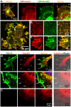
Figure 3. TRPV4 co-localizes with actin and microtubule cytoskeleton.
a–c. Shown are the live-cell confocal images of F11 cells expressing TRV4-GFP (green) and RFP-actin (red). Presences of TRPV4-GFP specifically in actin-enriched structures are shown. a. Enlarged view of lamellipodia and at the tip of the actin filaments are shown. b–c. Enlarged view of focal adhesion point-like structures (b) and cell cortex with actin ribs (c) are shown. Arrows indicate the localization of TRPV4-GFP at filopodial tips. d–f. TRPV4 co-localizes with microtubule cytoskeleton. Shown are the confocal images of F11 cells immunostained for TRPV4 (green) and tyrosinated tubulin (red). Arrows indicate presence and acumulation of microtubules in thin filopodial structures (d, upper panel) and thin lamellipodial structures (e, middle panel). The status of the microtubules in the non-transfected cells are shown in below (f, lower panel).
Figure 4
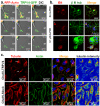
Figure 4. TRPV4 localizes in the growth cone and regulates axonal motility as activation of TRPV4 results in microtubule disassembly.
a. Shown are the confocal time series images of live F11 cell expressing TRPV4-GFP (green) and RFP-Actin (red). Fluorescence images were superimposed on the DIC images. Addition of 4αPDD (1 µM) results in growth cone retraction of the transfected cell (T) but not from the non-transfected (NT) cells. b. Prolonged activation of endogenous TRPV4 reduces neurite outgrowth. Shown are the images of cultured DRG neurons stained for IB4 (red) and βIII tubulin (green). IB4-positive neurons extend their neurites in control condition (upper panel). Majority of the IB4-positive neurons do not produce any neurite when 4αPDD at low dose (0.1 µM) was applied for 36 hours (middle panel). An enlarged view of an IB4-positive and an IB4-negative neuron is shown in the lower panel. Note that the majority of the IB4-negative neurons remain unaffected even in the presence of 4αPDD. c. CHO-KI-TRPV4 cells that express low level of TRPV4 or CHO-KI-Mock cells that do not express TRPV4 were activated with 4αPDD (1 µM). After activation, cells were extracted by detergent in isotonic buffer and fixed subsequently by PFA. Cells were immunostained for actin (green) and tubulin (red). CHO-KI-TRPV4 cells loose all the peripheral microtubules but retain filamentous actin after activation and extraction. The stable MTOC regions are marked with arrows. In contrast, CHO-KI-Mock cells remain unaffected. Intensity of the microtubule is shown (red and blue indicate highest and lowest intensity respectively).
Figure 5
Figure 5. Activation of TRPV4 results in reorganization of actin cytoskeleton.
Shown are the confocal images of live F11 cells expressing TRPV4-GFP (green) and Actin-RFP (red). a. Activation of TRPV4 results in merging of several actin-ribs at the tips and further transition of lamellipodial structures to filopodial structures. The arrow indicates the region and direction of the cell retraction at the same time. b. Activation of TRPV4 results in lateral initiation of filopodial structures from neurites and further elongation of them.
Figure 6
Figure 6. Microtubule cytoskeleton regulates Ca2+-influx via TRPV4.
a. Taxol (1 µM, 30 minutes) reduces TRPV4-mediated Ca2+-influx in Cos7 cells. Shown are the normalized average ratiometric Ca2+-influx in arbitrary units (AU). TRPV4 was activated by a pulse (indicated by a black line) of 4αPDD for 10 sec. The dark blue line (C1) represents average response due to first pulse in control conditions (n = 32), the light blue line (C2) represents average response due to second pulse in control conditions (n = 32), the blackish green line (T1) represents average response due to first pulse under the influence of Taxol-stabilized microtubules (n = 42) and the light green line (T2) represents the average response due to second pulse under the Taxol-stabilized microtubules (n = 30). b. Cells with Taxol-stabilized microtubules reveal reductions in total Ca2+-influx due to TRPV4 activation. Total Ca2+-influx was calculated from the total area appeared by the ratiometric calcium-influx graph (as shown in figure a) for each cell and was calculated by Origin 7G software. At the <0.05 level, the difference of the population means between 2nd pulse (untreated and taxol-treated, one sample t-test) is significant (*). The difference in case of 1st pulse is non-significant. c. Taxol stabilized cells reveal a trend for time delay to reach in maximum response. The time (in seconds) each cell took to reach its maximum response (indicated by filled triangles) was plotted. Times needed during first pulse in control conditions (dark blue), second pulse in control conditions (light blue), first pulse with Taxol (blackish green) and second pulse with Taxol (light green) are indicated. The average values (indicated by filled squares) and the standard deviations are also shown for each condition. The average time differences remain non-significant.

Similar articles

Cited by

References

    1. Gao X, Wu L, O'Neil RG. Temperature-modulated diversity of TRPV4 channel gating: activation by physical stresses and phorbol ester derivatives through protein kinase C-dependent and -independent pathways. J Biol Chem. 2003;278:27129–37. - PubMed
    1. Mizuno A, Matsumoto N, Imai M, Suzuki M. Impaired osmotic sensation in mice lacking TRPV4. Am J Physiol Cell Physiol. 2003;285:C96–101. - PubMed
    1. Alessandri-Haber N, Yeh JJ, Boyd AE, Parada CA, Chen X, et al. Hypotonicity induces TRPV4-mediated nociception in rat. Neuron. 2003;39:497–511. - PubMed
    1. Strotmann R, Harteneck C, Nunnenmacher K, Schultz G, Plant TD. OTRPC4, a nonselective cation channel that confers sensitivity to extracellular osmolarity. Nat Cell Biol. 2000;2:695–702. - PubMed
    1. Liedtke W, Tobin DM, Bargmann CI, Friedman JM. Mammalian TRPV4 (VR-OAC) directs behavioural responses to osmotic and mechanical stimuli in Caenorhabditis elegans. Proc Natl Acad Sci U S A. 2003;100:14531–14536. - PMC - PubMed

Publication types

LinkOut - more resources