

Nephrocalcinosis in animal models with and without stones
- PMID: 20658131
- PMCID: PMC2992101
- DOI: 10.1007/s00240-010-0303-4
Nephrocalcinosis in animal models with and without stones
Abstract
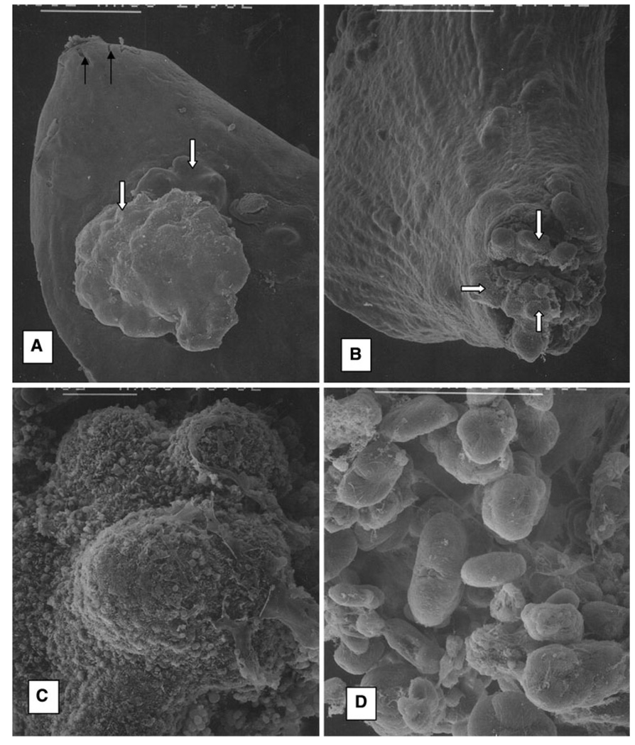
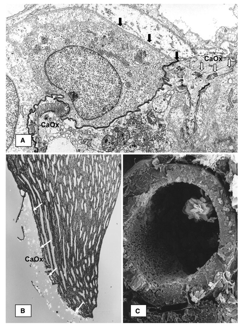
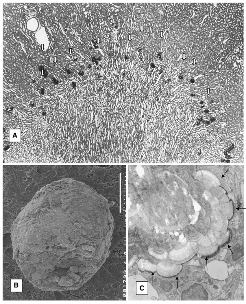
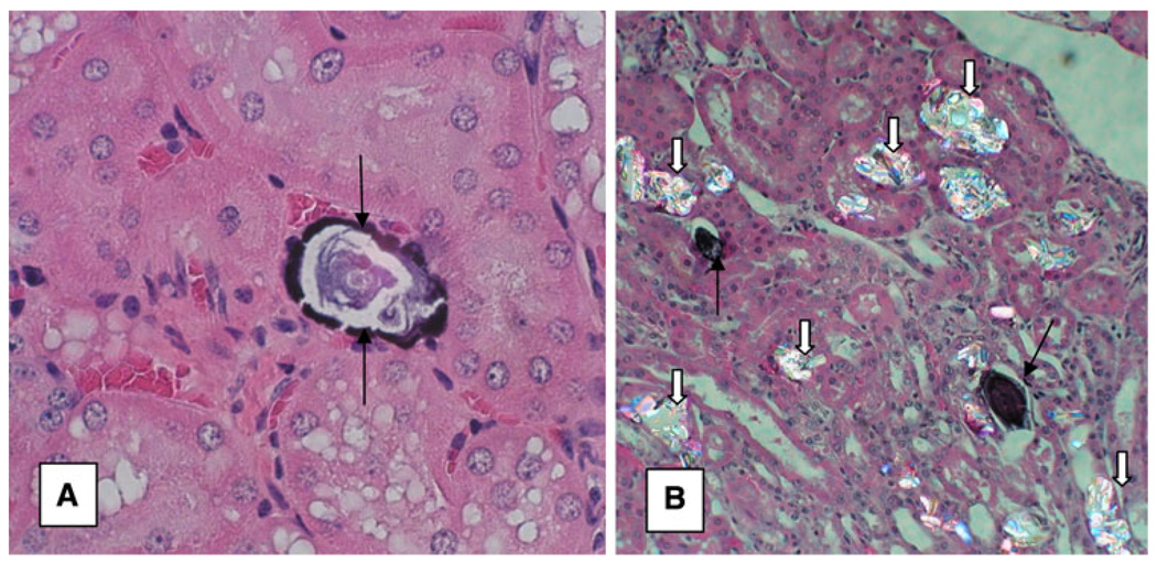
Nephrocalcinosis is the deposition of calcium salts in renal parenchyma and can be intratubular or interstitial. Animal model studies indicate that intratubular nephrocalcinosis is a result of increased urinary supersaturation. Urinary supersaturation with respect to calcium oxalate (CaOx) and calcium phosphate (CaP) are generally achieved at different locations in the renal tubules. As a result experimental induction of hyperoxaluria in animals with CaP deposits does not lead to growth of CaOx over CaP. Interstitial nephrocalcinosis has been seen in mice with lack of crystallization modulators Tamm-Horsfall protein and osteopontin. Sodium phosphate co-transporter or sodiumhydrogen exchanger regulator factor-1 null mice also produced interstitial nephrocalcinosis. Crystals plug the tubules by aggregating and attaching to the luminal cell surface. Structural features of the renal tubules also play a role in crystal retention. The crystals plugging the terminal collecting ducts when exposed to the metastable pelvic urine may promote the formation of stone.
Figures
Similar articles
-
Calcium oxalate crystal deposition in kidneys of hypercalciuric mice with disrupted type IIa sodium-phosphate cotransporter.Am J Physiol Renal Physiol. 2008 May;294(5):F1109-15. doi: 10.1152/ajprenal.00620.2007. Epub 2008 Mar 12. Am J Physiol Renal Physiol. 2008. PMID: 18337544 Free PMC article.
-
Renal calcinosis and stone formation in mice lacking osteopontin, Tamm-Horsfall protein, or both.Am J Physiol Renal Physiol. 2007 Dec;293(6):F1935-43. doi: 10.1152/ajprenal.00383.2007. Epub 2007 Sep 26. Am J Physiol Renal Physiol. 2007. PMID: 17898038
-
Potential role of fluctuations in the composition of renal tubular fluid through the nephron in the initiation of Randall's plugs and calcium oxalate crystalluria in a computer model of renal function.Urolithiasis. 2015 Jan;43 Suppl 1:93-107. doi: 10.1007/s00240-014-0737-1. Epub 2014 Nov 20. Urolithiasis. 2015. PMID: 25407799
-
A hypothesis of calcium stone formation: an interpretation of stone research during the past decades.Urol Res. 2011 Aug;39(4):231-43. doi: 10.1007/s00240-010-0349-3. Epub 2011 Jan 19. Urol Res. 2011. PMID: 21246193 Review.
-
Nephrolithiasis: a consequence of renal epithelial cell exposure to oxalate and calcium oxalate crystals.Mol Urol. 2000 Winter;4(4):305-12. Mol Urol. 2000. PMID: 11156696 Review.
Cited by
-
Genetic modulation of nephrocalcinosis in mouse models of ectopic mineralization: the Abcc6(tm1Jfk) and Enpp1(asj) mutant mice.Lab Invest. 2014 Jun;94(6):623-32. doi: 10.1038/labinvest.2014.52. Epub 2014 Apr 14. Lab Invest. 2014. PMID: 24732453 Free PMC article.
-
Nanocrystal-induced chronic tubular-nephropathy in tropical countries: diagnosis, mitigation, and eradication.Eur J Med Res. 2023 Jul 5;28(1):221. doi: 10.1186/s40001-023-01162-y. Eur J Med Res. 2023. PMID: 37408060 Free PMC article.
-
Kidney stone formation and antioxidant effects of Cynodon dactylon decoction in male Wistar rats.Avicenna J Phytomed. 2017 Mar-Apr;7(2):180-190. Avicenna J Phytomed. 2017. PMID: 28348973 Free PMC article.
-
Antiurolithic effects of medicinal plants: results of in vivo studies in rat models of calcium oxalate nephrolithiasis-a systematic review.Urolithiasis. 2021 Apr;49(2):95-122. doi: 10.1007/s00240-020-01236-0. Epub 2021 Jan 23. Urolithiasis. 2021. PMID: 33484322
-
Dianthi herba: a comprehensive review of its botany, traditional use, phytochemistry, and pharmacology.Chin Med. 2022 Jan 21;17(1):15. doi: 10.1186/s13020-022-00570-2. Chin Med. 2022. PMID: 35062995 Free PMC article. Review.
References
-
- Low RK, Stoller ML. Endoscopic mapping of renal papillae for Randall’s plaques in patients with urinary stone disease. J Urol. 1997;158:2062. - PubMed
-
- Low RK, Stoller ML, Schreiber CK. Metabolic and urinary risk factors associated with Randall’s papillary plaques. J Endourol. 2000;14:507. - PubMed
-
- Stoller ML, Low RK, Shami GS, et al. High resolution radiography of cadaveric kidneys: unraveling the mystery of Randall’s plaque formation. J Urol. 1996;156:1263. - PubMed
Publication types
MeSH terms
Substances
Grants and funding
LinkOut - more resources
Full Text Sources
Other Literature Sources
Research Materials
Miscellaneous
