

Glucosidase inhibition enhances presentation of de-N-glycosylated hepatitis B virus epitopes by major histocompatibility complex class I in vitro and in woodchucks
- PMID: 20658465
- PMCID: PMC2947625
- DOI: 10.1002/hep.23806
Glucosidase inhibition enhances presentation of de-N-glycosylated hepatitis B virus epitopes by major histocompatibility complex class I in vitro and in woodchucks
Abstract
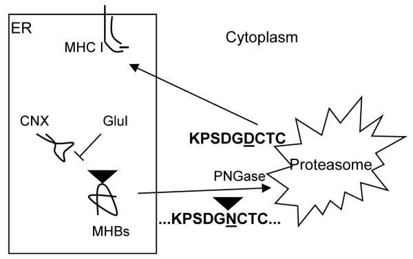
In this report, the possibility of pharmacologically altering the hepatitis B virus (HBV) epitopes presented by major histocompatibility complex class I on infected cells is demonstrated. The HBV middle envelope glycoprotein (MHBs) maturation appears to require calnexin-mediated folding. This interaction is dependent on glucosidases in the endoplasmic reticulum. Prevention of HBV envelope protein maturation in cultured cells through use of glucosidase inhibitors, such as 6-O-butanoyl castanospermine and N-nonyl deoxynorjirimycin, resulted in MHBs degradation by proteasomes. The de-N-glycosylation associated with polypeptide degradation was predicted to result in conversion of asparagine residues into aspartic acid residues. This prediction was confirmed by showing that peptides corresponding to the N-glycosylation sequons of MHBs, but with aspartic acid replacing asparagine, (1) can prime human cytotoxic T lymphocytes that recognize HBV-producing cells and (2) that the presentation of these envelope motifs by major histocompatibility complex class I is enhanced by incubation with glucosidase inhibitors. Moreover, although peripheral blood mononuclear cells isolated from woodchucks chronically infected with woodchuck hepatitis virus and vaccinated with woodchuck hepatitis virus surface antigen could be induced to recognize the natural MHBs asparagine-containing peptides, only cells isolated from animals treated with glucosidase inhibitor recognized the aspartic acid-containing peptides.
Conclusion: These data suggest that pharmacological intervention with glucosidase inhibitors can alter the MHBs epitopes presented. This editing of the amino acid sequence of the polypeptide results in a new epitope, or "editope", with possible medical significance.
Figures


Similar articles
-
Inhibition of cellular alpha-glucosidases results in increased presentation of hepatitis B virus glycoprotein-derived peptides by MHC class I.Virology. 2009 Feb 5;384(1):12-5. doi: 10.1016/j.virol.2008.11.027. Epub 2008 Dec 16. Virology. 2009. PMID: 19091367 Free PMC article.
-
Hepatitis B virus MHBs antigen is selectively sensitive to glucosidase-mediated processing in the endoplasmic reticulum.DNA Cell Biol. 2001 Oct;20(10):647-56. doi: 10.1089/104454901753340631. DNA Cell Biol. 2001. PMID: 11749723
-
Characterization of T-cell response to woodchuck hepatitis virus core protein and protection of woodchucks from infection by immunization with peptides containing a T-cell epitope.J Virol. 1997 Jan;71(1):65-74. doi: 10.1128/JVI.71.1.65-74.1997. J Virol. 1997. PMID: 8985324 Free PMC article.
-
Diverse Virus and Host-Dependent Mechanisms Influence the Systemic and Intrahepatic Immune Responses in the Woodchuck Model of Hepatitis B.Front Immunol. 2020 May 27;11:853. doi: 10.3389/fimmu.2020.00853. eCollection 2020. Front Immunol. 2020. PMID: 32536912 Free PMC article. Review.
-
T cell recognition of hepatitis B and C viral antigens.Eur J Clin Invest. 1994 Oct;24(10):641-50. doi: 10.1111/j.1365-2362.1994.tb01055.x. Eur J Clin Invest. 1994. PMID: 7531642 Review.
Cited by
-
Natural Products Database Screening for the Discovery of Naturally Occurring SARS-Cov-2 Spike Glycoprotein Blockers.ChemistrySelect. 2020 Nov 13;5(42):13309-13317. doi: 10.1002/slct.202003349. Epub 2020 Nov 16. ChemistrySelect. 2020. PMID: 33363254 Free PMC article.
-
Chronic hepatitis B: A wave of new therapies on the horizon.Antiviral Res. 2015 Sep;121:69-81. doi: 10.1016/j.antiviral.2015.06.014. Epub 2015 Jun 22. Antiviral Res. 2015. PMID: 26112647 Free PMC article. Review.
-
Characterization of Novel Hepatitis B Virus PreS/S-Gene Mutations in a Patient with Occult Hepatitis B Virus Infection.PLoS One. 2016 May 16;11(5):e0155654. doi: 10.1371/journal.pone.0155654. eCollection 2016. PLoS One. 2016. PMID: 27182775 Free PMC article.
-
Viral resistance of MOGS-CDG patients implies a broad-spectrum strategy against acute virus infections.Antivir Ther. 2015;20(3):257-9. doi: 10.3851/IMP2907. Epub 2014 Oct 15. Antivir Ther. 2015. PMID: 25318123 Free PMC article.
-
Present and future therapies of hepatitis B: From discovery to cure.Hepatology. 2015 Dec;62(6):1893-908. doi: 10.1002/hep.28025. Epub 2015 Oct 27. Hepatology. 2015. PMID: 26239691 Free PMC article. Review.
References
-
- Guidotti LG, Chisari FV. Immunobiology and pathogenesis of viral hepatitis. Annu. Rev. Pathol. Mech. Dis. 2006;1:23–61. - PubMed
-
- Rehermann B. Chronic infections with hepatotropic viruses: mechanisms of impairment of cellular immune responses. Sem. Liver Dis. 2007;27:152–160. - PubMed
-
- Bruss V. Envelopment of the hepatitis B virus nucleocapsid. Virus Res. 2004;106:199–209. - PubMed
Publication types
MeSH terms
Substances
Grants and funding
LinkOut - more resources
Full Text Sources