. 2011 Jan;226(1):132-40.
doi: 10.1002/jcp.22314.

Deficiency in core circadian protein Bmal1 is associated with a prothrombotic and vascular phenotype

Affiliations

Deficiency in core circadian protein Bmal1 is associated with a prothrombotic and vascular phenotype

Payaningal R Somanath et al. J Cell Physiol. 2011 Jan.

Abstract

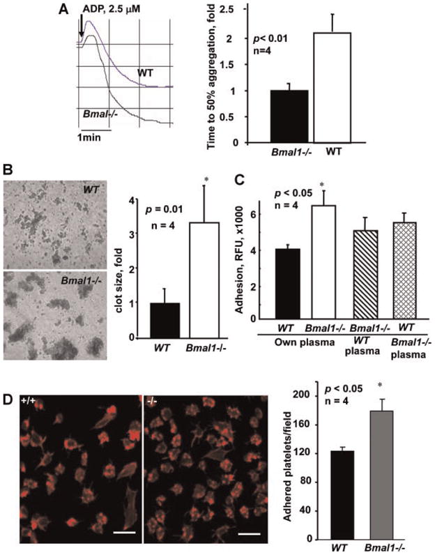
Aging is associated with both the disturbances of circadian rhythms and a prothrombotic phenotype. It remains poorly understood how the circadian system regulates thrombosis, a critical outcome of aging-related cardiovascular disease. Using multiple in vivo models, we now show that mice with genetic ablation of the core clock gene Bmal1, which display pre-mature aging, have a dramatic prothrombotic phenotype. This phenotype is mechanistically linked to changes in the regulation of key risk factors for cardiovascular disease. These include circulating vWF, fibrinogen, and PAI-1, all of which are significantly elevated in Bmal1(-/-) mice. We also show that major circadian transcriptional regulators CLOCK and Bmal1 directly regulate the activity of vWF promoter and that lack of Bmal1 results in upregulation of vWF both at mRNA and protein level. Here we report a direct regulation of vWF expression in endothelial cells by biological clock gene Bmal1. This study establishes a mechanistic connection between Bmal1 and cardiovascular phenotype.


Figures

Fig. 1
Fig. 1
Bmal1 regulates thrombosis in vivo. A–C: Mice of indicated genotypes were used in intravital thrombosis assay. Arterioles (50- to 80-μm diameter) and venules (60- to 80-μm diameter, part A) or carotid arteries (part B and C), were visualized, and in vivo thrombosis times were assessed as described in the Materials and Methods Section. Each point in parts A and B represents an individual animal. C: Progression of thrombus in carotid arteries is shown. Times after FeCl3-induced injury are indicated (min).
Fig. 2
Fig. 2
Bmal1 regulates hemostasis and wound healing in vivo. A: WT and Bmal1−/− null mice were anesthetized, then their tails were amputated at a position where the diameter of the tail was 2.5 mm and immersed in saline. The time from the amputation to cessation of bleeding was recorded. The difference between groups of mice was analyzed using nonparametric log-rank test. B,C: Dynamics of wound closure in WT and Bmal1−/− mice. Animals were anesthetized, backs shaved, and 15-mm incision wounds were made by excising the skin and panniculus carnosus. B: Representative images of wounds at day 7 after injury are shown. C: Quantification of wounds dimensions on days 3 and 7 after injury is presented as mean ± SE. [Color figure can be viewed in the online issue, which is available at wileyonlinelibrary.com.]
Fig. 3
Fig. 3
Platelets of Bmal1−/− mice exhibit enhanced aggregation and adhesion in the presence of plasma. A: Platelet aggregation in platelet rich plasma from WT and Bmal11−/− mice was induced by 2.5 μM ADP and optically monitored. Representative aggregation curves (left part) and bar graph of time to 50% aggregation is shown (mean ± SE, n = 4). B: Platelets from WT and Bmal1−/− mice in platelet rich plasma were activated as in (A) and thrombi were visualized using phase contrast microscopy. Clot size was analyzed by Image Pro software (mean ± SE, n = 4). C,D: WT and Bmal1−/− platelets were isolated, fluorescently labeled with calcein, reconstituted in autologuous or heterologuous plasma and used in adhesion assay on either rat tail collagen (C) or fibronectin (D). D: Adherent platelets were stained for F-actin (left part) and quantified (right part). [Color figure can be viewed in the online issue, which is available at wileyonlinelibrary.com.]
Fig. 4
Fig. 4
vWF level is increased in Bmal1−/− mice. A: Analysis of vWF multimer distribution in plasma of WT and Bmal1−/− mice. Representative gel (left part) and quantification of the band densities corresponding to low, medium, and large multimers (right) are shown. B: Analysis (B, left part) and quantification (B, right part) of vWF levels in plasma of WT and Bmal1−/− mice by Western blotting. C: Western Blot analysis of vWF in liver lysates of WT and Bmal1−/− mice. D: Immunohistochemical staining of vWF in peripheral vasculature of WT and Bmal1−/− mice in vivo. [Color figure can be viewed in the online issue, which is available at wileyonlinelibrary.com.]
Fig. 5
Fig. 5
Bmal1 regulates expression of vWF. A: Western blot analysis of the levels of vWF in plasma samples from WT and Bmal1−/− mice collected every 4 h during the 24-hr period. ZT—zeitgeber time, ZT0 corresponds to the lights-on time, ZT12—lights-off time. The graph shows densitometry analysis of the data. B: Expression of vWF mRNA in aortic tissue of WT and Bmal1−/−. C: Daily average levels of vWF mRNA in the liver of WT and Bmal1−/− mice. D: vWF promoter activity in HEK293 cells transfected with different combinations of CLOCK, Bmal1, and CRY1 expression plasmid. E: ChIP assay of vWF promoter and PAI-1 promoters in isolated murine lung endothelial cells demonstrating direct interaction of Bmal1 with the E-box sequence.

Similar articles

Cited by

References

    1. Anea CB, Zhang M, Stepp DW, Simkins GB, Reed G, Fulton DJ, Rudic RD. Vascular disease in mice with a dysfunctional circadian clock. Circulation. 2009;119:1510–1517. - PMC - PubMed
    1. Antoch MP, Gorbacheva VY, Vykhovanets O, Toshkov IA, Kondratov RV, Kondratova AA, Lee C, Nikitin AY. Disruption of the circadian clock due to the Clock mutation has discrete effects on aging and carcinogenesis. Cell Cycle (Georgetown, Tex) 2008;7:1197–1204. - PMC - PubMed
    1. Bunger MK, Walisser JA, Sullivan R, Manley PA, Moran SM, Kalscheur VL, Colman RJ, Bradfield CA. Progressive arthropathy in mice with a targeted disruption of the Mop3/Bmal-1 locus. Genesis. 2005;41:122–132. - PubMed
    1. Chen J, De S, Damron DS, Chen WS, Hay N, Byzova TV. Impaired platelet responses to thrombin and collagen in AKT-1-deficient mice. Blood. 2004;104:1703–1710. - PMC - PubMed
    1. Chen J, Somanath PR, Razorenova O, Chen WS, Hay N, Bornstein P, Byzova TV. Akt1 regulates pathological angiogenesis, vascular maturation and permeability in vivo. Nat Med. 2005;11:1188–1196. - PMC - PubMed

Publication types

MeSH terms