

Using ligand-based virtual screening to allosterically stabilize the activated state of a GPCR
- PMID: 20659113
- PMCID: PMC2911999
- DOI: 10.1111/j.1747-0285.2009.00944.x
Using ligand-based virtual screening to allosterically stabilize the activated state of a GPCR
Abstract
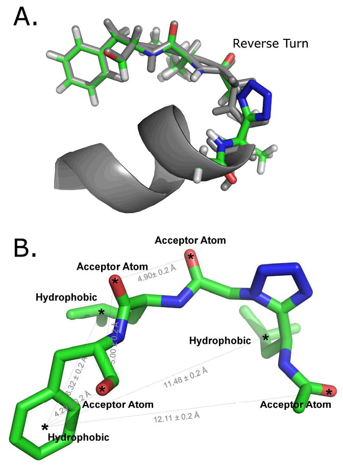
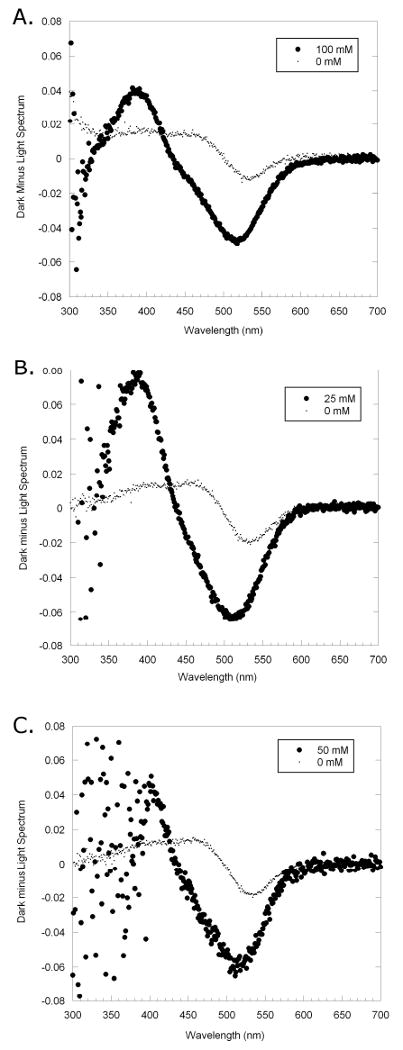
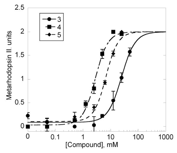
G-protein coupled receptors play an essential role in many biological processes. Despite an increase in the number of solved X-ray crystal structures of G-protein coupled receptors, capturing a G-protein coupled receptor in its activated state for structural analysis has proven to be difficult. An unexplored paradigm is stabilization of one or more conformational states of a G-protein coupled receptor via binding a small molecule to the intracellular loops. A short tetrazole peptidomimetic based on the photoactivated state of rhodopsin-bound structure of Gt(alpha)(340-350) was previously designed and shown to stabilize the photoactivated state of rhodopsin, the G-protein coupled receptor involved in vision. A pharmacophore model derived from the designed tetrazole tetrapeptide was used for ligand-based virtual screening to enhance the possible discovery of novel scaffolds. Maybridge Hitfinder and National Cancer Institute diversity libraries were screened for compounds containing the pharmacophore. Forty-seven compounds resulted from virtually screening the Maybridge library, whereas no hits resulted with the National Cancer Institute library. Three of the 47 Maybridge compounds were found to stabilize the MII state. As these compounds did not inhibit binding of transducin to photoactivated state of rhodopsin, they were assumed to be allosteric ligands. These compounds are potentially useful for crystallographic studies where complexes with these compounds might capture rhodopsin in its activated conformational state.
Figures

References
-
- Howard AD, McAllister G, Feighner SD, Liu Q, Nargund RP, Van der Ploeg LH, et al. Orphan G-protein-coupled receptors and natural ligand discovery. Trends Pharmacol Sci. 2001;22(3):132–40. - PubMed
-
- Gether U. Uncovering molecular mechanisms involved in activation of G protein-coupled receptors. Endocr Rev. 2000;21(1):90–113. - PubMed
-
- Kroeze WK, Sheffler DJ, Roth BL. G-protein-coupled receptors at a glance. J Cell Sci. 2003;116(Pt 24):4867–9. - PubMed
-
- Klabunde T, Hessler G. Drug design strategies for targeting G-protein-coupled receptors. Chembiochem. 2002;3(10):928–944. - PubMed
Publication types
MeSH terms
Substances
Grants and funding
LinkOut - more resources
Full Text Sources
Research Materials
Miscellaneous
