Review
. 2010 Sep;277(18):3622-36.
doi: 10.1111/j.1742-4658.2010.07754.x. Epub 2010 Jul 26.

Calcium-dependent mitochondrial function and dysfunction in neurons

Affiliations
Review

Calcium-dependent mitochondrial function and dysfunction in neurons

Natalia B Pivovarova et al. FEBS J. 2010 Sep.

Abstract

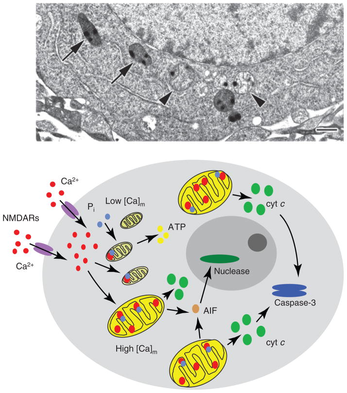
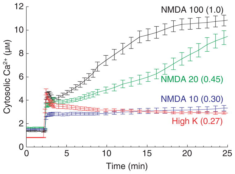
Calcium is an extraordinarily versatile signaling ion, encoding cellular responses to a wide variety of external stimuli. In neurons, mitochondria can accumulate enormous amounts of calcium, with the consequence that mitochondrial calcium uptake, sequestration and release play pivotal roles in orchestrating calcium-dependent responses as diverse as gene transcription and cell death. In this review, we consider the basic chemistry of calcium as a 'sticky' cation, which leads to extremely high bound/free ratios, and discuss areas of current interest or controversy. Topics addressed include methodologies for measuring local intracellular calcium, mitochondrial calcium buffering and loading capacity, mitochondrially directed spatial calcium gradients, and the role of calcium overload-dependent mitochondrial dysfunction in glutamate-evoked excitotoxic injury and neurodegeneration. Finally, we consider the relationship between delayed calcium de-regulation, the mitochondrial permeability transition and the generation of reactive oxygen species, and propose a unified view of the 'source specificity' and 'calcium overload' models of N-methyl-d-aspartate (NMDA) receptor-dependent excitotoxicity. Non-NMDA receptor mechanisms of excitotoxicity are discussed briefly.


Figures

Fig. 1
Fig. 1
Energy-filtered TEM (EFTEM) map of mitochondrial calcium distribution in a depolarization activated frog sympathetic neuron. Left panel: zero-loss structural EFTEM image of an ultra-thin cryosection prepared from a rapidly frozen frog sympathetic neuron 5 min after termination of a 2 min depolarization with 50 mM K+ [16]. Right panel: quantitative EFTEM map of the mitochondrial calcium distribution, recorded as described previously [26]. The field shown contains seven mitochondria, illustrating the general heterogeneity of mitochondrial calcium accumulation, i.e. some mitochondria (1, 2 and 3) have taken up little if any calcium, while others (4, 5, 6 and 7) have accumulated much more. The resolution of the map is high enough to reveal the punctate nature of mitochondrial calcium sequestration e.g. in mitochondria 6 and 7. Note that neither the dense pigment granule (G) nor the tear in the section (T) generate mass thickness artifacts in the calcium map. Note also the large field of view. Scale bar = 1 μm.
Fig. 2
Fig. 2
Mitochondrial calcium loads persist long after activation of the mitochondrial permeability transition. Electron micrographs of typical damaged in situ mitochondria of NMDA-treated cultured hippocampal neurons (upper panel) and calcium-loaded isolated rat brain mitochondria (lower panel). Isolated mitochondria were calcium-loaded in the presence of ATP using a continuous infusion protocol. After the abrupt onset of MPT, mitochondria were further exposed to the protonophore carbonyl cyanide 4-(trifluoromethoxy)-phenylhydrazone (FCCP) and frozen 200 s later (see [75] for experimental details). Both preparations were high pressure-frozen and freeze-substituted in order to preserve the calcium-rich precipitates, whose continued presence demonstrates the longevity of mitochondrial calcium loads. The lower panel also illustrates the variable response of individual mitochondria to calcium challenge. Scale bar = 500 nm (both panels).
Fig. 3
Fig. 3
Heterogeneous calcium accumulation within and among individual mitochondria. Electron micrograph of a high pressure-frozen, freeze-substituted, cultured hippocampal neuron demonstrating that neighboring mitochondria respond differently to NMDA exposure (100 μM for 30 min). Although all mitochondria took up significant amounts of calcium, as indicated by the electron-dense calcium- and phosphorus-rich precipitates, some (arrowheads) have presumably undergone MPT, becoming swollen and releasing matrix material and apoptogenic proteins, while others (arrows) have not. Scale bar = 500 nm. Modified from [38]. The diagram below illustrates schematically how this heterogeneity can account for delayed cell death. Specifically, damaged mitochondria early on release factors necessary to activate downstream death signaling, but undamaged mitochondria that are not dysfunctional maintain energy production and other essential mitochondrial functions for the time period between injury and death.
Fig. 4
Fig. 4
The variability of mitochondrial calcium loading capacity. Electron micrographs of high pressure-frozen, freeze-substituted, hippocampal neurons from naïve (left panel) and NMDA-tolerant (‘pre-conditioned’ [84]) cultures (right panel) after exposure to 100 μM NMDA. There are few swollen, damaged mitochondria in the tolerant cell, even though the calcium load is large and comparable to that in naive cells. This damage resistance reflects a general, experimentally induced increase in the calcium loading capacity. Scale bar = 500 nm (both panels).
Fig. 5
Fig. 5
Ca2+ entry and cell death are much higher after NMDAR activation than after depolarization-evoked VGCC activation. The traces show the dose–response of cytosolic Ca2+ in cultured hippocampal neurons to increasing concentrations of NMDA (μM, as indicated) in comparison with the strong depolarization-induced Ca2+ entry via VGCCs (90 mM K+ plus 1 μM Bay K 8644 (a calcium channel activator) in 10 mM Ca2+ saline). The relative death rates at 24 h are given in parentheses. Free Ca2+ was measured using the low-affinity ratiometric probe fura-4FF. Near-maximal VGCC activation and NMDA at the lowest dose used (10 μM) elicit similarly small Ca2+ elevations and minimal cell death.

References

    1. Clapham DE. Calcium signaling. Cell. 2007;131:1047–1058. - PubMed
    1. Friel DD. Mitochondria as regulators of stimulus-evoked calcium signals in neurons. Cell Calcium. 2000;28:307–316. - PubMed
    1. Verkhratsky A. Physiology and pathophysiology of the calcium store in the endoplasmic reticulum of neurons. Physiol Rev. 2005;85:201–279. - PubMed
    1. Rizzuto R, Pozzan T. Microdomains of intracellular Ca2+: molecular determinants and functional consequences. Physiol Rev. 2006;86:369–408. - PubMed
    1. Berridge MJ. Neuronal calcium signaling. Neuron. 1998;21:13–26. - PubMed

Publication types

MeSH terms