. 2010 Jul 26:10:199.
doi: 10.1186/1471-2180-10-199.

Damage of Streptococcus mutans biofilms by carolacton, a secondary metabolite from the myxobacterium Sorangium cellulosum

Affiliations

Damage of Streptococcus mutans biofilms by carolacton, a secondary metabolite from the myxobacterium Sorangium cellulosum

Brigitte Kunze et al. BMC Microbiol. .

Abstract

Background: Streptococcus mutans is a major pathogen in human dental caries. One of its important virulence properties is the ability to form biofilms (dental plaque) on tooth surfaces. Eradication of such biofilms is extremely difficult. We therefore screened a library of secondary metabolites from myxobacteria for their ability to damage biofilms of S. mutans.

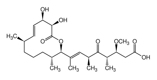
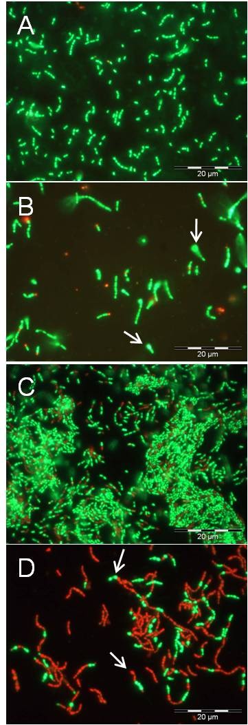
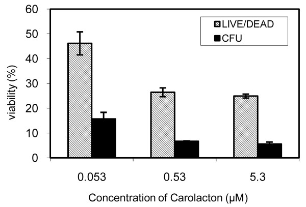
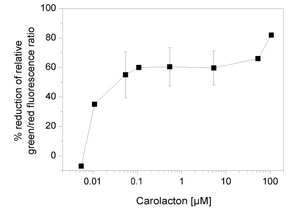
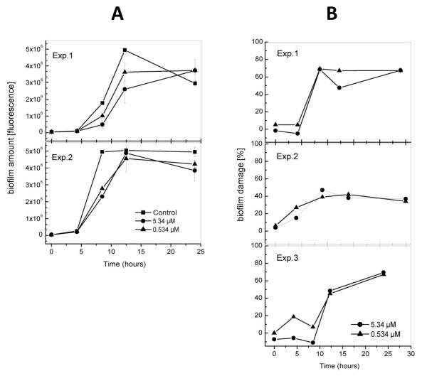
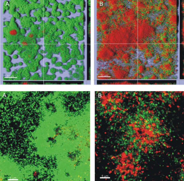
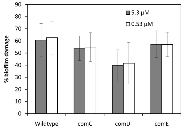
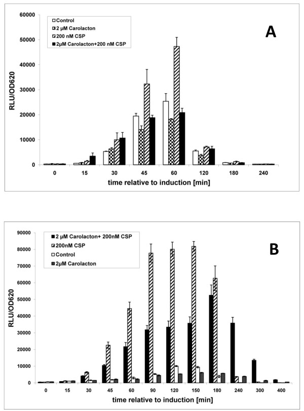
Results: Here we show that carolacton, a secondary metabolite isolated from Sorangium cellulosum, has high antibacterial activity against biofilms of S. mutans. Planktonic growth of bacteria was only slightly impaired and no acute cytotoxicity against mouse fibroblasts could be observed. Carolacton caused death of S. mutans biofilm cells, elongation of cell chains, and changes in cell morphology. At a concentration of 10 nM carolacton, biofilm damage was already at 35% under anaerobic conditions. A knock-out mutant for comD, encoding a histidine kinase specific for the competence stimulating peptide (CSP), was slightly less sensitive to carolacton than the wildtype. Expression of the competence related alternate sigma factor ComX was strongly reduced by carolacton, as determined by a pcomX luciferase reporter strain.

Conclusions: Carolacton possibly interferes with the density dependent signalling systems in S. mutans and may represent a novel approach for the prevention of dental caries.


Figures

Figure 1
Figure 1
Chemical structure of carolacton (from Ref. [30], with permission).
Figure 2
Figure 2
Effect of carolacton on cell morphology and viability. Fluorescent phase-contrast images of planktonically grown cultures (A, B) and biofilm cells of S. mutans (C, D) after LIVE/DEAD staining without (A, C) and in the presence of 5.3 μM carolacton (B, D). Planktonic cultures were grown in THB. Biofilms were grown in THB supplemented with 0.5% sucrose on microtitre plates for 24 h hours. Cultivation was at 37°C under anaerobic conditions (80% N2, 10% H2, 10% CO2). For microscopy, biofilm cells were scraped off from the bottom of the wells using pipette tips. Samples (100 μl) were stained with LIVE/DEAD BacLight bacterial viability staining kit L13152 (Molecular Probes; Eugene, OR, US) as recommended by the manufacturer and analysed using an Olympus BX60 microscope equipped with fluorescence filters U-MWB and U-MNUA2 and the Olympus digital camera Color View II (Olympus Optical Co., Ltd. Germany). Arrows (B, D) indicate bulging cells.
Figure 3
Figure 3
Quantification of the viability of carolacton treated S. mutans biofilms determined by counting colony forming units (CFU) and by measuring membrane damage, calculated as the green/red ratio after LIVE/DEAD BacLight Bacterial Viability staining in percent of untreated controls. Biofilms were grown for 24 h under anerobic conditions. Each data point is the average +/- standard deviation of triplicate to fourfold determinations. The CFU in the control without carolacton was 2.1 × 10⁷ml-1.
Figure 4
Figure 4
Effect of carolacton concentration on the membrane damage of S. mutans biofilms. Biofilms were grown for 24 h under anaerobic conditions and stained using the LIVE/DEAD BacLight Bacterial Viability kit. Green and red fluorescence was determined, and biofilm damage was calculated as reduction of the fluorescence ratio green/red compared to untreated controls. Each data point is the average of triplicate samples. Standard deviations are given for data points determined in at least three independent experiments.
Figure 5
Figure 5
Time course of biofilm growth of S. mutans in the presence and absence of carolacton. A Biofilm volume, determined by using only the SYTO9 (green) component of the LIVE/DEAD BacLight Bacterial Viability stain. Two representative experiments are shown. Green fluorescence, which is a measure of total biomass, is shown in absolute units. B Biofilm membrane damage, determined using the LIVE/DEAD BacLight Bacterial Viability stain. Green and red fluorescence was measured, and biofilm damage was calculated as reduction of the ratio of green/red fluorescence compared to controls without carolacton. Error values were calculated from the standard deviations of the green/red ratios of control and carolacton treated samples according to the error propagation formula of Gauss. Three representative experiments are shown. Biofilms were grown anaerobically. Mean and standard deviation are given for triplicate samples.
Figure 6
Figure 6
Confocal laser scanning microscope images of S. mutans biofilms in the absence (A) or presence (B) of 0.5 μM carolacton after 12 h of anaerobic cultivation. Staining using the LIVE/DEAD BacLight Bacterial Viability Kit assessed bacterial viability: green areas indicate live cells; red areas indicate dead or damaged cells. The top panel shows a bird's eye view on the biofilm with lines indicating the position of the vertical sections shown at the lower and right margins of both images. Acquired using an UPLSAPO 20× objective lens, size of scale bar 50 μm. The bottom panel shows enlarged horizontal sections of S. mutans biofilms in the absence (A) or presence (B) of 0.5 μM carolacton, aacquired using an UPLSAPO 40× objective lens with 7× digital magnification, size of scale bar 5 μm.
Figure 7
Figure 7
Damage of biofilms of S. mutans wildtype and knock-out mutants for comC, comD and comE by carolacton. Biofilms were grown under anaerobic conditions for 24 h and stained with the LIVE/DEAD BacLight Bacterial Viability staining kit. Green and red fluorescence was determined in triplicate samples, and biofilm damage was calculated as reduction of the fluorescence ratio green/red compared to untreated controls. Standard deviations were calculated from 3 - 5 independent experiments.
Figure 8
Figure 8
Effect of carolacton on the comX-promoter activity of S. mutans. The CSP-induced comX-luc gene expression in the reporter strain construct was determined in the presence and absence of 2 μM carolacton. The luciferase activity was normalized against the optical density at 620 nm and measured for different time-points after induction of luciferase expression with 0.2 μM CSP. The expression of comX-luc in cultures which were not induced by externally added CSP and its inhibition by carolacton is also shown. Cultures were grown under anaerobic conditions as biofilms (A) or in suspension (B).

Similar articles

Cited by

References

    1. Costerton JW, Stewart PS, Greenberg EP. Bacterial biofilms: a common cause of persistent infections. Science. 1999;284:1318–1322. doi: 10.1126/science.284.5418.1318. - DOI - PubMed
    1. Costerton JW, Montanaro L, Arciola CR. Bacterial communications in implant infections: a target for an intelligence war. Int J Artif Organs. 2007;30:757–763. - PubMed
    1. Lynch AS, Robertson GT. Bacterial and fungal biofilm infections. Annu Rev Med. 2008;59:415–428. doi: 10.1146/annurev.med.59.110106.132000. - DOI - PubMed
    1. Hall-Stoodley L, Costerton JW, Stoodley P. Bacterial biofilms: from the natural environment to infectious diseases. Nat Rev Microbiol. 2004;2:95–108. doi: 10.1038/nrmicro821. - DOI - PubMed
    1. Parsek MR, Singh PK. Bacterial biofilms: an emerging link to disease pathogenesis. Annu Rev Microbiol. 2003;57:677–701. doi: 10.1146/annurev.micro.57.030502.090720. - DOI - PubMed