. 2010 Jul 26:7:41.
doi: 10.1186/1742-2094-7-41.

Gp91phox (NOX2) in classically activated microglia exacerbates traumatic brain injury

Affiliations

Gp91phox (NOX2) in classically activated microglia exacerbates traumatic brain injury

Kenji Dohi et al. J Neuroinflammation. .

Abstract

Background: We hypothesized that gp91phox (NOX2), a subunit of NADPH oxidase, generates superoxide anion (O2-) and has a major causative role in traumatic brain injury (TBI). To evaluate the functional role of gp91phox and reactive oxygen species (ROS) on TBI, we carried out controlled cortical impact in gp91phox knockout mice (gp91phox-/-). We also used a microglial cell line to determine the activated cell phenotype that contributes to gp91phox generation.

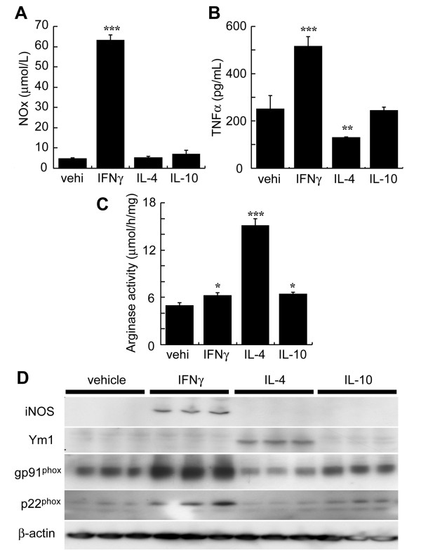
Methods: Unilateral TBI was induced in gp91phox-/- and wild-type (Wt) mice (C57/B6J) (25-30 g). The expression and roles of gp91phox after TBI were investigated using immunoblotting and staining techniques. Levels of O2- and peroxynitrite were determined in situ in the mouse brain. The activated phenotype in microglia that expressed gp91phox was determined in a microglial cell line, BV-2, in the presence of IFNgamma or IL-4.

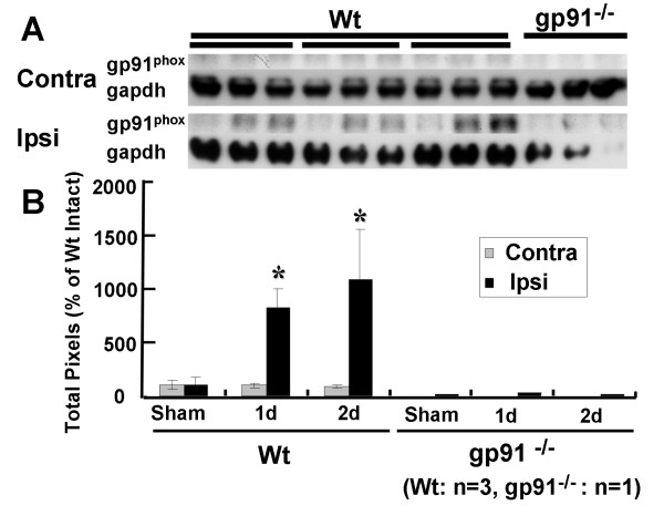
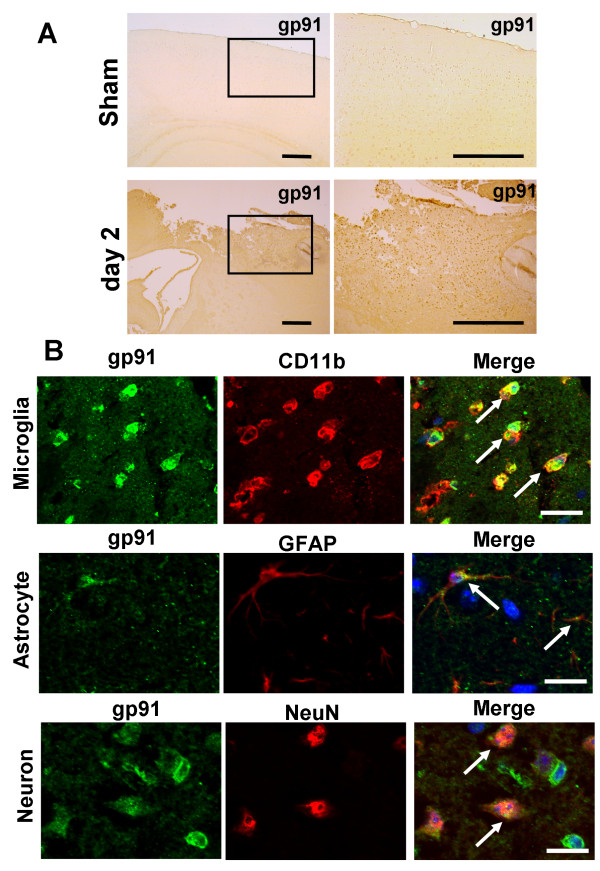
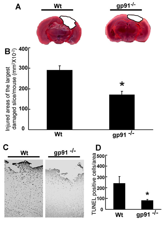
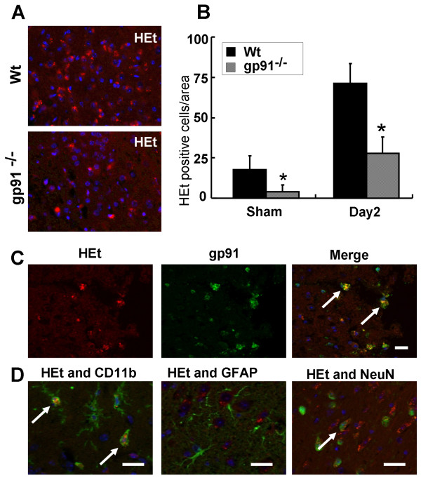
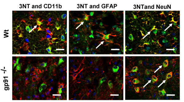
Results: Gp91phox expression increased mainly in amoeboid-shaped microglial cells of the ipsilateral hemisphere of Wt mice after TBI. The contusion area, number of TUNEL-positive cells, and amount of O2- and peroxynitrite metabolites produced were less in gp91phox-/- mice than in Wt. In the presence of IFNgamma, BV-2 cells had increased inducible nitric oxide synthase and nitric oxide levels, consistent with a classical activated phenotype, and drastically increased expression of gp91phox.

Conclusions: Classical activated microglia promote ROS formation through gp91phox and have an important role in brain damage following TBI. Modulating gp91phox and gp91phox -derived ROS may provide a new therapeutic strategy in combating post-traumatic brain injury.


Figures

Figure 1
Figure 1
Characterization of gp91phox after traumatic brain injury (TBI) using western immunoblots. (A) Immunoblotting signals before and after TBI in wild-type (Wt) mice (left) and gp91phox-/- mice (right). (B) In Wt mice, gp91phox levels were significantly increased on day 1 and day 2 after TBI (*p < 0.05 relative to sham). Gp91phox levels on the ipsilateral side were greater than on the contralateral side on day 1 and day 2 after TBI (*p < 0.05). Each value is the mean ± SE (n = 3). Note that the intensity of each sample was quantified and corrected relative to the labeling control, actin. Increasing levels of gp91phox were not seen in gp91phox-/- mice.
Figure 2
Figure 2
Expressions and cell identification of gp91phox in peri-contusional area after traumatic brain injury (TBI). (A) In sham-operated animals, weak immunoreactivity for gp91phox was observed in the cortex (upper panel). Two days after TBI, gp91phox immunoreactivity was dramatically increased in the peri-contusional area (lower panel). Scale bars = 400 μm. (B) Co-immunostaining of gp91phox with cell markers in the peri-contusional region. Immunostaining was carried out using antibodies for gp91phox (shown in green) together with Integrin alpha M (CD11b) (upper), glial fibrillary acidic protein (GFAP) (middle), and neuronal nuclear antigen (NeuN) (lower). Microglia, astrocyte, and neuron markers are shown in red. Gp91phox immunoreactive cells were co-labeled with all cell markers. Particularly strong expression of gp91phox was detected in microglial-like cells (CD11b-positive cells). Cells were counter-stained with DAPI to show nuclei (blue). Scale bars = 20 μm.
Figure 3
Figure 3
Brain damage and cell death after traumatic brain injury in wild type (Wt) mice and gp91phox-/- mice. (A) 2,3,5-triphenyltetrazolium chloride (TTC)-stained coronal brain sections from Wt mice (left) and gp91phox-/- mice (right) 2 days after TBI. (B) The contusion area in gp91phox-/- mice (170.5 ± 71.5 mm2 × 10-2, n = 20) was significantly smaller than that in the Wt group (290.7 ± 94.0 mm2 × 10-2, n = 10, *p < 0.01, t test). (C) Terminal deoxynucleotidyl transferase dUTP nick end labeling (TUNEL)-staining in peri-contusional area of wild type (left) and gp91phox-/- (right) mice 48 hours after TBI. TUNEL-positive cells numbers in the gp91phox-/- mice (117 ± 69.3 cells/area, n = 6) were significantly smaller than those in the Wt group (252 ± 128.9 cells/area, n = 6, *p < 0.05, t test) (D). All values represent the mean ± SE.
Figure 4
Figure 4
The roles of gp91phox in superoxide radical production after traumatic brain injury). (A) Superoxide radical production (hydroethidium [HEt]-positive cells: shown in red) in the peri-contusional area of wild type (upper) and gp91phox-/- (lower) mice 2 days after TBI. (B) TBI-induced superoxide radical production (HEt-positive cells) in the peri-contusional area was significantly attenuated by gp91phox deficiency in sham-operated mice, and 2 days after TBI (*p < 0.05, t test). All values represent mean ± SE. (C, D) Co-localization of HEt-positivity with cell markers in the peri-contusional region. Cell identification was carried out using antibodies for gp91phox (C), CD11b (D, left), GFAP (D, middle), and Neu N (D, right) (shown in green). HEt-positive cells were co-localized with gp91phox-positive cells (C, right, arrows) and microglia-like cells (D, left, arrows). Although HEt-positive cells were also slightly co-localized with neurons (D, right, arrows), immunoreactivity was lower than for gp91phox-positive cells and microglia-like cells. Cells were counter-stained with DAPI to show nuclei (blue). Scale bars = 20 μm (C, D).
Figure 5
Figure 5
Production and cell identification of peroxynitrite (ONOO-) in wild type (Wt) (upper) and gp91phox-/- (lower) mice 48 hours after TBI. ONOO- in Wt mice was produced in microglia-like cells (left, arrows), astrocytes (middle, arrows), and degenerated neurons (right, arrows). In gp91phox-/- mice, ONOO- production was strongly suppressed. In neurons, only weak production of ONOO- was observed, but degeneration of neurons was not seen (right, arrows). Scale bars = 20 μm.
Figure 6
Figure 6
Expression of gp91phox and p22phox in IFNγ-exposed classically activated mouse microglial BV-2 cells. BV-2 cells were exposed to vehicle (vehi), IFNγ, IL-4, or IL-10. The levels of microglial phenotype markers (NOx, TNFα, arginase activity, iNOS, and Ym1), gp91phox, and p22phox were evaluated in medium or cell homogenates 24 hours later. Production of (A) NOx and (B) TNFα were determined in the media. (C) Expression of iNOS, Ym1, gp91phox, and p22phox was determined by immunoblotting with reduced samples. ß-Actin levels were used as an internal control. IFNγ-exposed BV-2 cells had increased NOx, TNFα, and iNOS levels, all of which are classical activating markers of the microglial phenotype. In contrast, IL-4-exposed BV-2 cells had increased arginase activity and Ym1 levels, which are alternative markers of activation. Gp91phox and p22phox levels were increased only in IFNγ-exposed BV-2 cells, suggesting induction by classically activated microglial cells.

Similar articles

Cited by

References

    1. Dohi K, Satoh K, Nakamachi T, Yofu S, Hiratsuka K, Nakamura S, Ohtaki H, Yoshikawa T, Shioda S, Aruga T. Does edaravone (MCI- 186) act as an antioxidant and a neuroprotector in experimental traumatic brain injury? Antioxid Redox Signal. 2007;9:281–287. doi: 10.1089/ars.2007.9.281. - DOI - PubMed
    1. Lewen A, Matz P, Chan PH. Free radical pathways in CNS injury. J Neurotrauma. 2000;17:871–890. doi: 10.1089/neu.2000.17.871. - DOI - PubMed
    1. Pratico D, Reiss P, Tang LX, Sung S, Rokach J, McIntosh TK. Local and systemic increase in lipid peroxidation after moderate experimental traumatic brain injury. J Neurochem. 2002;80:894–898. doi: 10.1046/j.0022-3042.2002.00777.x. - DOI - PubMed
    1. Tyurin VA, Tyurina YY, Borisenko GG, Sokolova TV, Ritov VB, Quinn PJ, Rose M, Kochanek P, Graham SH, Kagan VE. Oxidative stress following traumatic brain injury in rats: quantitation of biomarkers and detection of free radical intermediates. J Neurochem. 2000;75:2178–2189. doi: 10.1046/j.1471-4159.2000.0752178.x. - DOI - PubMed
    1. Deng-Bryant Y, Singh IN, Carrico KM, Hall ED. Neuroprotective effects of tempol, a catalytic scavenger of peroxynitrite-derived free radicals, in a mouse traumatic brain injury model. J Cereb Blood Flow Metab. 2008;28:1114–1126. doi: 10.1038/jcbfm.2008.10. - DOI - PubMed

Publication types

MeSH terms

LinkOut - more resources