. 2010;12(4):R55.
doi: 10.1186/bcr2614. Epub 2010 Jul 26.

MYB suppresses differentiation and apoptosis of human breast cancer cells

Affiliations

MYB suppresses differentiation and apoptosis of human breast cancer cells

Yvette Drabsch et al. Breast Cancer Res. 2010.

Abstract

Introduction: MYB is highly expressed in estrogen receptor positive (ER + ve) breast tumours and tumour cell lines. We recently demonstrated that MYB is essential for the proliferation of ER + ve breast cancer cells, and have now investigated its role in mammary epithelial differentiation.

Methods: MCF-7 breast cancer cells were treated with sodium butyrate, vitamin E succinate or 12-O-tetradecanoylphorbol-13-acetate to induce differentiation as measured by Nile Red staining of lipid droplets and β-casein expression. The non-tumorigenic murine mammary epithelial cell (MEC) line, HC11, was induced to differentiate with lactogenic hormones. MYB levels were manipulated by inducible lentiviral shRNA-mediated knockdown and retroviral overexpression.

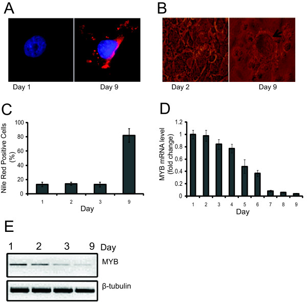
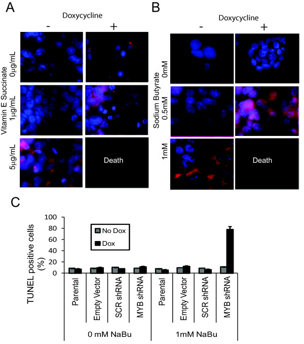
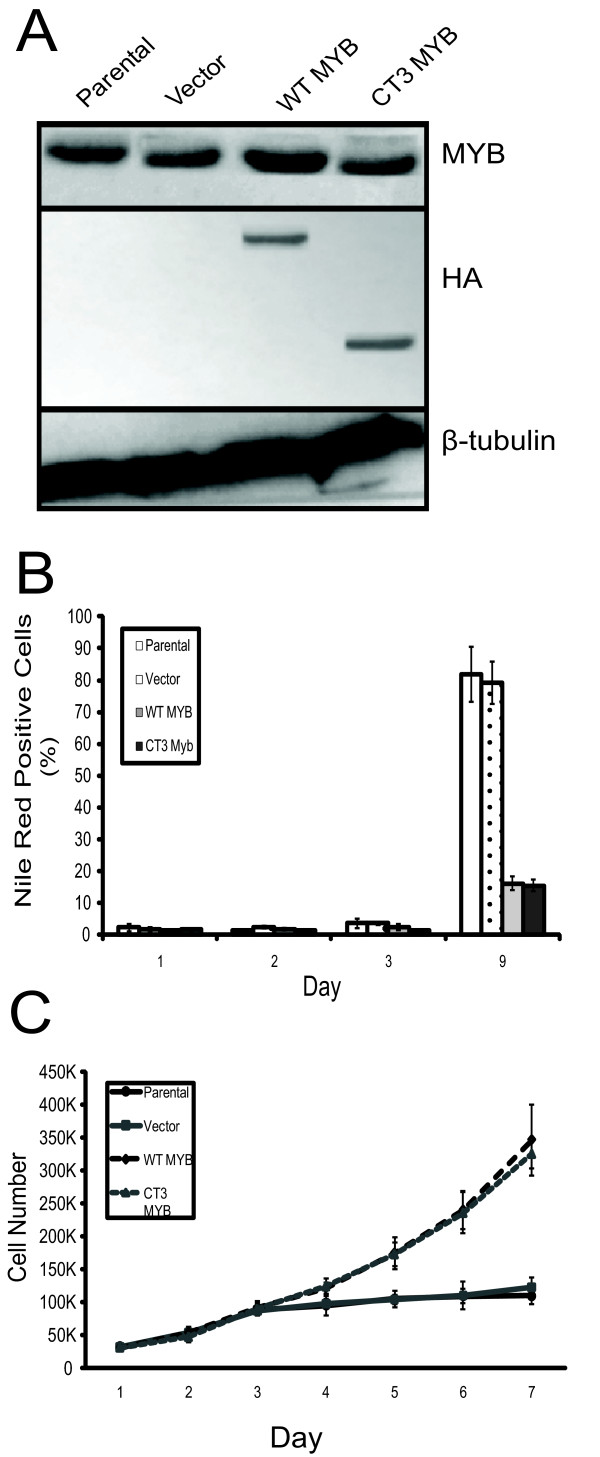
Results: We found that MYB expression decreases following chemically-induced differentiation of the human breast cancer cell line MCF-7, and hormonally-induced differentiation of a non-tumorigenic murine mammary epithelial cell (MEC) line, HC11. We also found that shRNA-mediated MYB knockdown initiated differentiation of breast cancer cells, and greatly sensitised them to the differentiative and pro-apoptotic effects of differentiation-inducing agents (DIAs). Sensitisation to the pro-apoptotic effects DIAs is mediated by decreased expression of BCL2, which we show here is a direct MYB target in breast cancer cells. Conversely, enforced expression of MYB resulted in the cells remaining in an undifferentiated state, with concomitant suppression of apoptosis, in the presence of DIAs.

Conclusions: Taken together, these data imply that MYB function is critical in regulating the balance between proliferation, differentiation, and apoptosis in MECs. Moreover, our findings suggest MYB may be a viable therapeutic target in breast cancer and suggest specific approaches for exploiting this possibility.


Figures

Figure 1
Figure 1
Differentiation induction and cells treated with the differentiation-inducing agent NaBu. (a) MCF-7 cells were grown on glass cover slips and treated with increasing amounts of sodium butyrate (NaBu), as indicated, for 72 hours. Morphological changes were evaluated by fluorescent microscopy (×63). Induction of biochemical differentiation was determined by using Nile Red stain for lipid vesicles in cells (red). Nuclear DNA was stained with 4',6-diamidino-2-phenylindole (DAPI) (blue). (b) Quantitative analysis of lipid induction. MCF-7 cells, as above, were stained with Nile Red and analyzed by flow cytometry as described in the Materials and methods. Standard deviations are shown as error bars (n = 6). (c) Quantitative (Q) PCR of ß-casein mRNA expression following treatment with increasing doses of NaBu. The induction of ß-casein is normalized against that seen in untreated cells. (d) Q-PCR of MYB mRNA from MCF-7. MCF-7 cells treated with the increasing dosage of NaBu show a dose dependent decrease in expression of MYB mRNA. Data from (c) and (d) represent mean values (n = 6), and standard deviations are shown as error bars. All Q-PCR data were normalized against cyclophilin A controls. (e) Western blot analysis of MYB from total cell lysates as above. A ß-tubulin loading control is also shown. Results are indicative of two independent experiments.
Figure 2
Figure 2
Myb expression during confluence/hormone-induced differentiation of HC11 cells. HC11 were proliferating (Day 1), grown to confluence (Day 2), and then differentiated by removing epidermal growth factor (EGF) for 24 hours (Day 3), followed by addition of differentiation medium containing lactogenic hormones (Days 4 to 9). (a) HC11 cells grown on glass cover slips were stained for DNA (4',6-diamidino-2-phenylindole [DAPI]; blue), lipid vesicles (Nile Red; red), and viewed under fluorescence microscopy at Day 1 and Day 9. (b) HC11 cells were grown in 24-well dishes, and induced to differentiate as described. Shown are light micrographs of confluent cells and 'domes' (arrow) which form in the presence of differentiation inducing medium. (c) Quantitative analysis of Nile Red staining by flow cytometry. HC11 cells were analyzed for lipid vesicle formation during the different stages of differentiation. (d) Quantitative PCR of MYB mRNA from HC11 cells undergoing differentiation. HC11 cells were analyzed each day during the differentiation process. Standard deviation is represented by error bars. (e) Western blot analysis of MYB protein from total cell lysates of HC11 cells during the differentiation process. A ß-tubulin loading control is also shown. Results are indicative of two independent experiments.
Figure 3
Figure 3
MYB knockdown sensitizes MCF-7 to differentiation-inducing agents. (a) Vitamin E succinate (VES) was used to treat MCF-7 cells with a doxycycline (Dox)-inducible shRNA targeting MYB [19] MCF-7 cells grown on a glass coverslip, and treated with or without 5 mg/mL Dox for 24 hours before exposure to VES for three days, were then stained with Nile Red (red) for lipid vesicles and 4',6-diamidino-2-phenylindole (DAPI) (blue) for DNA detection. (b) Sodium butyrate (NaBu) was used to treat MCF-7 cells with the Dox-inducible shRNA targeted against MYB, as in (a). Results are indicative of at least two independent experiments. (c) Flow cytometric analysis of TUNEL staining for apoptotic cells. MCF-7 cells stably transfected with inducible shRNA treated for 24 hours with or without Dox, were grown in the presence or absence of 1 mM NaBu for a further 24 hours, and were then assayed for apoptosis using TUNEL. Standard deviation is represented by error bars.
Figure 4
Figure 4
Expression, MYB regulation and function of BCL2 in MCF-7 cells. (a) BCL2 expression in MCF-7 cells treated with increasing doses sodium butyrate (NaBu). After 72 hours of treatment, cells were harvested and the mRNA was analyzed for expression of BCL2 by quantitative (Q) PCR. (b) MCF-7 cells stably transduced with an inducible MYB shRNA vector were grown in the presence of doxycycline (Dox) for three days, after which BCL2 was analyzed by Q-PCR. Standard deviation is represented by error bars. (c) A schematic of the BCL2 gene (not to scale) showing the relative locations of the primers (MBS-1-4) used for ChIP assays. (d) Q-PCR of MCF-7 cells assessed by ChIP assays using anti-MYB Ab. Rabbit IgG was used as a negative control. MYC is used as a positive control, and GAPDH and an upstream primer set are used as negative controls. (e) Western blot analysis of BCL2 knockdown by four siRNAs (#1-4), parental cells, and control siRNA. MCF-7 s were transiently treated with the siRNAs and at 24 hours, assessment of their ability to reduce BCL2 was undertaken. A ß-tubulin loading control is also shown. Results are indicative of two independent experiments. (f) MCF-7 cells treated with four independent BCL2 siRNAs were grown for 24 hours with 0, 0.1, or 1 mM NaBu as indicated. TUNEL assays were analyzed by flow cytometry to quantify the number of apoptotic cells. Standard deviation is represented by error bars.
Figure 5
Figure 5
MYB overexpression in MCF-7 cells prevents differentiation-inducing agent-induced differentiation and growth arrest. (a) MCF-7 cells were stably transduced with empty pMYS-IRES-GFP vector, or vectors encoding HA-tagged WT-or truncated, activated CT3-MYB. Western blotting with anti-MYB Ab and anti-HA Ab confirmed the expression of exogenous, HA-tagged MYB proteins. Note that the anti-MYB antibody used does not detect CT3-MYB. The Western blot was also probed for BCL2 expression (see text); a ß-tubulin loading control is also shown. (b) MYB overexpression blocks lipid accumulation associated with differentiation. Cells were treated with 0.5 mM or 1 mM sodium butyrate (NaBu) for 72 hours, stained with Nile Red and analyzed by flow cytometry. (c) Proliferation of MCF-7 cells overexpressing WT or CT3-MYB. MCF-7 cells overexpressing the WT or CT3-MYB, and controls, were grown in normal medium over seven days. (d) Proliferation of MCF-7 cells overexpressing the WT or CT3-MYB grown in medium containing 1 mM NaBu over seven days. In (c) and (d) cells were counted on days one, three, five, and seven. Standard deviation is represented by error bars (n = 3).
Figure 6
Figure 6
MYB overexpression in HC11 cells prevents differentiation and growth arrest. (a) HC11 cells were stably transduced with empty vector, WT-MYB, or CT3-MYB, as in Figure 5. Western blot with anti-MYB Ab and anti-HA Ab confirmed the expression of exogenous and HA-tagged MYB proteins. A ß-tubulin loading control is also shown. (b) MYB overexpression blocks lipid accumulation associated with differentiation. Cells were stained with Nile Red at the various stages of differentiation shown, and analyzed by flow cytometry. Standard deviation is indicated by error bars. (c) HC11 cells overexpressing WT or CT3-MYB continue to proliferate in differentiation-inducing conditions. HC11 cells as indicated were grown in complete media for two days, after which epidermal growth factor (EGF) was removed for 24 hours and subsequent lactogenic hormones were added to the medium each day Standard deviation is indicated by error bars (n = 3).
Figure 7
Figure 7
MYB overexpression prevents differentiation-inducing agents-induced apoptosis in a BCL2-dependent manner. (a) MCF-7 cells were treated with the indicated doses of sodium butyrate (NaBu) for 24 hours before being assayed with TUNEL, which was quantified by flow cytometry. (b) MCF-7 cells stably overexpressing WT-MYB, CT3-MYB, or vector, or control parental, were treated with 10 mM or 100 mM NaBu for 24 hours prior to TUNEL assay. (c) MCF-7 cells stably overexpressing WT MYB, or control vector, were transiently transfected with siRNAs targeting BCL2. They were then treated with 0 or 100 mM NaBu for 24 hours before being assayed with TUNEL. Standard deviation is represented by error bars (n = 6).
Figure 8
Figure 8
Fulvestrant synergizes with NaBu to enhance the DIA sensitivity of MCF-7 cells. (a) Western blot analysis of MYB expression in MCF-7 cells treated with fulvestrant or sodium butyrate (NaBu), alone or in combination, as indicated. (The combination of fulvestrant with 1 mM NaBu is not included because of extensive cell loss due to apoptosis. A ß-tubulin loading control is also shown. (b) MCF-7 cells grown on glass slides were treated with fulvestrant alone or in combination with 0.5 mM or 1 mM NaBu for 72 hours. The cells were then stained with Nile Red (red) for lipid vesicles and 4',6-diamidino-2-phenylindole (DAPI) (blue) for DNA. (c) Apoptosis and (d) lipid accumulation were detected by TUNEL assay and Nile Red staining, respectively, and quantitated by flow cytometry standard deviation is represented by error bars (n = 3).

Similar articles

Cited by

References

    1. Mucenski M, McLain K, Kier A, Swerdlow S, Schreiner C, Miller T, Pietryga D, Scott WJ, Potter S. A functional c-myb gene is required for normal murine fetal hepatic hematopoiesis. Cell. 1991;65:677–689. - PubMed
    1. Westin EH, Gallo RC, Arya SK, Eva A, Souza L, Baluda M, Aaronson S, Wong-Staal F. Differential expression of the amv gene in human hematopoietic cells. Proc Natl Acad Sci USA. 1982;79:2194–2198. - PMC - PubMed
    1. Gonda T, Metcalf D. Expression of myb, myc and fos proto-oncogenes during the differentiation of a murine myeloid leukaemia. Nature. 1984;310:249–251. - PubMed
    1. Wolff L. Myb-induced transformation. Crit Rev Oncog. 1996;7:245–260. - PubMed
    1. Ganter B, Lipsick J. Myb and oncogenesis. Adv Cancer Res. 1999;76:21–60. - PubMed

Publication types

MeSH terms

LinkOut - more resources