. 2010 Jul 26:11:59.
doi: 10.1186/1471-2121-11-59.

Endoplasmic reticulum stress response in an INS-1 pancreatic beta-cell line with inducible expression of a folding-deficient proinsulin

Affiliations

Endoplasmic reticulum stress response in an INS-1 pancreatic beta-cell line with inducible expression of a folding-deficient proinsulin

Taila Hartley et al. BMC Cell Biol. .

Abstract

Background: Cells respond to endoplasmic reticulum stress (ER) stress by activating the unfolded protein response. To study the ER stress response in pancreatic beta-cells we developed a model system that allows for pathophysiological ER stress based on the Akita mouse. This mouse strain expresses a mutant insulin 2 gene (C96Y), which prevents normal proinsulin folding causing ER stress and eventual beta-cell apoptosis. A double-stable pancreatic beta-cell line (pTet-ON INS-1) with inducible expression of insulin 2 (C96Y) fused to EGFP was generated to study the ER stress response.

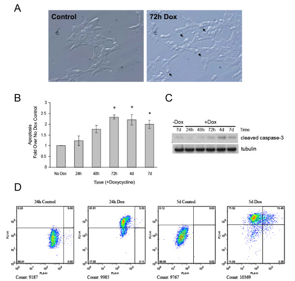
Results: Expression of Ins 2 (C96Y)-EGFP resulted in activation of the ER stress pathways (PERK, IRE1 and ATF6) and caused dilation of the ER. To identify gene expression changes resulting from mutant insulin expression we performed microarray expression profiling and real time PCR experiments. We observed an induction of various ER chaperone, co-chaperone and ER-associated degradation genes after 24 h and an increase in pro-apoptotic genes (Chop and Trib3) following 48 h of mutant insulin expression. The latter changes occurred at a time when general apoptosis was detected in the cell population, although the relative amount of cell death was low. Inhibiting the proteasome or depleting Herp protein expression increased mutant insulin levels and enhanced cell apoptosis, indicating that ER-associated degradation is maintaining cell survival.

Conclusions: The inducible mutant insulin expressing cell model has allowed for the identification of the ER stress response in beta-cells and the repertoire of genes/proteins induced is unique to this cell type. ER-associated degradation is essential in maintaining cell survival in cells expressing mutant insulin. This cell model will be useful for the molecular characterization of ER stress-induced genes.


Figures

Figure 1
Figure 1
Induction of insulin 2 (C96Y)-EGFP protein expression in pTet-ON INS-1 cells by doxycyline. A-B. pTet-ON INS-1 #46 cells were transiently transfected with pTRE-Tight Ins2 (C96Y)-EGFP construct in the presence or absence of 2 μg/ml doxycycline (Dox) for 48 h. In (A), cells were washed in PBS, lysed and 10 μg of protein were resolved by SDS-PAGE and immunoblotted using antibodies to insulin and GFP. In (B) the cells were washed in PBS, fixed and mounted. GFP fluorescence was visualized using a laser confocal fluorescence microscope. C-D. A stable Ins2 (C96Y)-EGFP expressing INS1 clone (Clone #4S2), generated as described in the methods, was untreated (-Dox) or treated with 2 μg/ml doxycycline (+Dox) for the times indicated. The cells were washed in PBS, lysed and an equal amount of protein per condition were resolved by SDS-PAGE and immunoblotted using antibodies to GM130 and GFP (C). In (D), EGFP fluorescence in fixed cells was visualized by confocal fluorescence microscopy.
Figure 2
Figure 2
Insulin 2 (C96Y)-EGFP is localized to the ER while degradation products appear in the cytosol. A. Clone #4S2 cells were untreated (-Dox) or treated with 2 μg/ml doxycycline (+Dox) for 48 h. Following the treatments, the cells were washed in PBS and lysed in either 1% TX-100 lysis buffer (with protease inhibitors) or directly in SDS sample buffer with β-mercaptoethanol. Equal amounts of protein (or equal volumes) were resolved by SDS-PAGE and immunoblotted using antibodies to GM130 or GFP. B. Cells were untreated (Control) or treated for 48 h with 2 μg/ml doxycycline before lysis and immunoprecipitation with anti-GFP or control mouse IgG antibodies. The entire precipitate (pellets; P) and 10 μg of the supernatant (SN) were resolved by SDS-PAGE and immunoblotted using an anti-GFP (rabbit, polyclonal) antibody. C. Cells were treated with 2 μg/ml doxycycline for 72 h, washed in PBS and homogenized in sucrose buffer as described in the methods. The homogenate was fractionated into membrane (M) and cytosol (C) fractions. D. Cells were treated with 2 μg/ml doxycycline for 72 h, washed in PBS and homogenized in sucrose buffer as described in the methods. The homogenate was fractionated on a linear sucrose density gradient and an equal volume of each fraction was resolved by NuPAGE and immunoblotted using antibodies to HSP90, GRP78, GFP and insulin.
Figure 3
Figure 3
Effect of insulin 2 (C96Y)-EGFP expression on ER morphology. Clone #4S2 cells untreated (A, B) or treated with 2 μg/ml doxycycline for 3 days (C, D) or 6 days (E, F) were washed in PBS and fixed. The cells were processed for transmission electron microscopy as described in the methods. ER structures in uninduced cells (A, B, arrows), and distended ER structures in mutant insulin expressing cells following doxycyline-induction (D-F, arrows) are indicated. The presence of apoptotic cells was evident in doxycyline-induced cells (C, arrowheads).
Figure 4
Figure 4
ER stress signaling pathways are activated by insulin 2 (C96Y)-EGFP expression. Clone #4S2 cells were untreated (-Dox) or treated with 2 μg/ml doxycycline (+Dox) for the times indicated. Control INS-1 cells exposed to thapsigargin (Tg) or dithiothreitol (DTT) were used as a positive control. A. RNA was isolated from the cells and XBP-1 cDNA was amplified by RT-PCR. The unspliced form of XBP-1 (uXBP-1, 480 bp) and spliced form of XBP-1 (sXBP-1, 454 bp) are indicated. B. Clone #4S2 cells were treated as in (A), washed in PBS and lysed. Equal amounts of protein were resolved by SDS-PAGE and immunoblotted with anti-phospho-eIF2α and GM130 antibodies. C. Clone #4S2 cells were treated with 1 mM dithiothreitol (DTT) for 30 min. or doxycycline as indicated and fixed for immunofluorescence labeling with anti-ATF6 antibody. Nuclear fluorescence of ATF6 was quantified as described in the methods.
Figure 5
Figure 5
Expression of insulin 2 (C96Y)-EGFP induces the mRNA and protein of known ER stress response genes. Clone #4S2 cells were untreated (Control) or treated with 2 μg/ml doxycycline (+Dox) for the times indicated (h, hours; d, days). A-D. Total RNA was isolated and the mRNA levels relative to cellular β-actin for Grp78 (A), Pdi (B), Chop (C), Sel1 (D) were analyzed by real-time PCR as outlined in the methods. Shown are the mean ± SE of 3 independent experiments. *p < 0.05 relative to control. E, F. Clone #4S2 cells were treated as described above, washed in PBS, lysed and equal amounts of protein were resolved by SDS-PAGE and immunoblotted as indicated. Control INS-1 cells exposed to thapsigargin (Tg) were used as a positive control in (F).
Figure 6
Figure 6
Expression of insulin 2(C96Y)-EGFP results in the induction of selected genes. Clone #4S2 cells were untreated (Control) or treated with 2 μg/ml doxycycline (+Dox) for the times indicated. Total RNA was isolated and the mRNA levels relative to cellular β-actin for Erdj4/Dnajb9 (A), Erdj5/Dnajc10 (B), Ero1β (C), Ero1α (D) were analyzed by real-time PCR as outlined in the methods. Shown are the mean ± SE of 3 independent experiments. *p < 0.05 relative to control.
Figure 7
Figure 7
Expression of insulin 2 (C96Y)-EGFP induces apoptosis. A. Clone #4S2 cells were treated or not with 2 μg/ml doxycycline for 72 h, fixed and imaged by differential interference contrast microscopy. Potentially apoptotic cells with a rounded morphology are indicated (arrows). B-D. Clone #4S2 cells were treated with 2 μg/ml doxycycline for the indicated times. B. Following the treatments, the cells were lysed and apoptosis was measured using a cell death detection ELISA kit (Roche) as described in the methods. Shown are the mean ± SE of 4 independent experiments. *p < 0.05. C. Cells were washed in PBS, lysed and equal amounts of protein were resolved by NuPAGE and immunoblotted using antibodies to cleaved caspase-3 and γ-tubulin. D. Cells were fixed and labeled with Alexa Fluor 647 dye-labeled anti-BrdU antibody. Cells were analyzed by flow cytometry; the FL1 laser excites GFP, whereas FL4 excites Fluor 647. Cells in the upper left and right quadrants were classified as mutant insulin expressing, cells in the upper and lower right quadrants were classified as apoptotic. The total number of cells examined is shown below each chart, and the percentage of cells in each quadrant is indicated.
Figure 8
Figure 8
The proteasome inhibitor lactacystin increases susceptibility of the mutant insulin expressing clone to apoptosis. A. Clone #4S2 cells were untreated or treated with 2 μg/ml doxycycline for 48 h. Cells were then untreated or treated with 10 μM of lactacystin (LC) or an equivalent volume of DMSO for the times indicated. Apoptosis was measured using a cell death detection ELISA kit as described in the Methods. Shown are the mean ± SE of 3 independent experiments. *p < 0.05. B. Following 42 h of doxycycline treatment, cells were untreated, or treated with 10 μM of lactacystin (LC) or an equivalent volume of DMSO for 6 h in the presence of doxycycline. The cells were washed in PBS and lysed. Cell lysates (left panel) or TX-100 insoluble material (right panel) were resolved by SDS-PAGE and immunoblotted using the indicated antibodies. Note the increase in the levels of degradation products detected by the GFP antibody with LC treatment (arrowheads).
Figure 9
Figure 9
Inhibiting Herp expression increases insulin 2 (C96Y)-EGFP levels and enhances apoptosis. A. Clone #4S2 cells were untreated or treated with 2 μg/ml doxycycline for the times indicated and cell lysates were prepared and immunoblotted. B. Clone #4S2 cells were transfected with control siRNA or an siRNA directed against Herp and treated or not with doxycycline for 48 h. Cells were lysed and immunoblotted as indicated. A low and high exposure of the anti-GFP western blot is shown. Results from two of four independent experiments are shown. C. Immunoblots were quantified and the mean relative expression of Herp from 4 independent experiments is presented. D. Clone #4S2 cells were transfected with control or Herp siRNA and 48 h later apoptosis was measured by immunobloting for cleaved caspase 3. Results from two experiments are shown. E. Clone #4S2 cells were transfected as in (D) and 24 h later doxycycline was added or not for 48 h and apoptosis was measured by immunobloting for cleaved caspase 3. INS-1 cells treated or not with 0.3 mM straurosporine (St.) was included as a positive control for cleaved caspase 3.

Similar articles

Cited by

References

    1. Ren J, Jin P, Wang E, Liu E, Harlan DM, Li X, Stroncek DF. Pancreatic islet cell therapy for type I diabetes: understanding the effects of glucose stimulation on islets in order to produce better islets for transplantation. J Transl Med. 2007;5 doi: 10.1186/1479-5876-5-1. - DOI - PMC - PubMed
    1. Ron D, Walter P. Signal integration in the endoplasmic reticulum unfolded protein response. Nat Rev Mol Cell Biol. 2007;8:519–529. doi: 10.1038/nrm2199. - DOI - PubMed
    1. Eizirik DL, Cardozo AK, Cnop M. The role for endoplasmic reticulum stress in diabetes mellitus. Endocr Rev. 2008;29:42–61. doi: 10.1210/er.2007-0015. - DOI - PubMed
    1. Oyadomari S, Mori M. Roles of CHOP/GADD153 in endoplasmic reticulum stress. Cell Death and Diff. 2004;11:381–389. doi: 10.1038/sj.cdd.4401373. - DOI - PubMed
    1. Harding HP, Zeng H, Zhang Y, Jungries R, Chung P, Plesken H, Sabatini DD, Ron D. Diabetes Mellitus and exocrine pancreatic dysfunction in Perk-/- mice reveals a role for translational control in secretory cell survival. Mol Cell. 2001;7:1153–1163. doi: 10.1016/S1097-2765(01)00264-7. - DOI - PubMed

Publication types

MeSH terms