Transactivation of PDGFRbeta by dopamine D4 receptor does not require PDGFRbeta dimerization
- PMID: 20659339
- PMCID: PMC2919524
- DOI: 10.1186/1756-6606-3-22
Transactivation of PDGFRbeta by dopamine D4 receptor does not require PDGFRbeta dimerization
Abstract
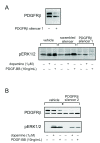
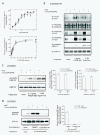
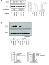
Growth factor-induced receptor dimerization and cross-phosphorylation are hallmarks of signal transduction via receptor tyrosine kinases (RTKs). G protein-coupled receptors (GPCRs) can activate RTKs through a process known as transactivation. The prototypical model of RTK transactivation involves ligand-mediated RTK dimerization and cross-phosphorylation. Here, we show that the platelet-derived growth factor receptor beta (PDGFRbeta) transactivation by the dopamine receptor D4 (DRD4) is not dependent on ligands for PDGFRbeta. Furthermore, when PDGFRbeta dimerization is inhibited and receptor phosphorylation is suppressed to near basal levels, the receptor maintains its ability to be transactivated and is still effective in signaling to ERK1/2. Hence, the DRD4-PDGFRbeta-ERK1/2 pathway can occur independently of a PDGF-like ligand, PDGFRbeta cross-phosphorylation and dimerization, which is distinct from other known forms of transactivation of RTKs by GPCRs.
Figures


Similar articles
-
The dopamine D4 receptor activates intracellular platelet-derived growth factor receptor beta to stimulate ERK1/2.Cell Signal. 2010 Feb;22(2):285-90. doi: 10.1016/j.cellsig.2009.09.031. Epub 2009 Sep 24. Cell Signal. 2010. PMID: 19782129
-
Potentiation of platelet-derived growth factor receptor-β signaling mediated by integrin-associated MFG-E8.Arterioscler Thromb Vasc Biol. 2011 Nov;31(11):2653-64. doi: 10.1161/ATVBAHA.111.233619. Arterioscler Thromb Vasc Biol. 2011. PMID: 21868707 Free PMC article.
-
Mutation of tyrosine residue 857 in the PDGF beta-receptor affects cell proliferation but not migration.Cell Signal. 2010 Sep;22(9):1363-8. doi: 10.1016/j.cellsig.2010.05.004. Epub 2010 May 18. Cell Signal. 2010. PMID: 20494825
-
The Ubiquitin Ligases c-Cbl and Cbl-b Negatively Regulate Platelet-derived Growth Factor (PDGF) BB-induced Chemotaxis by Affecting PDGF Receptor β (PDGFRβ) Internalization and Signaling.J Biol Chem. 2016 May 27;291(22):11608-18. doi: 10.1074/jbc.M115.705814. Epub 2016 Apr 5. J Biol Chem. 2016. PMID: 27048651 Free PMC article.
-
Receptor tyrosine kinase transactivation: fine-tuning synaptic transmission.Trends Neurosci. 2003 Mar;26(3):119-22. doi: 10.1016/S0166-2236(03)00022-5. Trends Neurosci. 2003. PMID: 12591212 Review.
Cited by
-
Platelet-derived growth factor receptor-β and epidermal growth factor receptor in pulmonary vasculature of systemic sclerosis-associated pulmonary arterial hypertension versus idiopathic pulmonary arterial hypertension and pulmonary veno-occlusive disease: a case-control study.Arthritis Res Ther. 2011 Apr 14;13(2):R61. doi: 10.1186/ar3315. Arthritis Res Ther. 2011. PMID: 21492463 Free PMC article.
-
The PDGF family in renal fibrosis.Pediatr Nephrol. 2012 Jul;27(7):1041-50. doi: 10.1007/s00467-011-1892-z. Epub 2011 May 21. Pediatr Nephrol. 2012. PMID: 21597969 Review.
-
Dopamine receptors - IUPHAR Review 13.Br J Pharmacol. 2015 Jan;172(1):1-23. doi: 10.1111/bph.12906. Br J Pharmacol. 2015. PMID: 25671228 Free PMC article. Review.
-
The cellular model for Alzheimer's disease research: PC12 cells.Front Mol Neurosci. 2023 Jan 4;15:1016559. doi: 10.3389/fnmol.2022.1016559. eCollection 2022. Front Mol Neurosci. 2023. PMID: 36683856 Free PMC article.
References
-
- Heldin CH, Ostman A, Ronnstrand L. Signal transduction via platelet-derived growth factor receptors. Biochim Biophys Acta. 1998;1378:F79–113. - PubMed
Publication types
MeSH terms
Substances
LinkOut - more resources
Full Text Sources
Miscellaneous

