. 2010 Oct 1;346(1):113-26.
doi: 10.1016/j.ydbio.2010.07.020. Epub 2010 Jul 24.

C. elegans CAND-1 regulates cullin neddylation, cell proliferation and morphogenesis in specific tissues

Affiliations

C. elegans CAND-1 regulates cullin neddylation, cell proliferation and morphogenesis in specific tissues

Dimple R Bosu et al. Dev Biol. .

Abstract

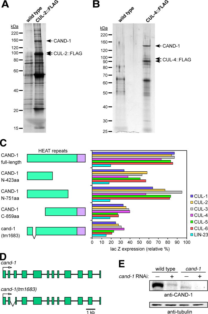
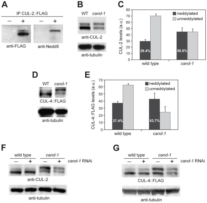
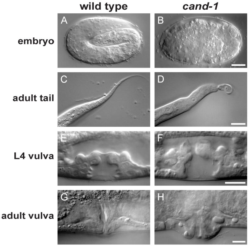
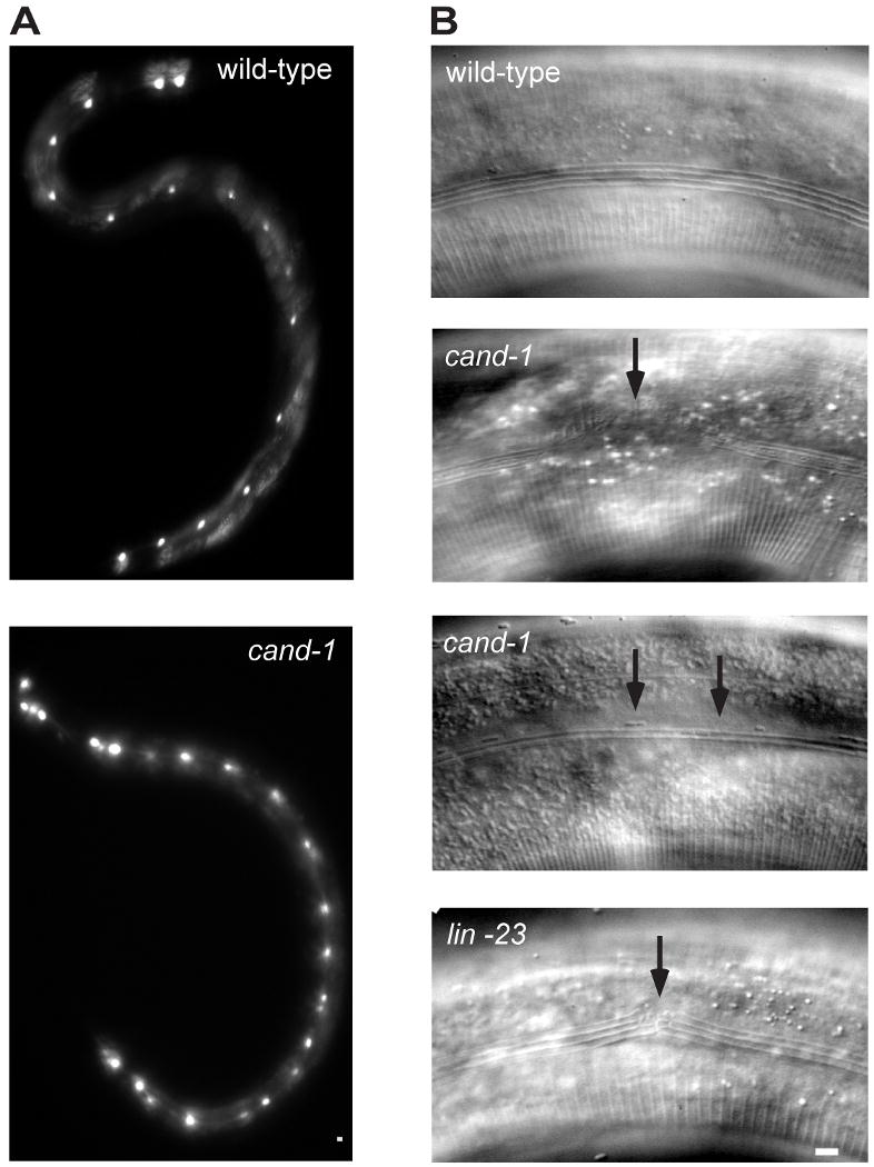
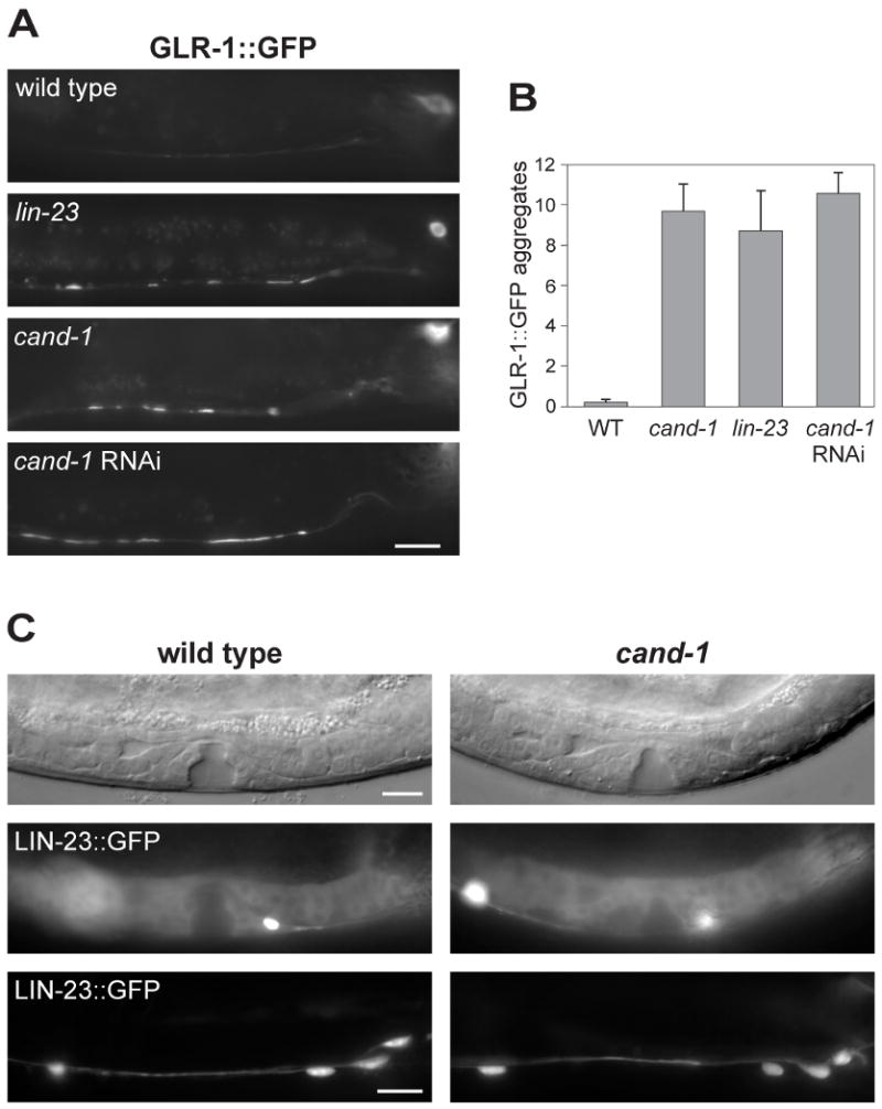
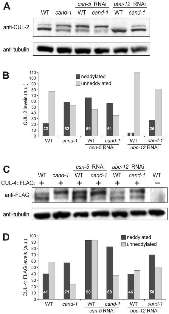
Cullin-RING ubiquitin ligases (CRLs) are critical regulators of multiple developmental and cellular processes in eukaryotes. CAND1 is a biochemical inhibitor of CRLs, yet has been shown to promote CRL activity in plant and mammalian cells. Here we analyze CAND1 function in the context of a developing metazoan organism. Caenorhabditis elegans CAND-1 is capable of binding to all of the cullins, and we show that it physically interacts with CUL-2 and CUL-4 in vivo. The covalent attachment of the ubiquitin-like protein Nedd8 is required for cullin activity in animals and plants. In cand-1 mutants, the levels of the neddylated isoforms of CUL-2 and CUL-4 are increased, indicating that CAND-1 is a negative regulator of cullin neddylation. cand-1 mutants are hypersensitive to the partial loss of cullin activity, suggesting that CAND-1 facilitates CRL functions. cand-1 mutants exhibit impenetrant phenotypes, including developmental arrest, morphological defects of the vulva and tail, and reduced fecundity. cand-1 mutants share with cul-1 and lin-23 mutants the phenotypes of supernumerary seam cell divisions, defective alae formation, and the accumulation of the SCF(LIN-23) target the glutamate receptor GLR-1. The observation that cand-1 mutants have phenotypes associated with the loss of the SCF(LIN-23) complex, but lack phenotypes associated with other specific CRL complexes, suggests that CAND-1 is differentially required for the activity of distinct CRL complexes.


Figures

Figure 1
Figure 1. Interaction of CAND-1 with C. elegans cullins
CAND-1 co-immunoprecipitates with CUL-2∷FLAG (A) and CUL-4∷FLAG (B). Silver-stained SDS-PAGE gels are shown for anti-FLAG affinity purifications from strains containing CUL-2∷FLAG or CUL-4∷FLAG, and from control wild-type animals. The CAND1 protein band (labeled) was identified by mass spectrometry. (C) Two-hybrid analysis of interaction between CAND-1 and the C. elegans cullins. On the left are diagrams of full-length, truncated, and tm1683 mutant CAND-1 proteins (the names reflect the number of N- or C-terminal amino acids remaining in the truncations; the HEAT-repeat region is in green). On the right is a graph of quantitation of interactions between the six cullin proteins and the CAND-1 proteins using a two-hybrid lacZ expression assay. CAND-1 was expressed from the pACT2 vector (fused to the Gal4 activation domain), and cullins or the negative control LIN-23 (Kipreos et al., 2000) are in the pAS1-CYH2 vector (fused to the Gal4 DNA binding domain). The scale derives from the level of the positive control (interaction between pACT2/SKR-1 and pAS1-CYH2/CUL-1; not shown), which is set at 100%. (D) Schematic of the cand-1 genomic region on chromosome V for wild type and the tm1683 deletion mutant. Exons are represented as boxes and lines represent introns. An arrow indicates the translational start point. The region deleted in the tm1683 mutant allele is encompassed by a ‘V-shaped’ lower line. (E) Effect of RNAi on CAND-1 protein levels in wild type and cand-1 mutants. cand-1 RNAi depletion is denoted by a plus above the lanes. Note that cand-1 mutants have lower CAND-1 levels than wild type, and that cand-1 RNAi further reduces CAND-1 levels.
Figure 2
Figure 2. CAND-1 expression pattern
Images of wild-type animals stained with anti-CAND-1 antibody and DAPI. (A, B) in situ staining of early embryos. The one-cell stage zygote is denoted by an arrowhead, and has maternal and paternal pronuclei visible. (C, D) Early embryo at ∼100-cell stage. (E, F) A pretzel-stage embryo, which is the last of the defined embryo stages and has largely completed embryonic cell divisions and morphogenesis. Note that CAND-1 staining decreases in the older embryo. (G, H) The posterior of an L1-stage larva with CAND1 expression in the rectal epithelia and intestinal cells (I). (I, J) An L2-stage larva with CAND-1 staining in somatic gonadal cells (s.g.), germ cells, and intestine cells. (K, L) A wild-type adult with CAND-1 staining in germ cells, oocytes, and intestinal cells. (M-P) Dissected adult gonads from wild-type (M, N) and cand-1(RNAi) (O, P) hermaphrodite adults. Note that CAND-1 staining is significantly decreased in the cand-1(RNAi) gonad. Scale bars, 10 μm.
Figure 3
Figure 3. CAND-1 negatively regulates CUL-2 and CUL-4 neddylated isoform levels
(A) Anti-FLAG immunoprecipitation from wild type (-) or animals expressing CUL-2∷FLAG (+), probed by Western blot with anti-FLAG and anti-Nedd8 antibodies. (B) Whole-worm lysates from wild type and cand-1 mutants blotted with anti-CUL-2 and anti-tubulin antibodies. (C) Graph of the average level of covalently-modified (neddylated) or unneddylated CUL-2 isoforms in wild type and cand-1 mutants from nine independent experiments. The levels of CUL-2 isoforms in cand-1 mutants are normalized to the total CUL-2 signal of wild-type animals, which is set to 100 arbitrary units (a.u.). The percentage of CUL-2 that is in the neddylated isoform relative to the total CUL-2 within a genotype is provided in white lettering within the neddylated isoform bar. This percentage is significantly different between wild type and cand-1 mutants (p < 0.0001, n=9; Student's T-test). (D) Whole-worm lysates prepared from wild type and cand-1 mutants expressing CUL-4∷FLAG and probed with anti-FLAG and anti-tubulin antibodies. (E) Graph of the average covalently-modified (neddylated) and unneddylated CUL-4∷FLAG isoforms in wild type and cand-1 mutants from seven independent experiments. Normalization and labeling are as in (C). The percentage of the neddylated CUL-4∷FLAG isoform is significantly different between wild type and cand-1 mutants (p < 0.0005, n=7). (F) Whole-worm lysates from wild type and cand-1 mutants with (+) or without (-) cand-1 RNAi, probed with anti-CUL-2 and anti-tubulin antibodies. (G) Whole worm lysates from wild type and cand-1 mutants expressing CUL-4∷FLAG with or without cand-1 RNAi, probed with anti-FLAG and anti-tubulin antibodies.
Figure 4
Figure 4. cand-1 mutant phenotypes
DIC images of a pretzel-stage wild-type embryo (A) and an arrested cand-1 mutant embryo (B). DIC images for wild type and cand-1 mutants of the adult tail (C, D), L4-stage vulva (E, F), and adult vulva (G, H). Scale bars, 10 μm.
Figure 5
Figure 5. cand-1 mutant has more seam cells and defective alae
(A) Epifluorescence images of seam cell marker scm∷GFP signal in seam cells from wild type (top) and cand-1 mutant (bottom). In these images, the wild-type adult has 16 seam cells on its lateral side, and the cand-1 adult has 22 seam cells. (B) DIC images of adult alae in wild-type (top), cand-1 mutant (two middle images), and lin-23 mutant (bottom). Note that wild-type alae have four ridges. In cand-1 mutants, alae are often missing from sections (second panel) or have defective morphology (third panel). The lin-23 mutant, which also exhibits excessive seam cell numbers, has similar defects in alae formation (bottom panel and data not shown). Scale bars, 10 μm.
Figure 6
Figure 6. The glutamate receptor GLR-1 accumulates in cand-1 mutants
(A) Epifluorescence images of GLR-1∷GFP signal in the posterior ventral nerve cord of wild-type, lin-23(e1883) mutant, or cand-1 (tm1683) mutant young adults. Note that the signal from nerve cell bodies (upper right) is out of focus in several of the images. (B) A graph of the number of GLR-1∷GFP aggregates per ventral nerve cord that are ≥ 2 μm in length, n=10 animals. (C) Analysis of LIN-23∷GFP levels in wild type and cand-1(tm1683) mutants. The vulva and gonadal regions of early L4-stage stage animals are shown as DIC images (top) or LIN-23∷GFP epifluorescence images (middle). LIN-23∷GFP epifluorescence in the posterior ventral nerve cord of young adults are shown in the bottom panels. Posterior is to the right in all images. Scale bars, 10 μm.
Figure 7
Figure 7. Co-inactivation of CAND-1 and neddylation pathway components
(A) Whole-worm lysates from wild type and cand-1 mutants with or without RNAi depletion of csn-5 (COP9 Signalosome deneddylase) or ubc-12 (Nedd8 E2), and probed with anti-CUL-2 and anti-tubulin antibodies. (B) Quantitation of the neddylated and unneddylated CUL-2 isoform levels in (A) relative to tubulin levels. The levels of CUL-2 isoforms in cand-1 mutants are normalized to the total CUL-2 signal of wild-type animals (set to 100 a.u.); and the percentage of neddylated isoform within a genotype is provided in white lettering. (C) Total-worm lysates prepared from wild type and cand-1 mutants expressing CUL-4∷FLAG with or without csn-5 or ubc-12 RNAi, and probed with anti-FLAG or anti-tubulin antibodies. (D) Quantitation of slower-migrating covalently-modified and unmodified CUL-4∷FLAG isoforms in (C). Normalization and labeling are as in (B).

References

    1. Bosu DR, Kipreos ET. Cullin-RING ubiquitin ligases: global regulation and activation cycles. Cell Div. 2008;3 - PMC - PubMed
    1. Cheng Y, Dai X, Zhao Y. AtCAND1, a HEAT-repeat protein that participates in auxin signaling in Arabidopsis. Plant Physiol. 2004;135:1020–6. - PMC - PubMed
    1. Chew EH, Hagen T. Substrate-mediated regulation of cullin neddylation. J Biol Chem. 2007;282:17032–40. - PubMed
    1. Chew EH, Poobalasingam T, Hawkey CJ, Hagen T. Characterization of cullin-based E3 ubiquitin ligases in intact mammalian cells--evidence for cullin dimerization. Cell Signal. 2007;19:1071–80. - PubMed
    1. Chuang HW, Zhang W, Gray WM. Arabidopsis ETA2, an apparent ortholog of the human cullin-interacting protein CAND1, is required for auxin responses mediated by the SCF(TIR1) ubiquitin ligase. Plant Cell. 2004;16:1883–97. - PMC - PubMed

Publication types

MeSH terms

LinkOut - more resources