. 2010 Oct 1;346(1):68-79.
doi: 10.1016/j.ydbio.2010.07.018. Epub 2010 Jul 24.

Division of labor: subsets of dorsal-appendage-forming cells control the shape of the entire tube

Affiliations

Division of labor: subsets of dorsal-appendage-forming cells control the shape of the entire tube

Michael J Boyle et al. Dev Biol. .

Abstract

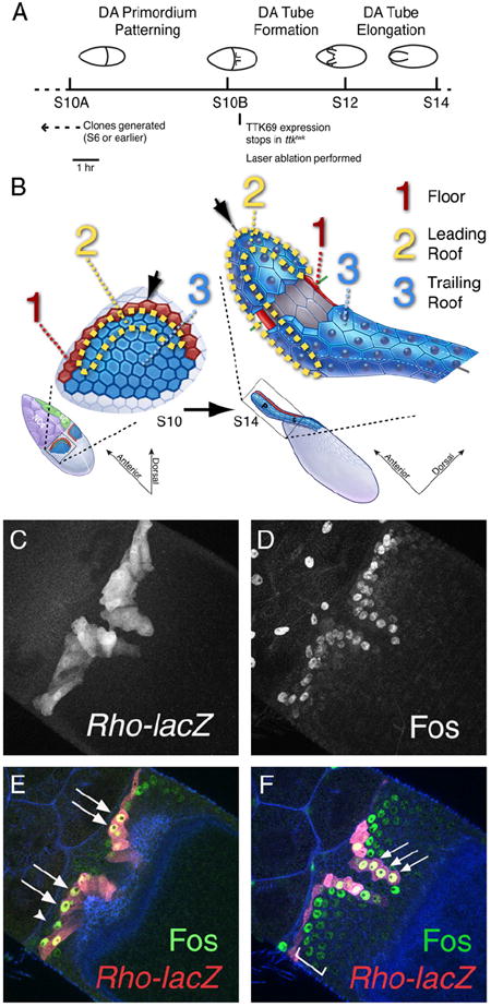
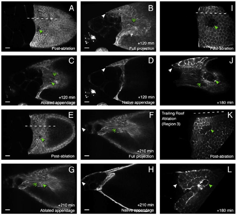
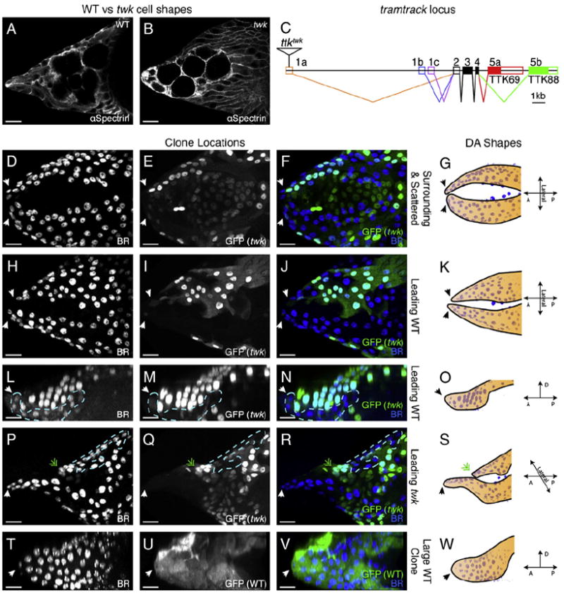
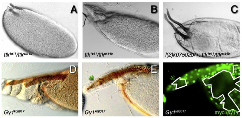
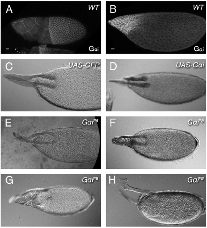
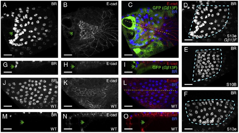
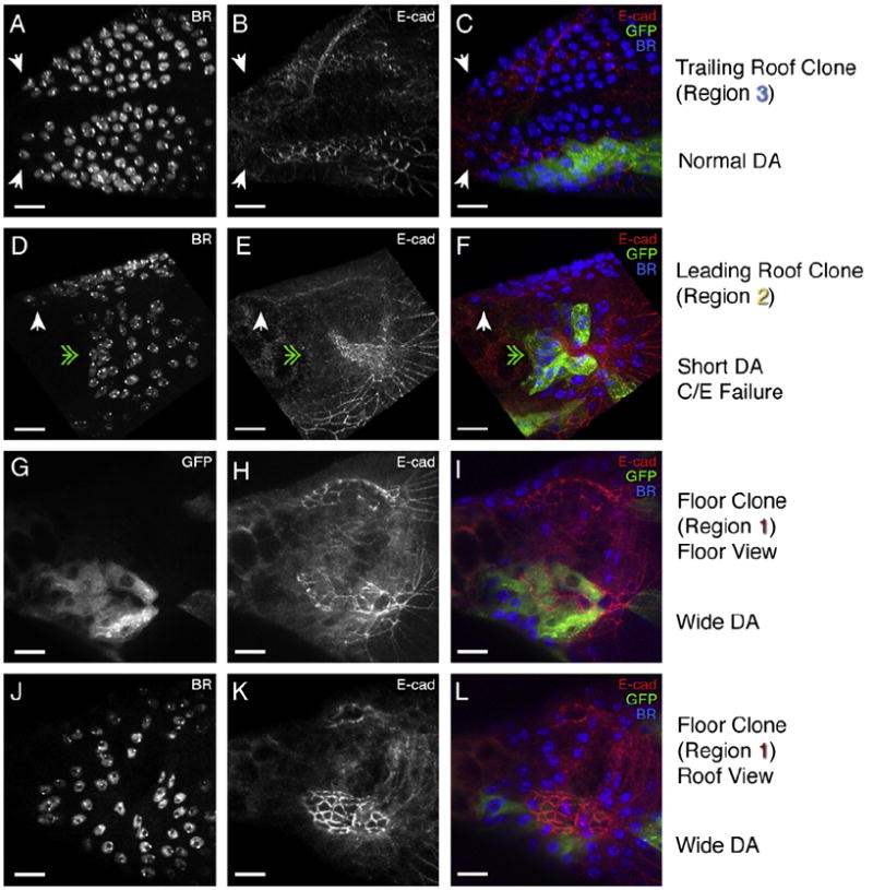
The function of an organ relies on its form, which in turn depends on the individual shapes of the cells that create it and the interactions between them. Despite remarkable progress in the field of developmental biology, how cells collaborate to make a tissue remains an unsolved mystery. To investigate the mechanisms that determine organ structure, we are studying the cells that form the dorsal appendages (DAs) of the Drosophila melanogaster eggshell. These cells consist of two differentially patterned subtypes: roof cells, which form the outward-facing roof of the lumen, and floor cells, which dive underneath the roof cells to seal off the floor of the tube. In this paper, we present three lines of evidence that reveal a further stratification of the DA-forming epithelium. Laser ablation of only a few cells in the anterior of the region causes a disproportionately severe shortening of the appendage. Genetic alteration through the twin peaks allele of tramtrack69 (ttk(twk)), a female-sterile mutation that leads to severely shortened DAs, causes no such shortening when removed from a majority of the DA-forming cells, but rather, produces short appendages only when removed from cells in the very anterior of the tube-forming tissue. Additionally we show that heterotrimeric G-protein function is required for DA morphogenesis. Like TTK69, Gbeta 13F is not required in all DA-forming follicle cells but only in the floor and leading roof cells. The different phenotypes that result from removal of Gbeta 13F from each region demonstrate a striking division of function between different DA-forming cells. Gbeta mutant floor cells are unable to control the width of the appendage while Gbeta mutant leading roof cells fail to direct the elongation of the appendage and the convergent-extension of the roof-cell population.


Figures

Fig. 1
Fig. 1
An overview of dorsal-appendage morphogenesis and preliminary evidence of subdivision within the roof-cell region. (A) A timeline of DA morphogenesis. Mosaics are generated at S6 or earlier, before entry into the endocycle, and well before the DA primordium is patterned at S10. In the case of ttktwk, however, due to the selective nature of the allele, TTK69 expression does not cease until S10B, delaying the onset of defects. We performed laser ablations at S10B or early S11. By S12 the DA tube has formed and it elongates during S12, completing elongation in early S13. (B) Subregions of the DA-forming epithelium before (S10) and after (S14) DA tube formation and elongation. The roof cells (blue; regions 2 and 3) express high levels of the transcription factor Broad (BR) and form the roof of the DA lumen. The gray region in the cut-away in the right half of the panel defines the apical surfaces of the roof cells. The floor cells (red; region 1) dive beneath the roof to seal off the floor of the tube. Floor cells eventually contact the stretch cells (green) overlying the nurse cells (purple). We further divide the roof cells conceptually into two regions: the “leading roof” (region 2), those roof cells that contact the floor cells and span the “hinge” (black arrowhead), and the “trailing roof” (region 3), which consists of the rest of the roof-cell population. As the DA tube forms and elongates, the hinge closes in a convergent-extension movement and the cells at the corner of the hinge (black arrowhead) lead the anterior migration. Schematics modified with permission from Dorman et al., 2004. (C-F) FOS expression in relation to the floor cells in a S11 egg chamber; dorsal view, anterior at the top left. (C) Optical projection shows rho-lacZ-expressing floor cells. (D) FOS is expressed in stretch cells, floor cells, and the first row of roof cells. (E,F) Samples are stained for FOS (green), rho-lacZ (red), and F-actin (phalloidin, blue). In ascending optical sections, both anterior (E) and medial (F) rows of rho-lacZ floor cells express high levels of FOS (arrows). FOS is expressed in a gradient within the columnar FCs, with highest levels in the floor cells and decreasing levels in the roof cells (bracket in F).
Fig. 2
Fig. 2
Ablation of cells in the anterior of the DA-forming region blocks DA elongation (A–L). Four cultured egg chambers (A–D, E–H, I–J, and K–L) with laser-ablated cells in the DA primordium (green solid-arrowheads). The egg chambers pictured in A–D and E–H have 3–5 ablated cells in the vicinity of the hinge point of the DA primordium (green solid-arrowheads in A and E). The egg chamber pictured in I–J contains a large patch of ~8–11 ablated cells including the hinge region. The egg chamber pictured in K–L contains a large patch of ~11 ablated cells in the last two rows (most posterolateral) of roof cells. Anterior is to the left and dotted white lines in A, E, I, and K indicate the approximate position of the dorsal midline. A, E, I, and K show S10B egg chambers just after ablation. Ablated cells exhibit reduced GFP–moesin due to photobleaching. After ablation the egg chambers were allowed to develop and imaged again at various time points. After 120 min, the egg chamber shown in A has matured to S12 (B–D). After 210 min, the egg chamber shown in E has matured to S13 (F–H). After 180 min, the egg chambers shown in I and K have matured to S12 (J,K). E and F are confocal projections revealing both the ablated and native DA-forming tubes. C and G are confocal sections showing the blocked migration of the affected appendage (green double-arrowheads). D and H are confocal sections showing the elongated lumen of the native (untreated) DA tube (white arrowheads). J is a confocal section showing both the blocked migration of the affected appendage (green double-arrowhead) and the migration of the native one (white arrowhead). L is a confocal section showing the elongation of the affected appendage (white arrowhead). When the location of the ablation includes the hinge region (green arrowheads; A–J) the affected appendage fails to elongate (green double-arrowheads). When the ablated cells (green solid-arrowhead) are located in the posterior (region 3), the appendage elongates (white arrowhead; K–L). Scale bars are 20 μm.
Fig. 3
Fig. 3
TTK69 is required for DA elongation only in the anterior of the tube. (A–B) Wild-type (A) and ttktwk mutant (B) DA-forming cell shapes as revealed by αSpectrin staining, which primarily localizes to the lateral membranes. ttktwk cells become thin and elongated and remain apically constricted, resulting in the characteristic “twk” phenotype of short DAs (Boyle and Berg, 2009). (C) A gene diagram depicting the exons of the ttk locus (filled boxes are coding regions, open boxes are non-coding). The ttk gene contains three promoters that produce alternative first exons, and two alternatively spliced final exons containing distinct zinc-finger domains, of which only the TTK69 zinc finger is active in the follicle cells during oogenesis (French et al., 2003). The twk allele results from a P-element insertion into one of the three alternative first exons, 1a. After the start of S10B, only transcripts containing this exon continue to be expressed, meaning that ttktwk is effectively a temporally selective allele, removing TTK69 expression starting during S10B (Boyle and Berg, 2009; French et al., 2003). (D–W) Four examples of S13 egg chambers bearing MARCM clones homozygous for ttktwk (marked by GFP expression) differing by clone position (D–S), and one wild-type clone (T–W). Anterior is to the left and DA-roof cells are marked by nuclear BR expression (D, H, L, P, and T; blue in F, J, N, R, and V). G, K, O, S, and W diagram the shapes of the DA primordia at S13 that result from clones in these locations and indicate the orientation of the egg chamber pictured. (D–G) A dorsal view of an egg chamber bearing two normally elongated DA tubes (white arrowheads). Many cells are mutant for ttktwk, including floor, centripetal, and stretch cells (cells green, but not blue, in F) as well as about 1/3 of the roof of the right appendage (cyan cells in F). In spite of these ttktwk mutant cells, the appendages have elongated normally (diagrammed in G). (H–K) Another dorsal view of an egg chamber bearing two normally elongated DA tubes (white arrowheads). The top appendage is composed of more than half ttktwk mutant cells while the bottom appendage contains very few mutant cells. Critically, the anterior-most DA-forming cells are wild type. The top appendage is ~3–4 μm shorter than the bottom appendage which may result from the clone, but is likely within natural variation at this stage. (L–O) A lateral view of a normally extended DA tube (white arrowhead). About 2/3 of the BR cells forming the roof of the tube are ttktwk mutants, but the elongation is unaffected. The dotted line outlines the patch of wild-type cells; note that this patch includes the leading roof cells at the tip (white arrowhead). (P–S) A dorsolateral view of an egg chamber with one elongated (white arrowhead) and one short appendage (green double-arrowhead). The short appendage is ~66 μm shorter than the wild-type one and contains approximately the same proportion of wild-type and mutant cells as the elongated tube pictured in L–N(~1/3 of the cells are wild-type and are outlined by the dotted line), but their location differs. While the wild-type cells in L–N (as well as in D–F and H–J) include the leading roof, the leading roof cells of the short appendage pictured in P–R are ttktwk mutant (green double-arrowhead). (T–W) Unlike ttktwk, a large wild-type clone covering the entire appendage results in a normally elongated tube. Scale bars are 20 μm.
Fig. 4
Fig. 4
Gγ1 is a Suppressor of the twk phenotype and results in shortened appendages. A and B demonstrate the range of DA phenotypes present in ttk1e11/ttkex14b transheterozygotes used to screen for genetic modifiers of the ttktwk phenotype. C shows an example of lengthened DAs present when this background is combined with a dominant suppressor (in this case l(2)k07502b). (D) DAs of unequal length generated by females bearing clones of Gγ1k08017. (E–F) A rare late stage egg chamber where the negatively marked clone of Gγ1k08017 can be visualized (absence of myc tag defines homozygous cells; F) resulting in a shortened DA (green double-arrowhead). (E) The jagged edge of the mutant DA is silhouetted against the wild-type appendage that lies behind it. (F) Mutant cells are outlined in white. The green nuclei to the anterior of the small clone mark the anterior tip of the wild-type appendage that bends into focus in E.
Fig. 5
Fig. 5
Gαi is required for proper DA morphogenesis. (A–B) Expression of Gαi protein during late oogenesis. Gαi is initially expressed throughout the follicular epithelium through Stage 10 (A and data not shown) but then becomes elevated in the anterior and reduced in the posterior late in oogenesis (B). (C) Normal DAs on an egg chamber expressing UAS-GFP under the control of CY2-GAL4, which is expressed in all columnar follicle cells. (D) CY2-GAL4-driven expression of UAS-Gαi results in variable short, lumpy DAs. (E–H) The range of abnormal DA phenotypes produced in 22.9% of egg chambers formed by rare surviving adult females homozygous for the Gαi null, GαiP8, ranging from almost normal, but with lumpy tips (E), to short and lumpy (F), extremely short (G), or broad and flat (H). Scale bars are 20 μm.
Fig. 6
Fig. 6
Gβ13F is required for DA elongation and roof-cell convergent extension. (A–C) A S13 egg chamber bearing a large clone mutant for the null allele of Gβ13F, Gβ13FΔ1–96A, which results in short, wide, and misshapen appendages. Anterior is to the left and the green double-arrowhead indicates the tip of the DA tube. A part of the adjacent appendage is visible at the top of the frame. (A) Nuclear BR staining labels the roof cells, which are distributed in an abnormally wide and short configuration. (B) E-cadherin staining reveals the roof-cell apices that line the short DA lumen, which has a flat, jagged anterior face. (C) These phenotypes result from a large clone of Gβ13FΔ1–96A (marked by GFP expression) in the DA primordium. (D–F) A comparison of the roof-cell distribution in the early-S13 Gβ13FΔ1–96A mutant DA primordium pictured in A (D) to the normal S10B roof-cell distribution (E) and the normal roof-cell distribution at early S13 (F). Gβ13FΔ1–96A clones cause a failure in convergent extension, resulting in a roof-cell population distributed like that of a S10B DA primordium: short and wide with flat anterior and medial surfaces and a curved posterolateral surface. (G–I) A re-slice through the lumen in C along the dotted line. Rather than migrating over the nurse cells as they normally would, the roof cells of this DA tube have become blocked at the nurse cells and even dive ventrally one or two cell-lengths with the centripetal cells (green double-arrowhead). (J–L) A DA composed of wild-type cells at a similar stage (13) and orientation to the one shown in A–I. (M–O) A re-slice through the lumen in L along the dotted line. Note that the roof cells migrate over the top of the nurse cells rather than stopping at the nurse-cell boundary (green double-arrowhead) and diving down as occurs in the Gβ13FΔ1–96A clone pictured in I. Scale bars are 20 μm.
Fig. 7
Fig. 7
Gβ13F is required for distinct aspects of DA formation in the leading roof and floor cells, but is dispensable from the trailing roof cells. Three examples of moderately sized Gβ13FΔ1–96A clones at early S13 differing in clone position, each viewed from a dorsal or dorsolateral perspective. Anterior is to the left. (A–C) Normally shaped and elongated DA primordia (white arrowheads). BR (A) and E-cadherin (B) staining show none of the Gβ13FΔ1–96A defects seen in Fig. 6, in spite of a clone in the lower DA primordium occupying >1/3 of the trailing roof-cell population (region 3 from Fig. 1B) (C). (D–F) An extremely short DA lumen (green double-arrowhead), as compared to the length of the normal DA primordium at the top of the panel (white arrowhead). BR staining shows roof cells (D) that have failed to converge and extend yet E-cadherin staining (E) reveals a slim lumen of normal width. These phenotypes result from a Gβ13FΔ1–96A clone almost entirely in the leading roof (region 2). (G–L) Confocal projections emphasizing different depths of a single egg chamber. (G–I) A confocal projection through the floor of two DA lumens. The lower one contains a Gβ13FΔ1–96A clone through most of the floor (G, I). Loss of Gβ13F results in a disorganized floor (G, H). Compare the orderly arrangement of the floor cells in the top lumen to those of the bottom lumen (H). In addition, the lumen is abnormally wide in the middle, although it comes back inward to a point at the anterior. A projection through the roof of DA lumens from the same egg chamber (J–L) shows that only 2 or 3 roof cells are included in the clone, at least one of which is a leading roof cell (which may explain why this lumen has a mild elongation defect). The widening is also visible in the roof (K), even though few roof cells are included in the clone (L). The narrowing back to a point may result from successful convergent extension of the roof cells. BR staining in the roof cells (J) shows an abnormally wide distribution at the posterior of the population, but the anterior of the roof is curved and comes to a point, unlike the roof-cell populations that bear large leading roof-cell clones. Scale bars are 20 μm.

Similar articles

Cited by

References

    1. Althauser C, Jordan KC, Deng WM, Ruohola-Baker H. Fringe-dependent Notch activation and Tramtrack function are required for specification of the polar cells in Drosophila oogenesis. Dev Dyn. 2005;232:1013–1020. - PubMed
    1. Bloor JW, Kiehart DP. zipper nonmuscle myosin-II functions downstream of PS2 integrin in Drosophila myogenesis and is necessary for myofibril formation. Dev Biol. 2001;239:215–228. - PubMed
    1. Boyle MJ, Berg CA. Control in time and space: Tramtrack69 cooperates with Notch and Ecdysone to repress ectopic fate and shape changes during Drosophila egg chamber maturation. Development. 2009;136:4187–4197. - PMC - PubMed
    1. Caussinus E, Colombelli J, Affolter M. Tip-cell migration controls stalk-cell intercalation during Drosophila tracheal tube elongation. Curr Biol. 2008;18:1727–1734. - PubMed
    1. Chen Y, Schüpbach T. The role of brinker in eggshell patterning. Mech Dev. 2006;123:395–406. - PubMed

Publication types

Substances

LinkOut - more resources