. 2010 Oct;153(1):134-42.
doi: 10.1016/j.virusres.2010.07.021. Epub 2010 Jul 24.

Evaluation of innate immunity and vector toxicity following inoculation of bovine, porcine or human adenoviral vectors in a mouse model

Affiliations

Evaluation of innate immunity and vector toxicity following inoculation of bovine, porcine or human adenoviral vectors in a mouse model

Anurag Sharma et al. Virus Res. 2010 Oct.

Abstract

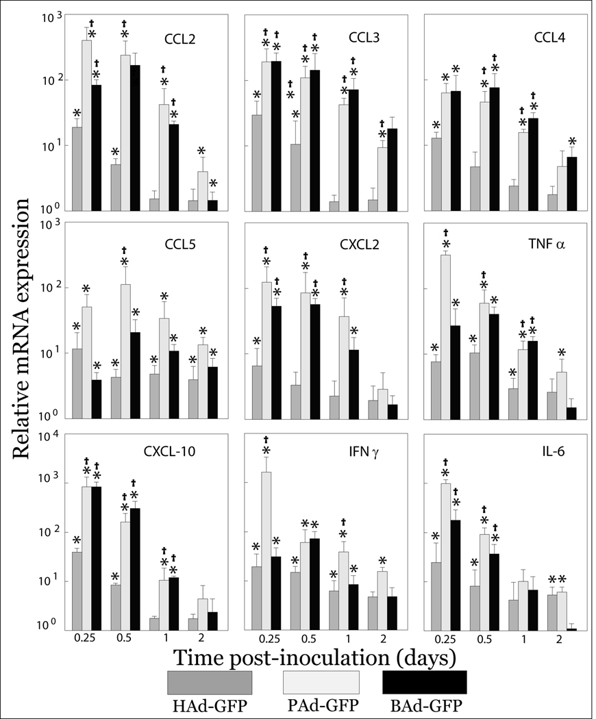
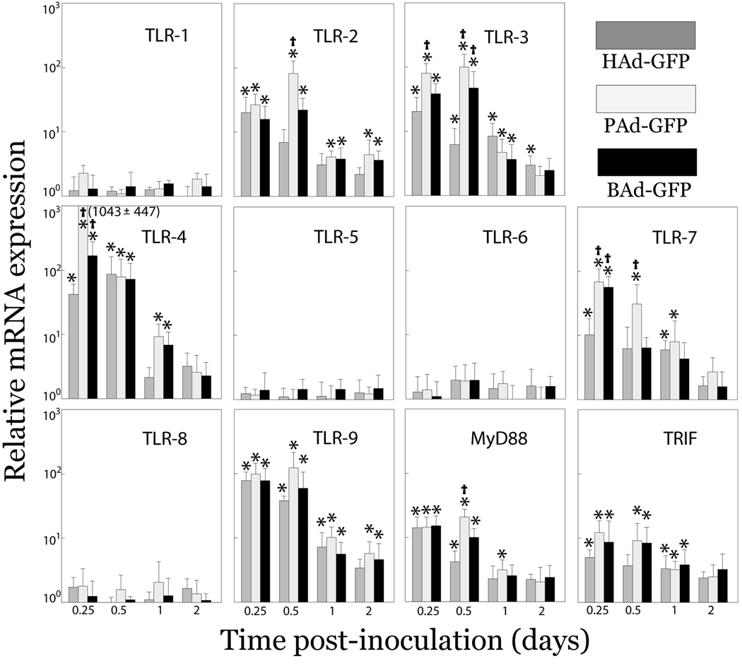
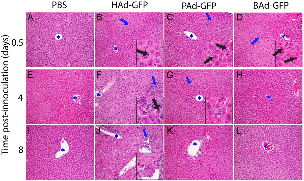
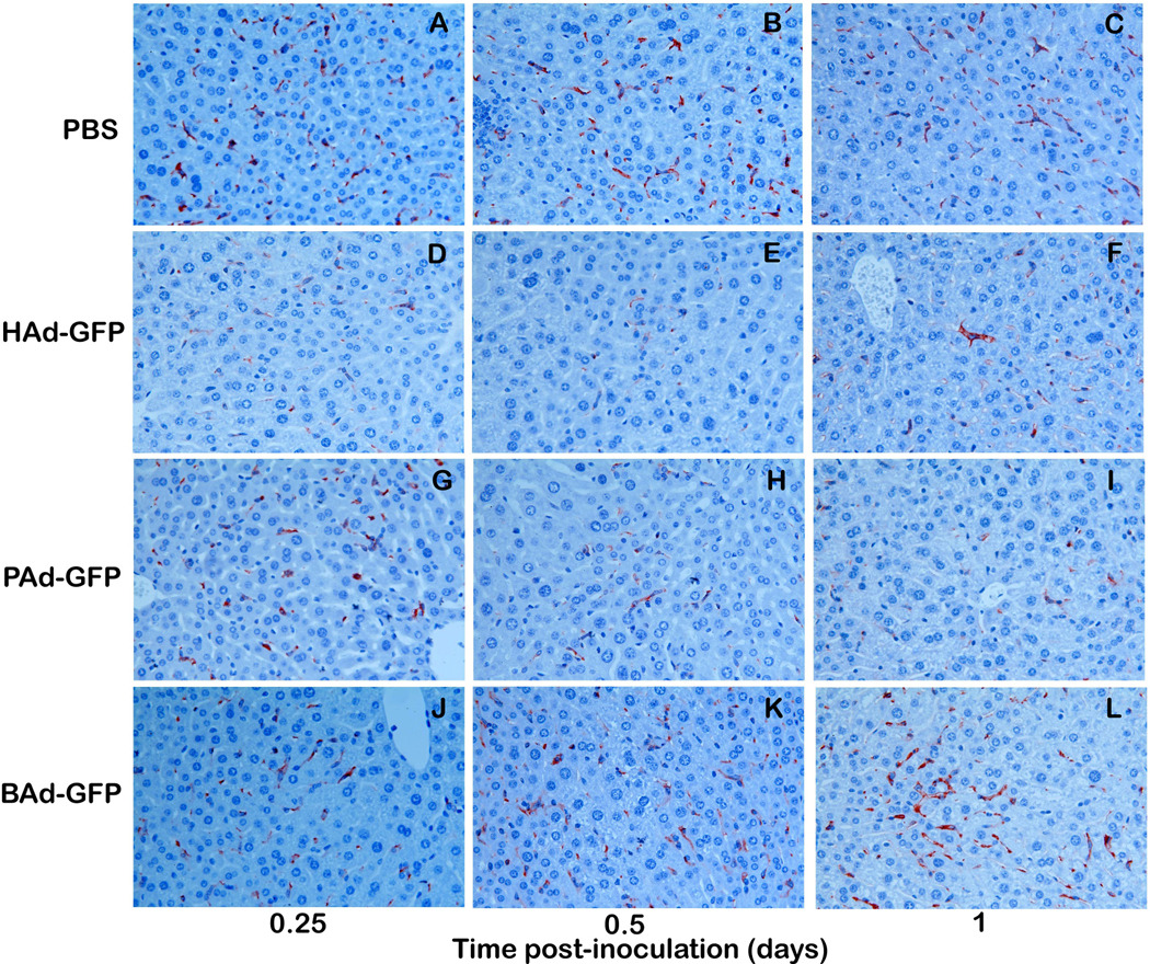
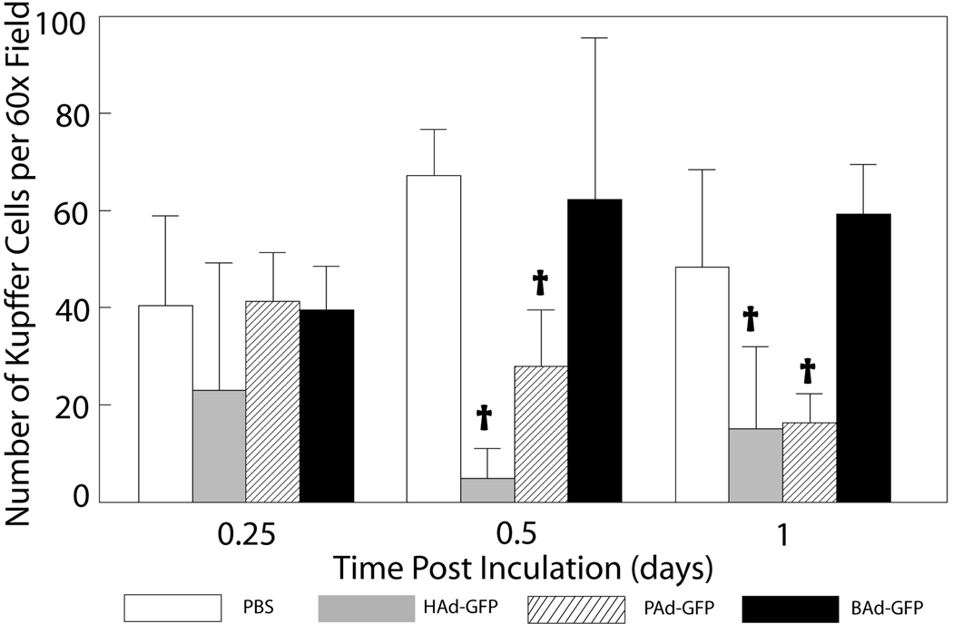
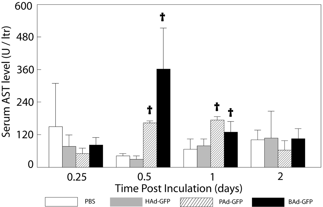
Nonhuman adenovirus (Ad) vectors derived from bovine Ad serotype 3 (BAd3) or porcine Ad serotype 3 (PAd3) can circumvent pre-existing immunity against human Ad (HAd). We have previously reported differential transduction of human and nonhuman cells by these Ad vectors, and their distinct receptor usage and biodistribution. To compare the induction of innate immunity, vector toxicity and vector uptake by Kupffer cells (KCs) following intravenous administration of PAd3, BAd3, or HAd5 vectors in mice, we determined mRNA expression levels of proinflammatory chemokines and cytokines, and Toll-like receptors (TLRs) in the liver and spleen. Tissue toxicity of these vectors was assessed by comparing serum levels of liver-specific enzymes, histopathology and Kupffer cell (KC) depletion. Compared to the HAd5 vector, PAd3 and BAd3 vectors were more potent stimulators of innate immune responses as indicated by enhanced mRNA expression of TLRs and proinflammatory chemokines and cytokine genes. Histopathological changes in the liver were most pronounced in HAd5-inoculated mice while BAd3- or PAd3-inoculated mice revealed mild histologic changes that were confined to early time points. Inoculation with HAd5 or PAd3 vectors resulted in a significant (P<0.05) decline of the number of KCs in the liver. Together, these results extend our previous observations regarding distinct in vivo biology of nonhuman and human Ad vectors.


Figures

Figure 1
Figure 1. Expression levels of proinflammatory cytokines and chemokines mRNA at different time points post-inoculation in the liver of mice inoculated with HAd-GFP, PAd-GFP, or BAd-GFP
Real-time RT-PCR assays for various cytokines and chemokines together with 18S rRNA (as an endogenous control for normalization) were performed using 200 ng of total RNA and comparative quantification of cytokines and chemokines expression levels was achieved. Values are reported relative to mean expression levels observed with mock-inoculated mice at each time point post-inoculation. Values are shown as the mean ± standard deviation for three mice at each time point. *P < 0.05 versus expression level in PBS-inoculated mice. †P < 0.05 for PAd-GFP or BAd-GFP versus HAd-GFP at each time point.
Figure 2
Figure 2. mRNA expression levels of various TLRs and adaptor molecules (Myd88 and TRIF) at different time points post-inoculation in the liver of mice inoculated with HAd-GFP, PAd-GFP, or BAd-GFP
Real-time RT-PCR assays for various TLRs, Myd88, or TRIF together with 18S rRNA (as an endogenous control for normalization) were performed using 200 ng of total RNA and comparative quantification of TLRs, Myd88 and TRIF expression levels was achieved. Values are reported relative to mean expression levels observed with mock-inoculated mice at each time point post-inoculation. Values are shown as the mean ± standard deviation for three mice at each time point. *P < 0.05 versus expression level in PBS-inoculated mice. †P < 0.05 for PAd-GFP or BAd-GFP versus HAd-GFP at each time point.
Figure 3
Figure 3. Representative photomicrographs of the liver sections of mice inoculated with PBS, HAd-GFP, PAd-GFP, or BAd-GFP
Blue asterisks indicate lumen of portal veins and insets represent a high magnification of area indicated by blue arrows. Note scattered apoptotic hepatocytes (black arrows) in Ad-vector-inoculated mice liver at 0.5 day post-inoculation. Multifocal, random lymphocytic infiltration and hepatocellular degeneration/apoptosis as well as portal inflammation were observed only in HAd-GFP-inoculated mice at 4 and 8 days post-inoculation (panels F and J). Rare clusters of macrophages (inset, panel G) were occasionally found in mice from all groups. Stain: hematoxylin and eosin. Original magnification: 200 × (insets 600 ×)
Figure 4
Figure 4
a) Immunohistochemistry for Kupffer cells (KCs) in the liver sections of mice inoculated with PBS, HAd-GFP, PAd-GFP, or BAd-GFP. Formalin-fixed, paraffin-embedded liver tissue sections were processed for immunohistochemical analysis as described in material and methods. Anti-F4/80 labeled KCs stained in brown color could be observed (600 × magnification). b) Quantitative estimation of KCs in the liver sections at various time points. KCs in seven randomly selected non-overlapping fields were counted from each mouse liver section. Values are reported as the mean ± standard deviation for three mice at each time point. †P < 0.05 for PAd-GFP, BAd-GFP or HAd-GFP versus PBS-inoculated mice at each time point.
Figure 4
Figure 4
a) Immunohistochemistry for Kupffer cells (KCs) in the liver sections of mice inoculated with PBS, HAd-GFP, PAd-GFP, or BAd-GFP. Formalin-fixed, paraffin-embedded liver tissue sections were processed for immunohistochemical analysis as described in material and methods. Anti-F4/80 labeled KCs stained in brown color could be observed (600 × magnification). b) Quantitative estimation of KCs in the liver sections at various time points. KCs in seven randomly selected non-overlapping fields were counted from each mouse liver section. Values are reported as the mean ± standard deviation for three mice at each time point. †P < 0.05 for PAd-GFP, BAd-GFP or HAd-GFP versus PBS-inoculated mice at each time point.
Figure 5
Figure 5. Serum levels of AST enzyme in mice inoculated with PBS, HAd-GFP, PAd-GFP, or BAd-GFP
Values are reported as the mean ± standard deviation for three mice at each time point. †P < 0.05 for PAd-GFP, BAd-GFP or HAd-GFP versus PBS-inoculated mice at each time point.

Similar articles

Cited by

References

    1. Mouse Phenome Database. http://www.jax.org/phenome.
    1. Alemany R, Suzuki K, Curiel DT. Blood clearance rates of adenovirus type 5 in mice. J.Gen.Virol. 2000;81:2605–2609. - PubMed
    1. Appledorn DM, Kiang A, McBride A, Jiang H, Seregin S, Scott JM, Stringer R, Kousa Y, Hoban M, Frank MM, Amalfitano A. Wild-type adenoviruses from groups A-F evoke unique innate immune responses, of which HAd3 and SAd23 are partially complement dependent. Gene Ther. 2008a;15:885–901. - PubMed
    1. Appledorn DM, Patial S, McBride A, Godbehere S, van RN, Parameswaran N, Amalfitano A. Adenovirus vector-induced innate inflammatory mediators, MAPK signaling, as well as adaptive immune responses are dependent upon both TLR2 and TLR9 in vivo. J.Immunol. 2008b;181:2134–2144. - PubMed
    1. Bangari DS, Mittal SK. Porcine adenoviral vectors evade preexisting humoral immunity to adenoviruses and efficiently infect both human and murine cells in culture. Virus Res. 2004;105:127–136. - PubMed

Publication types

MeSH terms