. 2010 Dec;40(3):503-17.
doi: 10.1016/j.nbd.2010.07.010. Epub 2010 Jul 24.

Impaired dopaminergic neurotransmission and microtubule-associated protein tau alterations in human LRRK2 transgenic mice

Affiliations

Impaired dopaminergic neurotransmission and microtubule-associated protein tau alterations in human LRRK2 transgenic mice

H L Melrose et al. Neurobiol Dis. 2010 Dec.

Abstract

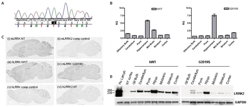
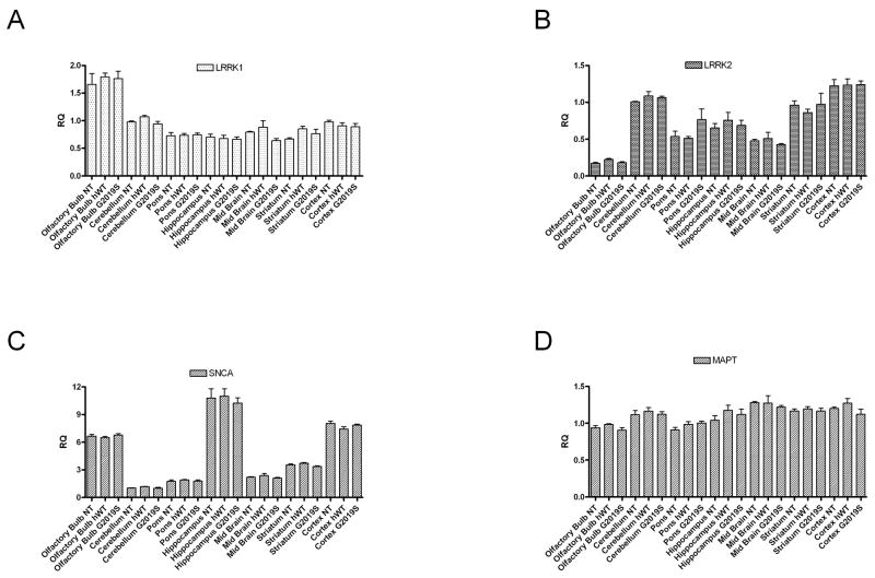
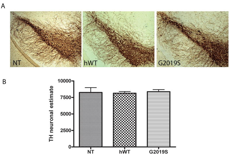
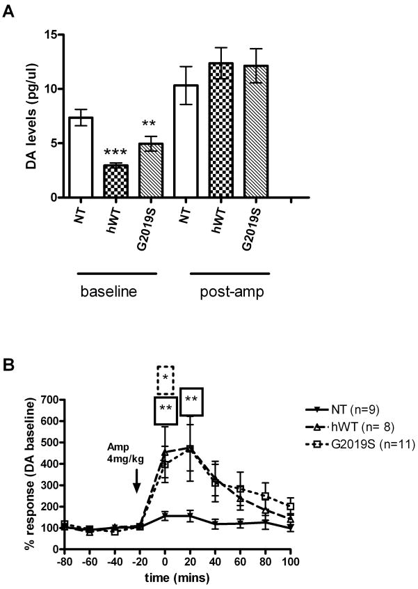
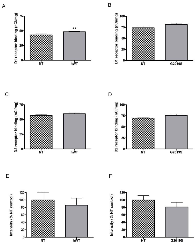
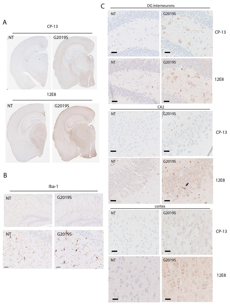
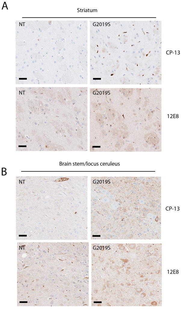
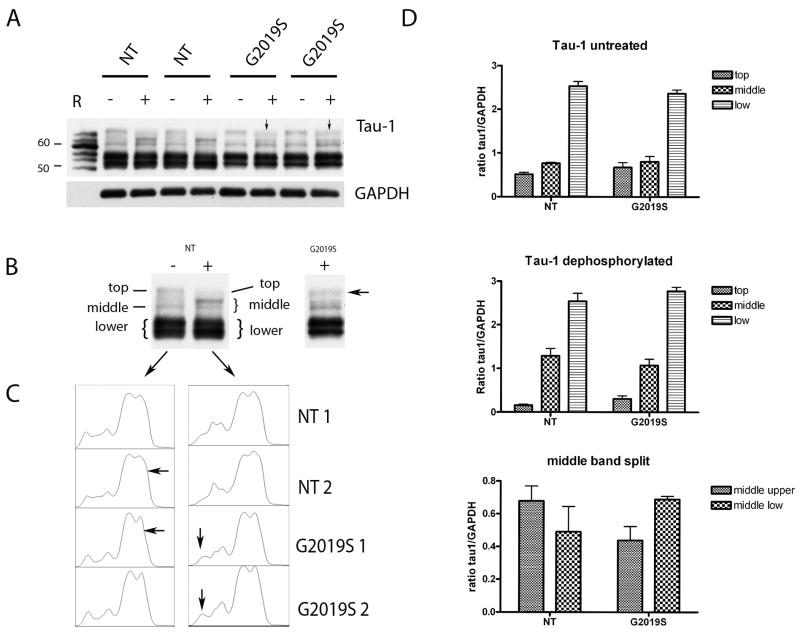
Mutations in the Leucine Rich Repeat Kinase 2 (LRRK2) gene, first described in 2004 have now emerged as the most important genetic finding in both autosomal dominant and sporadic Parkinson's disease (PD). While a formidable research effort has ensued since the initial gene discovery, little is known of either the normal or the pathological role of LRRK2. We have created lines of mice that express human wild-type (hWT) or G2019S Lrrk2 via bacterial artificial chromosome (BAC) transgenesis. In vivo analysis of the dopaminergic system revealed abnormal dopamine neurotransmission in both hWT and G2019S transgenic mice evidenced by a decrease in extra-cellular dopamine levels, which was detected without pharmacological manipulation. Immunopathological analysis revealed changes in localization and increased phosphorylation of microtubule binding protein tau in G2019S mice. Quantitative biochemical analysis confirmed the presence of differential phospho-tau species in G2019S mice but surprisingly, upon dephosphorylation the tau isoform banding pattern in G2019S mice remained altered. This suggests that other post-translational modifications of tau occur in G2019S mice. We hypothesize that Lrrk2 may impact on tau processing which subsequently leads to increased phosphorylation. Our models will be useful for further understanding of the mechanistic actions of LRRK2 and future therapeutic screening.


Figures

Figure 1
Figure 1
(A) Confirmation that transgenic mRNA contains the G2019S mutation. RNA was extracted from F1 G2019S transgenic mouse brain, cDNA synthesized and sequencing with human specific primers to LRRK2 exon 41 performed (the G to A transition highlighted in black). (B) Real time PCR to examine regional expression of human mRNA expression in transgenic mice. Real-time PCR was performed with ABI (gene human LRRK2 specific TaqMan® probe Hs00417273_ml using mouse GAPDH (Mm99999915_ml) as the endogenous reference gene. Data is plotted as mean ± SEM. (C) In situ hybridization reveals resemblance between endogenous murine LRRK2 expression and transgenic expression. (i) Murine LRRK2 was visualized with a probe to mouse exon 15 in NT mice. (ii) Excess unlabeled probe was used as control. (iii) Human LRRK2 in hWT (iv) G2019S mice was visualized with a probe to human exon 41. (v) Excess unlabeled exon 41 probe on transgenic mouse section was used as an additional control. (vi) Specificity of the human probe was confirmed by the absence of signal in NT mice. (D) hWT and G2019S transgenic mice expression human Lrrk2 protein in multiple brain regions. Human Lrrk2 transgenic protein expression in the hWT line and the highest expressing G2019S line was evident throughout the mouse brain. 50μg of protein lysate was loaded per sample and following electrophoresis, membranes were immunoblotted with antibody PA0362. Human lymphoblast was used as a positive control. GAPDH antibody was used as a loading control.
Figure 2
Figure 2. Endogenous LRRK1, LRRK2, SNCA and MAPT levels unchanged in hWT and G2019S BAC mice
No compensatory changes are observed in the expression levels of murine LRRK1, LRRK2, α-synuclein (SNCA) or tau (MAPT) genes in G2019S mice compared to non-transgenic littermates. Real-time PCR was performed with ABI TaqMan® probes to murine (A) LRRK1 (Mm00713303_ml), (B) murine LRRK2 (Mm00481934_ml), (C) murine SNCA (Mm00447333_ml) and (D) murine MAPT (Mm00521988_ml). Mouse GAPDH (Mm99999915_ml) as the endogenous reference gene. Data is plotted as mean ± SEM.
Figure 3
Figure 3. Stereological estimates of TH neurons in the substantia nigra reveal no differences between NT, hWT and G2019S mice
Counting was performed using the optical fractionator probe in tissue processed for TH staining from hWT, G2019S and NT mice aged 22–24 months. (A) TH immunostaining in the nigra of NT, hWT and G2019S mice (B) Neuronal estimates in NT, hWT and G2019S mice. Data is plotted as mean ± SEM.
Figure 4
Figure 4. In vivo microdialysis reveals significantly lower baseline levels of dopamine and elevated response to amphetamine in hWT and G2019S transgenic mice
(A) Dopamine levels pre and post amphetamine treatment in hWT and G2019S mice versus NT. Dopamine levels were measured by HPLC from microdialysis samples collected from the striatum. (B) % response plots for NT, hWT and G2019S following amphetamine challenge. Dopamine levels were measured by HPLC from microdialysis samples collected from the striatum and plotted as a percentage of each individual animals mean baseline levels to compare response over time. Data is plotted as mean ± SEM. * p<0.05, ** p<0.01, ***p<0.001 Mann Whitney Test.
Figure 5
Figure 5. A modest compensatory increase is observed in Dopamine D1 receptors in hWT but not G2019S mice, whereas D2 receptors and DAT levels remain unchanged in hWT and G2019S mice
Quantitative autoradiography was performed with (A, B) D1 receptor ligand [3H] SCH 23390 and (C, D) D2 receptor ligand [3H] methylspiperone in serial striatal sections. (E, F) Quantitative immunohistochemistry with anti-DAT antibody. Data is plotted as mean ± SEM. ** p<0.01
Figure 6
Figure 6. Aged G2019S mice have increased tau phosphorylation and microglial staining compared to age matched NT controls
Immunohistochemistry was performed on paraffin embedded sections using phospho-tau antibodies CP-13 (pSer202) and 12E8 (pSer262/365). (A) Low power images of NT and G2019S mice (B) Upper panel: low power image of microglial staining in the hippocampus of G2019S and NT mice. Lower panel: Higher power image to demonstrate the activated morphology of microglia in G2019S mice (C) CP-13 and 12E8 staining in upper panel is dentate gyrus (DG) interneurons, middle panel CA2 region of hippocampus (arrow denotes tau immoreative glia) and lower panel the cortex of G2019S and NT mice. Scale bars 25μm.
Figure 7
Figure 7. Aged G2019S mice have increased tau phosphorylation in the striatum and brainstem compared to age matched NT controls
Immunohistochemistry was performed on paraffin embedded sections using phospho-tau antibodies CP-13 (pSer202) and 12E8 (pSer262/365). (A) in the striatum and (B) the brain stem at the locus ceruleus. Scale bars 25μm.
Figure 8
Figure 8. Immunoblotting with CP-13 antibody reveals that increased levels of phospho-tau, but not hyperphosphorylated tau, are present in G2019S mice
Lysates were prepared from 18–22 month old G2019S mice sacrificed by craniotomy to preserve phosphorylation. (A) CP-13 (pSer202) immunoblot from cortical and hippocampal lysates. JNP3L mice that overexpress tau-P301L (Lewis et al., 2000) were used as a positive control for hyperphosphorylated tau. Arrows denote bands of interest for quantification. (B) Quantitative analysis of immunoblots. The average ratio of the bands of interest (indicated by arrows) of the two mice analyzed for each region was plotted as mean ± SEM.
Figure 9
Figure 9. Immunoblotting with Tau-1 and PHF-1 antibodies reveals that the ratio of tau species is altered in G2019S mice
(A) PHF-1 (pSer396/404) and Tau-5 (total tau) and immunoblots from multiple brain regions. Arrows denote bands of interest for quantification. (B) Quantitative analysis of immunoblots. The average ratio of the bands of interest (indicated by arrows) of the two mice analyzed for each region was plotted as mean ± SEM.
Figure 10
Figure 10. Tau species composition is altered following desphosphorylation in G2019S mice
(A) Immunoblot with tau-1 antibody in non-treated (−) and dephosphorylated (+) samples. Arrows denote the heavier upper band around 65kDa. Control “R” denotes human recombinant tau isoforms (recombinant murine isoforms were not available). Of note adult mice produce only 4-repeat tau. (B) Example to show how the bands were measured for quantification. (C) Histogram plots from Image J analysis package show the appearance of an additional peak in the G2019S samples. (D) Graphical representation of the tau-1 immunoblots. The mean intensity of the bands of interest of the two mice analyzed for each region was plotted as mean ± SEM.
Figure 11
Figure 11. Modest increases in tau immunostaining were observed in hWT mice in hippocampus
Upper panel shows CP-13 (pSer202) immunostaining in NT and hWT mice. Lower panel shows 12E8 (pSer 262/365) immunostaining in NT and hWT mice. Scale bars 25μm.
Figure 12
Figure 12. Open Field Test reveals abnormal exploratory behavior in G2019S mice
(A) Mean path length in G2019S was found to be significantly different from NT and hWT mice (p<0.006). (B) Overall activity in hWT and G2019S mice was comparable to NT controls. (C) G2019S mice display and almost significant trend towards increased thigmotaxis (wall hugging) tending to (D) take less turns than hWT and NT mice (E) spend less time in innermost zone and (F) at the novel object.

References

    1. Allen JP, et al. Somatostatin receptor 2 knockout/lacZ knockin mice show impaired motor coordination and reveal sites of somatostatin action within the striatum. Eur J Neurosci. 2003;17:1881–95. - PubMed
    1. Andres-Mateos E, et al. Unexpected lack of hypersensitivity in LRRK2 knock-out mice to MPTP (1-methyl-4-phenyl-1,2,3,6-tetrahydropyridine) J Neurosci. 2009;29:15846–50. - PMC - PubMed
    1. Augustinack JC, et al. Specific tau phosphorylation sites correlate with severity of neuronal cytopathology in Alzheimer's disease. Acta Neuropathol. 2002;103:26–35. - PubMed
    1. Biskup S, et al. Localization of LRRK2 to membranous and vesicular structures in mammalian brain. Ann Neurol. 2006;60:557–69. - PubMed
    1. Clelland CD, et al. A functional role for adult hippocampal neurogenesis in spatial pattern separation. Science. 2009;325:210–3. - PMC - PubMed

Publication types

MeSH terms