. 2010 Oct 15;285(42):32557-67.
doi: 10.1074/jbc.M110.141960. Epub 2010 Jul 21.

Mapping the catalytic cycle of Schistosoma mansoni thioredoxin glutathione reductase by X-ray crystallography

Affiliations

Mapping the catalytic cycle of Schistosoma mansoni thioredoxin glutathione reductase by X-ray crystallography

Francesco Angelucci et al. J Biol Chem. .

Abstract

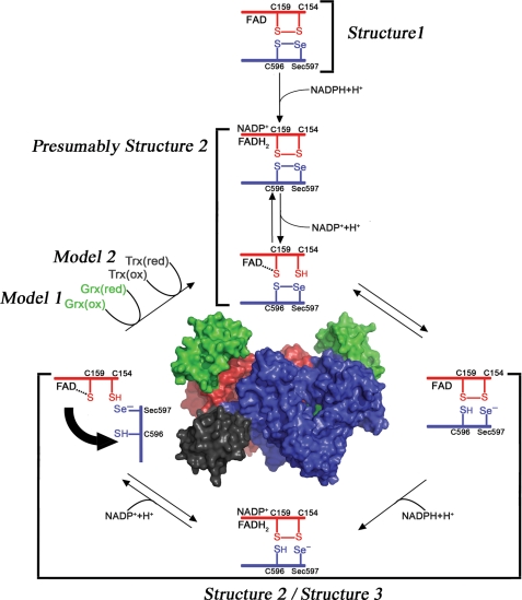
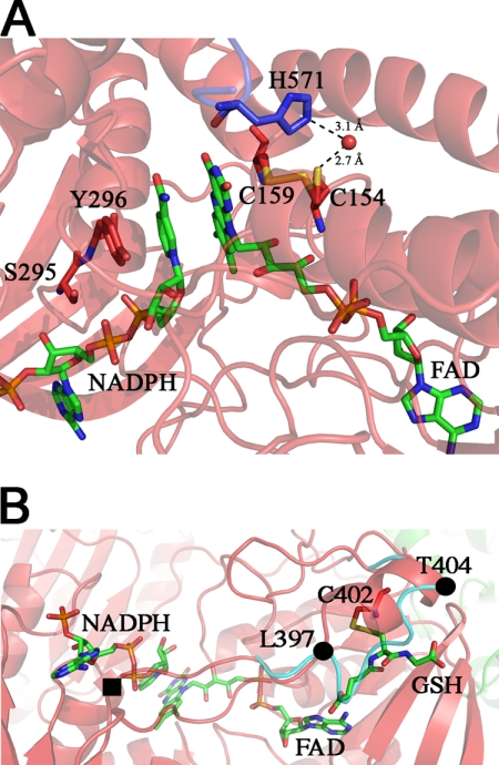
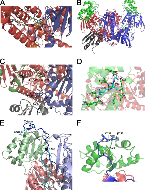
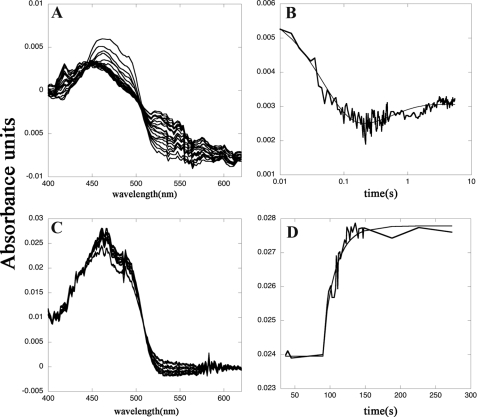
Schistosomiasis is the second most widespread human parasitic disease. It is principally treated with one drug, praziquantel, that is administered to 100 million people each year; less sensitive strains of schistosomes are emerging. One of the most appealing drug targets against schistosomiasis is thioredoxin glutathione reductase (TGR). This natural chimeric enzyme is a peculiar fusion of a glutaredoxin domain with a thioredoxin selenocysteine (U)-containing reductase domain. Selenocysteine is located on a flexible C-terminal arm that is usually disordered in the available structures of the protein and is essential for the full catalytic activity of TGR. In this study, we dissect the catalytic cycle of Schistosoma mansoni TGR by structural and functional analysis of the U597C mutant. The crystallographic data presented herein include the following: the oxidized form (at 1.9 Å resolution); the NADPH- and GSH-bound forms (2.3 and 1.9 Å, respectively); and a different crystal form of the (partially) reduced enzyme (3.1 Å), showing the physiological dimer and the entire C terminus of one subunit. Whenever possible, we determined the rate constants for the interconversion between the different oxidation states of TGR by kinetic methods. By combining the crystallographic analysis with computer modeling, we were able to throw further light on the mechanism of action of S. mansoni TGR. In particular, we hereby propose the putative functionally relevant conformational change of the C terminus after the transfer of reducing equivalents from NADPH to the redox sites of the enzyme.


Figures

FIGURE 1.
FIGURE 1.
Electron transfer in and out of the TR domain of SmTGR. The reaction scheme has been drawn following the experimental evidence reported in this work and elsewhere (18). When NADPH binds to the oxidized state of the enzyme (Structure 1), electrons are transferred from the nicotinamide ring to the FAD; reduced FADH2 donates electrons to the Cys154–Cys159 couple (in red) that is close to the isoalloxazine ring of the cofactor (as observed in Structure 2), and finally electrons reach the Cys596–Sec597 redox couple on the C-terminal segment of the other subunit (in blue, as observed in Structure 3). The C terminus acts as a flexible arm, which might donate electrons either internally to the Cys28–Cys31 redox active couple on the oxidized Grx domain (in green) or externally to various oxidized substrates such as SmTrx (in dark gray).
FIGURE 2.
FIGURE 2.
Structure 2, NADPH-binding site in SmTGRfl. Panel A, zoom in the FAD active site. The NADPH and FAD are shown as green sticks; Tyr296, Ser295, Cys154, and Cys159 are shown as red sticks, and His571 of the partner subunit is shown as blue sticks. Upon NADPH binding, the loop 295–297 changes conformation re-orienting the side chains of Ser295 and Tyr296 with respect to Structure 1 to make room for the reductant. The Cys159–Cys154 couple is partially reduced, and accordingly, Cys154 is found in a double conformation. In the reduced conformation (50% occupancy), the sulfur of Cys154 points toward the solvent and is in contact with a water molecule (2.7 Å, shown as red ball), which is kept in place by His571. Panel B, GSH-binding site on TR domain of Structure 2. The GSH (green sticks) is found in a position that in other TGR structures is usually occupied by the loop 397–404 (cyan ribbon). The ligand is found in a pocket formed by one α-helix (Leu397–Thr404), a β-strand (Lys227–Leu230), and the nucleotide moiety of the FAD. GSH makes a mixed disulfide with Cys402 and forces the loop 397–404 to adopt a α-helical secondary structure (red ribbon) by turning around two pivot residues Leu397 and Thr404 (shown as full black circles). One of the gates of the NADPH-binding site (full black square) is four residues upstream of Leu397.
FIGURE 3.
FIGURE 3.
Structure 3 and 4 and Models 1 and 2. The three-dimensional structure of the C-terminal region of SmTGRfl and the computed model of SmTGRfl-SmTrx complex are shown. The GSH-binding site of the Grx domain and a model of the C terminus pointing in its direction are shown. Panel A, Structure 3, ribbon representation of the C-terminal region of SmTGRfl. The two catalytic Cys belonging to the C-terminal arm (Cys596 and Cys597), shown as blue sticks, are both reduced; Cys597 is located about 13 Å from the Cys154–Cys159 couple (see text). The positively charged residues Lys124, Lys128, and Arg450 of one subunit involved in the stabilization of the C-terminal arm of the partner subunit are shown as red sticks. Panel B, Model 2, relative location of SmTrx and SmTGR in the modeled complex is shown. Components are as follows: SmTrx (gray); subunit A (red); subunit B of SmTGR (blue); Grx domains (green). Panel C, SmTrx-binding site on SmTGR in Model 2. The magnification allows us to visualize the relative topology of the redox sites (FAD, Cys154/Cys159, C terminus (Cys596/Cys597), and SmTrx (Cys34–Cys37)) involved in the electron transfer chain. The redox sites of both enzymes and the main side chains involved in the contact between SmTrx and SmTGR are shown as sticks. Panel D, GSH-binding site on the Grx domain. GSH (cyan sticks) is in a pocket above the redox site of the Grx domain (Cys28–Cys32, green sticks). Polar contacts are shown by dotted lines. The γ-glutamyl moiety of GSH interacts with Asp84, Ser85, and Gln86 (green sticks). The GSH sulfur points toward Cys28, and its position is stabilized by contacts with Thr70 and Val71. The carboxylate of the glutamic acid of GSH is H-bonded to Gln60 and Lys25. Panel E, superposition between Structure 3 and Model 1. The ribbon representation depicts the movement of the C-terminal arm. A rotation on the pivot residue Lys586 (visualized as a full black circle) brings the C terminus from the position found in Structure 3 (cyan) to the Grx domain (from Model 1, blue). The superposition between the last two residues of the C terminus (Cys597–Gly598, blue sticks) and the analogue residues of GSH (cyan sticks, as bound in Structure 4) is also shown. The distances between Cα of Lys586 and Cα of Cys597 and that between the former and the Cα of the GSH Cys are shown in the figure. Panel F, zoom of the superposition between Cys597–Gly598 (blue sticks) of the C-terminal arm and the GSH (cyan sticks) onto the Grx redox site (green sticks).
FIGURE 4.
FIGURE 4.
Reductive and oxidative half-reactions of SmTGRfl, spectrophotometric experiments. Panel A, spectra of 2.5 μm SmTGRfl reduced with 5 μm NADPH recorded in a stopped-flow apparatus. The decrease of the FAD peak at 460 nm and the increase of the absorbance around 550 nm due to the formation of the charge transfer complex are shown. Spectra were recorded every 5 ms from 5 to 500 ms and then every 50 ms up to 5500 ms. Panel B, time course recorded at 460 nm relative to the experiment reported in panel A fitted with a double exponential. The first process is assigned to the reduction of the FAD by NADPH (k = 4 × 10⁶ m−1 s−1) to form the two-electron reduced species EH2; the second process is the perturbation of the cofactor spectrum due to the formation of EH4 (k = 1 × 10⁶ m−1 s−1). These two processes are compatible with electrons transferred from NADPH to Structure 1 and from the reduced Cys154–Cys159 couple (as found in Structure 2) to the C-terminal redox center, allowing the formation of its reduced form as found in Structure 3. Panel C, spectra of reduced SmTGRfl (3.0 μm) oxidized with SmTrx (7 μm) were recorded in a spectrophotometer apparatus. The decrease of absorbance of the charge transfer complex around 550 nm and the increase of the FAD peak at 460 nm is shown. Panel D, time course at 460 nm relative to the experiment reported in panel C fitted to a single exponential. The second-order process is assigned to the oxidation of the FADH2 of SmTGRfl by SmTrx (k = 0.8 × 10⁴ m−1 s−1). This step is compatible with the electron transfer from the reduced C terminus, as found in Structure 3, to SmTrx (see Fig. 1).

Similar articles

Cited by

References

    1. Engels D., Chitsulo L., Montresor A., Savioli L. (2002) Acta Trop. 82, 139–146 - PMC - PubMed
    1. Angelucci F., Basso A., Bellelli A., Brunori M., Pica Mattoccia L., Valle C. (2007) Parasitology 134, 1215–1221 - PubMed
    1. Doenhoff M. J., Cioli D., Utzinger J. (2008) Curr. Opin. Infect. Dis. 21, 659–667 - PubMed
    1. Stothard J. R., Chitsulo L., Kristensen T. K., Utzinger J. (2009) Parasitology 136, 1665–1675 - PubMed
    1. Kuntz A. N., Davioud-Charvet E., Sayed A. A., Califf L. L., Dessolin J., Arnér E. S., Williams D. L. (2007) PLoS Med. 4, e206. - PMC - PubMed

Publication types

LinkOut - more resources