. 2010 Sep 15;21(18):3093-105.
doi: 10.1091/mbc.E10-04-0356. Epub 2010 Jul 21.

Functional relationship between protein disulfide isomerase family members during the oxidative folding of human secretory proteins

Affiliations

Functional relationship between protein disulfide isomerase family members during the oxidative folding of human secretory proteins

Lori A Rutkevich et al. Mol Biol Cell. .

Abstract

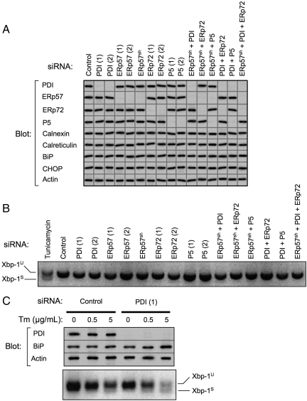
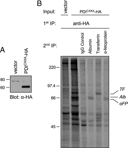
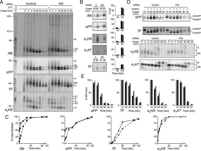
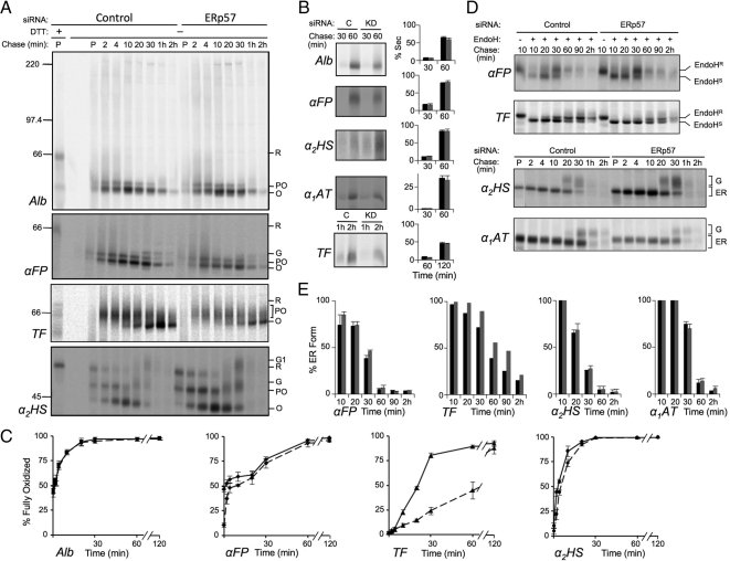
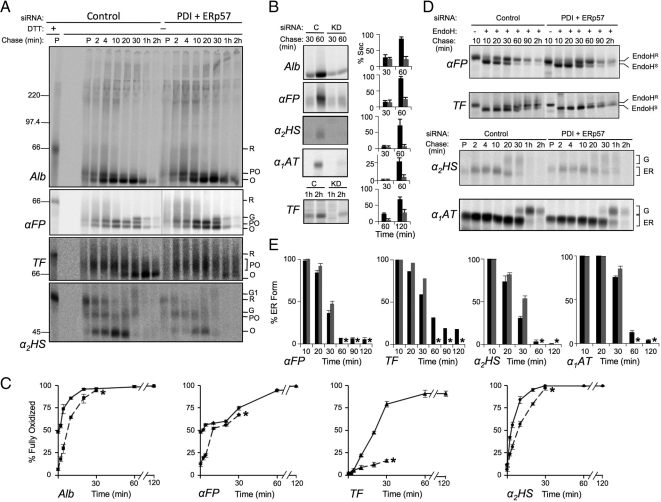
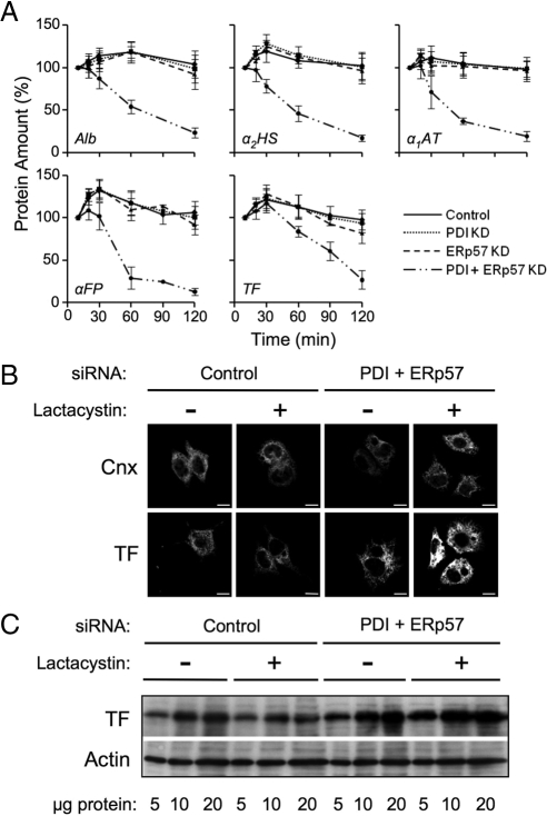
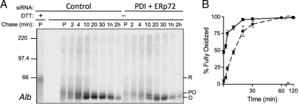
To examine the relationship between protein disulfide isomerase family members within the mammalian endoplasmic reticulum, PDI, ERp57, ERp72, and P5 were depleted with high efficiency in human hepatoma cells, either singly or in combination. The impact was assessed on the oxidative folding of several well-characterized secretory proteins. We show that PDI plays a predominant role in oxidative folding because its depletion delayed disulfide formation in all secretory proteins tested. However, the phenotype was surprisingly modest suggesting that other family members are able to compensate for PDI depletion, albeit with reduced efficacy. ERp57 also exhibited broad specificity, overlapping with that of PDI, but with preference for glycosylated substrates. Depletion of both PDI and ERp57 revealed that some substrates require both enzymes for optimal folding and, furthermore, led to generalized protein misfolding, impaired export from the ER, and degradation. In contrast, depletion of ERp72 or P5, either alone or in combination with PDI or ERp57 had minimal impact, revealing a narrow substrate specificity for ERp72 and no detectable role for P5 in oxidative protein folding.


Figures

Figure 1.
Figure 1.
Efficacy of PDI family member knockdown and assessment of UPR. (A) PDI family members were depleted from HepG2 cells by RNAi over a period of 6 d. Indicated single or multiple knockdowns were carried out transiently using two independent targeting siRNAs, designated (1) and (2), or in some instances using cells constitutively expressing shRNAmir targeting ERp57 (ERp57sh). Equivalent amounts of cell lysates were immunoblotted for PDI family members, ER chaperones, CHOP, and actin. Images shown are a composite from various experiments, all of which included actin as a loading control. Exposures were adjusted to an equivalent density of the actin control. Data are representative of a minimum of five replicate knockdowns. (B) After the indicated knockdowns or overnight treatment with 5 μg/ml tunicamycin, total RNA was isolated from HepG2 cells and analyzed for Xbp-1 splicing by reverse-transcription PCR. Xbp-1U, unspliced Xbp-1 product; Xbp-1S, spliced Xbp-1 product. (C) HepG2 cells were subjected to PDI knockdown for 10 d. On day 9, cells were treated overnight with the indicated concentrations of tunicamycin (Tm). The following day, cell lysates were immunoblotted to detect PDI, BiP, and actin. Lanes shown were assembled from a single gel. Total RNA was also isolated and analyzed for Xbp-1 splicing.
Figure 2.
Figure 2.
Identification of PDI substrates. (A) Lysates of HepG2 cells stably expressing either the pQCXIH expression vector (vector) or HA-tagged PDI carrying C56A and C400A mutations (PDICXXA-HA) were immunoblotted with anti-HA antibody. (B) HepG2 cells expressing either vector or PDICXXA-HA were radiolabeled with [35S]Met for 15 min, and treated with 20 mM NEM in PBS. Cell lysates were immunoisolated with anti-HA antibody, immune complexes were disrupted and then immunoisolated a second time with the indicated antibodies. Proteins were separated by reducing SDS-PAGE and visualized by fluorography. Alb, albumin; αFP, α-fetoprotein; TF, transferrin.
Figure 3.
Figure 3.
Effect of PDI knockdown on oxidative folding of secretory proteins. HepG2 cells were depleted of PDI or treated with control siRNA for 6 d, radiolabeled with [35S]Met for 3 min, then chased with unlabeled Met for the indicated times. The medium was removed, and then cells were treated on ice with 20 mM NEM in PBS and lysed in RIPA buffer containing 20 mM NEM. Both cell lysates and media were immunoisolated with antisera directed against the indicated proteins. (A) Kinetics of disulfide formation. Immune complexes from cell lysates were subjected to SDS-PAGE under reducing (first lane) or nonreducing conditions and proteins were visualized and quantified by phosphorimaging. Shown are representative gels of three independent experiments for substrates albumin (Alb), α-fetoprotein (αFP), transferrin (TF), and α2-HS-glycoprotein (α2HS). Reduced (R), partially oxidized (PO), oxidized (O), and Golgi (G, G1) forms of each protein are indicated. (B) Secretion kinetics. Immune complexes recovered from media samples at the indicated chase times were analyzed by SDS-PAGE and visualized and quantified by phosphorimaging. Histograms indicate the amount of protein observed in the medium as a percentage of the combined intra- and extracellular signal at each chase time. Black and gray bars represent control (C) and knockdown (KD) conditions, respectively. (C) Quantification of gels shown in panels A and B was carried out by expressing the amount of fully oxidized protein in lysate and media samples as a percentage of the total combined amount of reduced, partially oxidized, and oxidized forms at a given time point. Solid lines represent control conditions; dashed lines represent knockdown conditions. (D) ER to Golgi transport kinetics. The indicated proteins were immunoisolated and either digested or not with endo H as indicated and analyzed by reducing SDS-PAGE. Endo HS and Endo HR represent endo H–sensitive and -resistant species, respectively. G and ER represent Golgi-processed and ER forms which could be resolved without endo H digestion. (E) Quantification of the gels in panel D. Histograms represent the amount of protein remaining in the ER as a percentage of the total protein recovered from cells and medium at each time point. Black bars, control; gray bars, knockdown. Error bars represent the average of three independent experiments ± one SD, except in the case of TF which was examined in a single experiment.
Figure 4.
Figure 4.
ERp57 depletion delays oxidative folding of glycosylated substrates. HepG2 cells were depleted of ERp57 or treated with control siRNA and pulse-chase experiments were conducted and analyzed as described in Figure 3. (A) Kinetics of disulfide formation. (B) Secretion kinetics. Black and gray bars represent control and knockdown conditions, respectively. (C) Quantification of oxidative folding. Solid lines, control conditions; dashed lines, knockdown conditions. (D) ER to Golgi transport kinetics. (E) Quantification of the gels in panel D. Error bars represent the average of three independent experiments ± one SD, except in the case of TF which was examined in a single experiment.
Figure 5.
Figure 5.
Combined knockdown of PDI and ERp57 substantially impairs protein folding. HepG2 cells constitutively depleting ERp57 by shRNAmir were subjected to knockdown of PDI by siRNA for 6 d. HepG2 cells transfected with control siRNA were prepared in parallel. Pulse-chase experiments were conducted and analyzed as described in Figure 3. (A) Kinetics of disulfide formation. (B) Secretion kinetics. Amount secreted was quantified as a percentage of the total signal recovered at the 10 min chase time point. Black and gray bars represent control and knockdown conditions, respectively. (C) Quantification of oxidative folding. Solid lines, control conditions; dashed lines, knockdown conditions. (D) ER to Golgi transport kinetics. (E) Quantification of the gels in panel D. Error bars represent the average of three independent experiments ± one SD, except in the case of TF which was examined in a single experiment. Asterisks indicate quantifications were not included beyond 30 min of chase due to substrate degradation.
Figure 6.
Figure 6.
Misfolded transferrin is degraded by the proteasome in PDI/ERp57-depleted cells. (A) Pulse-chase data from Figures 3–5 was analyzed for changes in the total amount of radiolabeled Alb, αFP, TF, α2HS, and α1AT recovered from the combined intracellular and medium fractions. Total protein, as measured by densitometry, was defined as 100% at the 10 min chase time. Error bars represent the average of three independent experiments ± one SD. (B, C) HepG2 cells were treated with control siRNA or depleted of PDI and ERp57 for 6 d and then incubated in the absence or presence of 5 μg/ml lactacystin for 5 h. Cells were either plated on Cell-Tak-treated coverslips (BD Biosciences), fixed in paraformaldehyde, and permeabilized for confocal microscopy (B) or lysed in RIPA buffer for Western blot analysis (C). Immunoblot or immunofluorescence was carried out using the indicated antibodies (Scale bar = 10 μm).
Figure 7.
Figure 7.
Effect of combined PDI and ERp72 knockdown on oxidative folding of albumin. HepG2 cells were depleted of PDI and ERp72 by siRNA or treated with control siRNA for 6 d and then subjected to pulse-chase radiolabeling and immunoisolation of Alb as described for Figure 3. (A) Representative gel of Alb oxidation from three independent knockdown experiments. (B) Quantification of gel shown in panel A. Solid lines, control conditions; dashed lines, knockdown conditions. Error bars represent the average of three independent experiments ± one SD.

References

    1. Adeli K., Wettesten M., Asp L., Mohammadi A., Macri J., Olofsson S.-O. Intracellular assembly and degradation of apolipoprotein B-100-containing lipoproteins in digitonin-permeabilized HEP G2 cells. J. Biol. Chem. 1997;272:5031–5039. - PubMed
    1. Alanen H. I., Salo K. E., Pirneskoski A., Ruddock L. W. pH dependence of the peptide thiol-disulfide oxidase activity of six members of the human protein disulfide isomerase family. Antioxid. Redox. Signal. 2006;8:283–291. - PubMed
    1. Appenzeller-Herzog C., Ellgaard L. The human PDI family: versatility packed into a single fold. Biochim. Biophys. Acta. 2008;1783:535–548. - PubMed
    1. Appenzeller-Herzog C., Riemer J., Christensen B., Sorensen E. S., Ellgaard L. A novel disulphide switch mechanism in Ero1alpha balances ER oxidation in human cells. EMBO J. 2008;27:2977–2987. - PMC - PubMed
    1. Bass R., Ruddock L. W., Klappa P., Freedman R. B. A major fraction of endoplasmic reticulum-located glutathione is present as mixed disulfides with protein. J. Biol. Chem. 2004;279:5257–5262. - PubMed

Publication types

MeSH terms

LinkOut - more resources