. 2010 Sep 15;21(18):3182-92.
doi: 10.1091/mbc.E10-01-0066. Epub 2010 Jul 21.

Cortical mechanics and meiosis II completion in mammalian oocytes are mediated by myosin-II and Ezrin-Radixin-Moesin (ERM) proteins

Affiliations

Cortical mechanics and meiosis II completion in mammalian oocytes are mediated by myosin-II and Ezrin-Radixin-Moesin (ERM) proteins

Stephanie M Larson et al. Mol Biol Cell. .

Abstract

Cell division is inherently mechanical, with cell mechanics being a critical determinant governing the cell shape changes that accompany progression through the cell cycle. The mechanical properties of symmetrically dividing mitotic cells have been well characterized, whereas the contribution of cellular mechanics to the strikingly asymmetric divisions of female meiosis is very poorly understood. Progression of the mammalian oocyte through meiosis involves remodeling of the cortex and proper orientation of the meiotic spindle, and thus we hypothesized that cortical tension and stiffness would change through meiotic maturation and fertilization to facilitate and/or direct cellular remodeling. This work shows that tension in mouse oocytes drops about sixfold during meiotic maturation from prophase I to metaphase II and then increases ∼1.6-fold upon fertilization. The metaphase II egg is polarized, with tension differing ∼2.5-fold between the cortex over the meiotic spindle and the opposite cortex, suggesting that meiotic maturation is accompanied by assembly of a cortical domain with stiffer mechanics as part of the process to achieve asymmetric cytokinesis. We further demonstrate that actin, myosin-II, and the ERM (Ezrin/Radixin/Moesin) family of proteins are enriched in complementary cortical domains and mediate cellular mechanics in mammalian eggs. Manipulation of actin, myosin-II, and ERM function alters tension levels and also is associated with dramatic spindle abnormalities with completion of meiosis II after fertilization. Thus, myosin-II and ERM proteins modulate mechanical properties in oocytes, contributing to cell polarity and to completion of meiosis.


Figures

Figure 1.
Figure 1.
Changes in effective tension during meiotic maturation and fertilization. (A) Schematic diagram illustrating progression of the prophase I germinal vesicle-intact (GVI) oocytes through meiotic maturation, through germinal vesicle breakdown (GVBD) and metaphase I (MI) stages to metaphase II (MII) arrest, followed by fertilization-triggered exit from MII arrest. The MII egg is characterized by distinct domains of membrane and underlying cortex: the microvillar (MV) domain to which sperm bind and fuse, and the amicrovillar (AMV) domain, overlying the meiotic spindle and characterized by an actin-rich cap, contrasting the uniform distribution of actin and microvilli at prophase I stage. Gray lines indicate cortical actin. Polar bodies (PB1, first polar body; PB, second polar body) are the products of the asymmetric meiotic divisions. (B) Micropipette aspiration (MPA) of mouse eggs. The egg is aspirated until the equilibrium length of the cortex pulled into the pipette (Lp) equals the radius of the pipette (Rp). The aspiration pressure, where Lp = Rp, is defined as the critical pressure. (C) Effective tension (Teff) in oocytes, eggs, and zygotes. For eggs and zygotes in this panel, assessments were done on microvillar domains. Number of cells analyzed: GVI, 142; GVBD, 16; MI, 16; MII, 387; and zygote, 46. (D) Teff)in MII eggs in the microvillar (MV) and amicrovillar (AMV) domains. These data are the composite of all cells analyzed: total MV, 387; total AMV, 175. (E) Teff the microvillar (MV) and amicrovillar (AMV) domains of MII eggs that had been ovulated and matured in vivo (Ov) compared with eggs that had been matured to MII in vitro (IVM). Number of cells analyzed: ovulated MV, 200; ovulated AMV, 91; in vitro–matured MV, 187; in vitro–matured AMV, 84. (F) Spindle morphology, revealed by anti-tubulin staining, in ovulated and in vitro–matured M II eggs. Insets show a second example of a spindle in each type of egg. Scale bar, 15 μm.
Figure 2.
Figure 2.
Effects of actin manipulation on oocyte effective tension. (A) Phalloidin staining of a GVI oocyte (left) and a metaphase II (MII) egg (right). Polarity develops during meiotic maturation, such that the metaphase II egg has an actin-rich cap over the meiotic spindle. Red, actin; blue, DNA. Scale bar, (left) 20 μm; (right) 22 μm. (B) Effects on effective tension (Teff) of actin manipulation by cytochalasin D treatment of GVI oocytes (*p < 0.0001). Numbers of cells analyzed: cytochalasin D–treated GVI oocytes, 75; DMSO control GVI oocytes, 43. (C) Cellular concentration (in μM) of total actin, and actin in the phalloidin-stabilized, polymeric fraction (polymer) and in the soluble fraction in GVI oocytes (■) and MII eggs (□). Values for GVI oocytes show the mean ± range from two experiments and for MII eggs, mean ± range from three experiments.
Figure 3.
Figure 3.
Localization of pMRLC and effects of myosin-II manipulation on effective tension. (A–K) Immunofluorescence localization of phosphorylated myosin-II regulatory light chain (pMRLC) in oocytes and eggs. Stages shown are GVI oocyte (A–D) and metaphase II (MII) eggs (E–K). With the exception of A and B (which show nonimmune antibody staining), the top row of black and white images shows pMRLC, whereas the second row of images shows pMRLC in green and DNA in blue. Images are taken at different focal planes; most are focused at or near the cell equator, whereas J and K are focused toward the top of the egg to show the amicrovillar domain (AMV). The egg in J and K also has the first polar body nearby (PB1). Scale bar, 20 μm. (L and M) Effects on effective tension (Teff) of myosin manipulation by ConA treatment of MII egg microvillar domains (MV) and amicrovillar (AMV) domains (L) or ML-7-treatment on GVI oocytes (GVI), MII egg microvillar domains (MII MV), and MII egg amicrovillar domains (MII AMV; M). Values shown are mean Teff ± SEM in nN/μm. ■, control values (medium for L, DMSO for M); □, Teff values for treated eggs (ConA for L, 15 μM ML-7 for M). The difference between each control and treatment group is statistically significant (Mann Whitney U-test, p < 0.05). Number of cells analyzed: ConA-treated MII MV, 89; untreated MII MV controls, 61; ConA-treated MII AMV, 52; untreated MII AMV controls, 35; ML-7-treated GVI, 53; DMSO control GVI, 45; ML-7–treated-MII MV, 84; DMSO control MII MV, 79; ML-7–treated-MII AMV, 23; and DMSO control MII AMV, 36.
Figure 4.
Figure 4.
ERMs in mouse eggs. (A and B) Immunofluorescence analysis of radixin (A) and pERM (B) in GVI oocytes (Ai–iv; Bi–iii) and MII eggs (Av–xii and Biv–ix), showing anti-radixin (Aii, vi, and x), anti-pERM (Bii, v, and viii), DNA (Aiii, vii and xi and Biii, vi, and ix), or anti-β-tubulin (Aiv, viii, and xii). Scale bar, 15 μm. (C) Immunoblot analysis of ERM (i) or radixin (ii) expression in eggs. In Ci, the solid arrowhead indicates the electrophoretic mobility of ezrin and radixin, the open arrowhead indicates moesin. Radixin is detectable in lysates of five eggs (125 ng protein) with a band of comparable intensity to that detected in 1 μg of mouse liver lysate. (D) Representative blots of oocyte and egg lysates (20 cells per lane) probed with anti-pERM (i and iii) or anti-ERM (ii and iv) antibodies. Oocyte meiotic maturation (GVI to MII; i and ii) and fertilization (iii and iv) were analyzed in separate blots with different exposure times, to optimize capturing the range of signals (e.g., the blot in iii was exposed to show the increase in signal from unfertilized eggs to fertilized eggs). (E and F) Quantification of band intensities of anti-pERM (■) and ERM (□) levels during oocyte maturation (E) or the egg-to-embryo transition (F; zygote sample prepared at 90 min after insemination); sample blots are shown above in D. Values were normalized to metaphase II (MII) eggs.
Figure 5.
Figure 5.
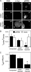
Perturbation of ERM action in eggs. (A) Analysis of DN-RDX-cmyc and pERM by immunofluorescence in uninjected (i–iii), water-injected (iv–vi), and DN-RDX-cRNA-injected eggs (vii–ix). DN-RDX-cmyc protein was detected in ∼33% of eggs injected with DN-RDX-cmyc cRNA. (viii and ix) Two eggs, one with detectable cmyc staining (viii) and reduced pERM staining (ix), and a second egg (*) that does not have detectable cmyc staining (viii) and has cortically localized pERM (ix). (B) Assessment of pERM and actin levels in control (uninjected and water-injected) and DN-RDX–expressing egg through quantification of Icort/Iwhole signals (described in Materials and Methods). This analysis shows that cortical pERM (■) is reduced in DN-RDX–expressing eggs (*p < 0.0001), whereas the ratio of cortical/whole actin polymers is unaffected. Numbers of cells analyzed: uninjected, pERM, 5; uninjected actin, 8; water-injected, pERM, 4; water-injected, 4; cRNA-injected, pERM, 22; and cRNA-injected, actin, 14. (A sample of phalloidin staining of a metaphase II egg is shown in Figure 2.) (C) Effects of DN-RDX expression and reduction of pERM on effective tension (Teff) levels in eggs. Eggs were subjected to Teff measurements, then recovered, and analyzed for c-myc expression (indicative of DN-RDX expression) and reduction of pERM. Number of cells analyzed: uninjected, 10; water-injected, 7; and DN-RDX-expressing, 8. The difference between uninjected and water-injected eggs is not statistically significant, whereas the differences between the cRNA-injected eggs and the other two groups is significant (*p < 0.001). Furthermore, a small number of cRNA-injected eggs deformed as soon as aspiration pressure was applied, making it impossible to obtain a Teff measurement but suggesting that these eggs had extremely low Teff.
Figure 6.
Figure 6.
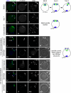
Spindle defects upon exit from metaphase II arrest with actin, myosin-II, or ERM disruption. Eggs were inseminated for 1.5 h (A–P) or 4 h (Q-BB). Control fertilized eggs (A–C, J–M, and Q–T) show normal spindle rotation and second polar body morphology, as illustrated in the schematic diagram. A failure in spindle rotation was observed in eggs treated with the MLCK inhibitor ML-7 (D–F; 73/75 eggs) and in eggs treated with the actin filament disruptor cytochalasin D (G–I; 34/34 eggs). Distorted, curved spindles were observed in DN-RDX–expressing eggs (N–P, U-BB; 26/36 eggs). In these embryos, two polar body-like (PBL) structures formed; these PBL structures did not resolve with increased time after insemination (U-BB). Arrowheads identify polar bodies (PB) and PBL structures. Maternal DNA is labeled m, and sperm DNA is labeled s; some eggs are polyspermic, although this is not uncommon with the insemination conditions used, particularly with cytochalasin D–treated eggs (McAvey et al., 2002). In several panels, DNA of the fertilizing sperm is out of the plane of focus. FC (T) identifies the fertilization cone containing the DNA of a fertilizing sperm. Scale bar, (DD) 10 μm.
Figure 7.
Figure 7.
Schematic diagram of mechanical properties in the egg driving asymmetric cell division. (A) The mechanical polarity in the metaphase II (MII) mouse egg between the microvillar domain (MV) and the actin- and myosin-II–enriched (red) amicrovillar domain over the spindle (AMV); the amicrovillar domain creates a microdomain in the egg containing the metaphase II spindle (green). The subregion of the egg cell defined by the amicrovillar domain with its increased local Teff likely serves to isolate the spindle and developing polar body and allows contraction mediated by actin and myosin-II (red) to locally deform the cortex. Myosin II accumulates at the polar cortex of the emerging polar body (B), facilitating asymmetric meiotic cell division (C). The change in gray shading in the cytoplasm through A–C illustrates the progression of the egg from the metaphase II arrest (A) through anaphase and telophase to embryonic interphase.

Similar articles

Cited by

References

    1. Azoury J., Lee K. W., Georget V., Rassinier P., Leader B., Verlhac M. H. Spindle positioning in mouse oocytes relies on a dynamic meshwork of actin filaments. Curr. Biol. 2008;18:1514–1519. - PubMed
    1. Barrett S. L., Albertini D. F. Allocation of gamma-tubulin between oocyte cortex and meiotic spindle influences asymmetric cytokinesis in the mouse oocyte. Biol. Reprod. 2007;76:949–957. - PubMed
    1. Bretscher A., Edwards K., Fehon R. G. ERM proteins and merlin: integrators at the cell cortex. Nat. Rev. Mol. Cell Biol. 2002;3:586–599. - PubMed
    1. Brunet S., Maro B. Cytoskeleton and cell cycle control during meiotic maturation of the mouse oocyte: integrating time and space. Reproduction. 2005;130:801–811. - PubMed
    1. Carreno S., Kouranti I., Glusman E. S., Fuller M. T., Echard A., Payre F. Moesin and its activating kinase Slik are required for cortical stability and microtubule organization in mitotic cells. J. Cell Biol. 2008;180:739–746. - PMC - PubMed

Publication types