. 2010 Sep 15;21(18):3232-46.
doi: 10.1091/mbc.E09-05-0408. Epub 2010 Jul 21.

The BNIP-2 and Cdc42GAP homology (BCH) domain of p50RhoGAP/Cdc42GAP sequesters RhoA from inactivation by the adjacent GTPase-activating protein domain

Affiliations

The BNIP-2 and Cdc42GAP homology (BCH) domain of p50RhoGAP/Cdc42GAP sequesters RhoA from inactivation by the adjacent GTPase-activating protein domain

Yi Ting Zhou et al. Mol Biol Cell. .

Abstract

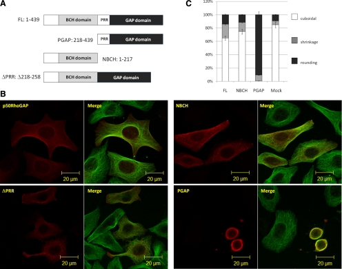
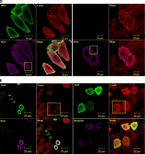
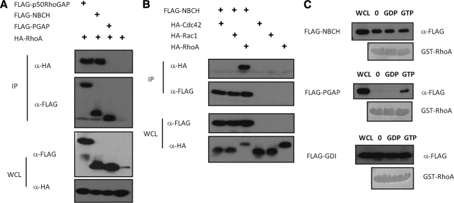
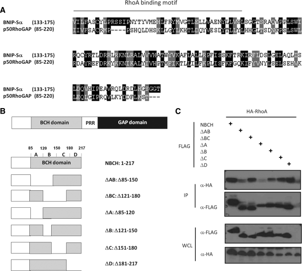
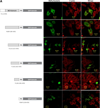
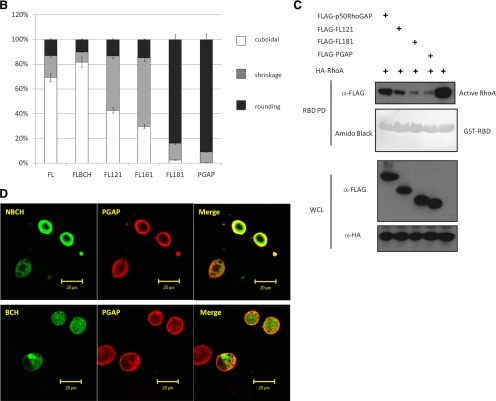
The BNIP-2 and Cdc42GAP homology (BCH) domain is a novel regulator for Rho GTPases, but its impact on p50-Rho GTPase-activating protein (p50RhoGAP or Cdc42GAP) in cells remains elusive. Here we show that deletion of the BCH domain from p50RhoGAP enhanced its GAP activity and caused drastic cell rounding. Introducing constitutively active RhoA or inactivating GAP domain blocked such effect, whereas replacing the BCH domain with endosome-targeting SNX3 excluded requirement of endosomal localization in regulating the GAP activity. Substitution with homologous BCH domain from Schizosaccharomyces pombe, which does not bind mammalian RhoA, also led to complete loss of suppression. Interestingly, the p50RhoGAP BCH domain only targeted RhoA, but not Cdc42 or Rac1, and it was unable to distinguish between GDP and the GTP-bound form of RhoA. Further mutagenesis revealed a RhoA-binding motif (residues 85-120), which when deleted, significantly reduced BCH inhibition on GAP-mediated cell rounding, whereas its full suppression also required an intramolecular interaction motif (residues 169-197). Therefore, BCH domain serves as a local modulator in cis to sequester RhoA from inactivation by the adjacent GAP domain, adding to a new paradigm for regulating p50RhoGAP signaling.


Figures

Figure 1.
Figure 1.
The BCH domain of p50RhoGAP inhibits GAP-induced cell rounding. (A) Schematic diagram of p50RhoGAP and its mutants: N-terminus without GAP domain (NBCH, amino acids 1-217), C-terminus without BCH domain (PGAP, amino acids 218-439), and a mutant without the proline-rich region, PPR (ΔPPR, Δ218-258). (B) HeLa cells were transfected with plasmids encoding FLAG-tagged full-length p50RhoGAP, NBCH, PGAP, or ΔPPR mutants. Cells were then fixed after 16–20 h and subjected to confocal fluorescence microscopy as described in Materials and Methods. Morphological changes and cytoskeletal rearrangements were revealed by indirect immunostaining with Alexa Fluor 488–conjugated goat anti-mouse IgG against anti-tubulin for microtubules and with FLAG antibody for expressed FLAG-tagged proteins. (C) For quantitative analysis, the ratio of cuboidal, protrusion/shrinkage, and round cells was scored with at least 150 transfected cells counted per sample per experiment. Data are means ± SD (n = 3).
Figure 2.
Figure 2.
The GAP domain of p50RhoGAP induces cell rounding by inactivating Rho GTPases (A) HeLa cells were treated with Rho inhibitor C3 Transferase followed by rhodamine-conjugated phalloidin staining and confocal fluorescence microscopy analysis. (B) Cells were transfected with FLAG-p50RhoGAP in the presence or absence of HA-tagged expression constructs of Cdc42, Rac1, and RhoA. Lysates were immunoprecipitated (IP) with anti-FLAG beads, and the associated proteins were separated on SDS-PAGE, blotted, and probed with HA antibody. Expression of FLAG-p50RhoGAP and HA-tagged Cdc42, Rac1, and RhoA were verified by Western blot analyses for the whole cell lysates (WCL) using anti-FLAG (third panel) and anti-HA (bottom panel), respectively. The bound GTPase was detected by anti-HA (top panel), and equal loading of IP beads were verified by anti-FLAG (second panel). (C) To determine the Rho GTPase activity, HeLa cells were transfected with FLAG-tagged wild-type RhoA in the presence and absence of HA-tagged p50RhoGAP, NBCH, or PGAP mutants. Cell were lysed and incubated with GST fusion of the Rho-binding domain of rhotekin immobilized on beads as described in Materials and Methods Bound active RhoA were resolved on SDS-PAGE and detected by immunoblotting with FLAG-antibody (top panel). Equal loading of GST fusion proteins is shown in the second panel. (D) HeLa cells were transfected with plasmids encoding HA-RhoA alone or with FLAG-tagged full-length p50RhoGAP, PGAP, or NBCH mutants. Cells were then fixed after 20 h and subjected to confocal fluorescence microscopy as described in Materials and Methods The actin filaments were detected by direct costaining with rhodamine-conjugated phalloidin. (E) HeLa cells were cotransfected with HA-tagged PGAP mutant and wild-type or constitutively active RhoA-G14V. Cells were then fixed and images analyzed by confocal fluorescence microscopy after direct staining with rhodamine-conjugated phalloidin for actin filaments.
Figure 2.
Figure 2.
The GAP domain of p50RhoGAP induces cell rounding by inactivating Rho GTPases (A) HeLa cells were treated with Rho inhibitor C3 Transferase followed by rhodamine-conjugated phalloidin staining and confocal fluorescence microscopy analysis. (B) Cells were transfected with FLAG-p50RhoGAP in the presence or absence of HA-tagged expression constructs of Cdc42, Rac1, and RhoA. Lysates were immunoprecipitated (IP) with anti-FLAG beads, and the associated proteins were separated on SDS-PAGE, blotted, and probed with HA antibody. Expression of FLAG-p50RhoGAP and HA-tagged Cdc42, Rac1, and RhoA were verified by Western blot analyses for the whole cell lysates (WCL) using anti-FLAG (third panel) and anti-HA (bottom panel), respectively. The bound GTPase was detected by anti-HA (top panel), and equal loading of IP beads were verified by anti-FLAG (second panel). (C) To determine the Rho GTPase activity, HeLa cells were transfected with FLAG-tagged wild-type RhoA in the presence and absence of HA-tagged p50RhoGAP, NBCH, or PGAP mutants. Cell were lysed and incubated with GST fusion of the Rho-binding domain of rhotekin immobilized on beads as described in Materials and Methods Bound active RhoA were resolved on SDS-PAGE and detected by immunoblotting with FLAG-antibody (top panel). Equal loading of GST fusion proteins is shown in the second panel. (D) HeLa cells were transfected with plasmids encoding HA-RhoA alone or with FLAG-tagged full-length p50RhoGAP, PGAP, or NBCH mutants. Cells were then fixed after 20 h and subjected to confocal fluorescence microscopy as described in Materials and Methods The actin filaments were detected by direct costaining with rhodamine-conjugated phalloidin. (E) HeLa cells were cotransfected with HA-tagged PGAP mutant and wild-type or constitutively active RhoA-G14V. Cells were then fixed and images analyzed by confocal fluorescence microscopy after direct staining with rhodamine-conjugated phalloidin for actin filaments.
Figure 3.
Figure 3.
Three essential residues for GAP domain in inducing cell rounding. (A) HeLa cells were transfected with HA-tagged PGAP or the various mutants indicated, fixed after 20 h, and subjected to confocal fluorescence microscopy with anti-HA and Alexa Fluor 488 dye–conjugated goat anti-rabbit IgG as described in Materials and Methods. (B) To determine the Rho GTPase activity, HeLa cells were transfected with FLAG-tagged RhoA alone or with various HA-tagged PGAP or its various mutants. Cell were lysed and incubated with GST fusion of the Rho-binding domain of rhotekin immobilized on beads as described in Materials and Methods. Bound active RhoA were resolved on SDS-PAGE and detected by immunoblotting with FLAG-antibody (top panel). Equal loading of GST fusion proteins is shown in the second panel.
Figure 4.
Figure 4.
Intramolecular interaction alone does not regulate GAP activity. HeLa cells were transfected with FLAG-tagged-FL53 or FLΔ151-217 mutants and subjected to confocal fluorescence microscopy after direct staining with rhodamine-conjugated phalloidin for actin filaments.
Figure 5.
Figure 5.
The BCH domain of p50RhoGAP displays distinctive RhoA binding profile. (A) HEK293T cells were transfected with plasmid encoding HA-RhoA alone or with FLAG-p50RhoGAP, NBCH or PGAP. Lysates were immunoprecipitated (IP) with anti-FLAG beads, and the associated proteins were detected with HA antibody (top panel). Expression of FLAG-tagged proteins and HA-RhoA were verified by Western blot analyses by anti-FLAG (third panel) and anti-HA (bottom panel), respectively. Equal loading of IP beads were verified by anti-FLAG (second panel). (B) Cells were transfected with plasmid encoding FLAG-NBCH in the presence or absence of HA-tagged Cdc42, Rac1, and RhoA. Lysates were immunoprecipitated (IP) with anti-FLAG beads, and the associated proteins were separated on SDS-PAGE, and probed with HA antibody (top panel). Expression of FLAG-tagged NBCH and HA-tagged GTPases were verified by Western blot analyses using anti-FLAG (third panel) and anti-HA (bottom panel), respectively. Equal loading of IP beads were verified by anti-FLAG (second panel). (C) HEK293T lysates expressing FLAG-NBCH, PGAP fragments or Rho-GDIα were incubated with unloaded GST-RhoA, GST-RhoA preloaded with GDP, or GTPγS as described in Materials and Methods. Bound proteins and whole cell lysates (WCL) input were analyzed with anti-FLAG and equal loading of GST beads verified by amido black staining.
Figure 6.
Figure 6.
Identification of a RhoA-binding motif within the BCH domain of p50RhoGAP. (A) Multiple sequence alignments of BCH domains from Homo sapiens BNIP-Sα (AY078983) and p50RhoGAP/Cdc42GAP (Q07960) using ClustalW and formatted using BOXSHADE. Identical residues are shaded black whereas similar or conserved ones are in gray. The region corresponding to the previously described Rho-binding motif in BNIP-Sα (Zhou et al., 2006) was underlined. (B) Schematic diagram of NBCH domain and its mutants. (C) Cells were cotransfected with HA-tagged RhoA and FLAG-tagged NBCH wild type or mutants as depicted in Figure 6B. Lysates were immunoprecipitated (IP) with anti-FLAG beads, and the associated proteins were separated on SDS-PAGE, blotted, and probed with HA antibody. Expression of FLAG-NBCH constructs and HA- RhoA were verified by Western blot analyses of the whole cell lysates (WCL) using anti-FLAG (third panel) and anti-HA (bottom panel), respectively. The bound RhoA was detected by anti-HA (top panel), and equal loading of IP beads were verified by anti-FLAG (second panel).
Figure 7.
Figure 7.
The BCH domain of p50RhoGAP inhibits the adjacent GAP function by sequestering RhoA. (A) HeLa cells were transfected for 20 h with FLAG-tagged p50RhoGAP wild type and mutants including FLBCH, FL121, FL161, FL181, or PGAP. Cells were then fixed and incubated with FLAG monoclonal antibodies, followed by Alexa Fluor 488-conjugated goat anti-mouse IgG. Cell morphology was monitored by direct staining with rhodamine-conjugated phalloidin for actin filaments. (B) For quantitative analysis, the ratio of cuboidal, protrusion/shrinkage, and round cells was scored and at least 150 transfected cells were counted per sample per experiment. Data are means ± SD (n = 3). (C) Cells were transfected with FLAG-tagged p50RhoGAP full-length, FL121, FL181, or PGAP in the presence of HA-RhoA. After 20 h, cell were lysed and incubated with GST fusion of the Rho-binding domain of rhotekin-immobilized on beads, in order to assess the impacts of p50RhoGAP and the mutants in regulating RhoA activity as described in Materials and Methods. Bound GTPases were resolved on SDS-PAGE and detected by immunoblotting with HA-antibody (top panel). Equal loading of GST fusion proteins is shown in the second panel. (D) HeLa cells were cotransfected with HA-tagged PGAP and FLAG-tagged-NBCH or BCH domain. Cells were then fixed and analyzed with confocal fluorescence microscopy as described in Materials and Methods.
Figure 7.
Figure 7.
The BCH domain of p50RhoGAP inhibits the adjacent GAP function by sequestering RhoA. (A) HeLa cells were transfected for 20 h with FLAG-tagged p50RhoGAP wild type and mutants including FLBCH, FL121, FL161, FL181, or PGAP. Cells were then fixed and incubated with FLAG monoclonal antibodies, followed by Alexa Fluor 488-conjugated goat anti-mouse IgG. Cell morphology was monitored by direct staining with rhodamine-conjugated phalloidin for actin filaments. (B) For quantitative analysis, the ratio of cuboidal, protrusion/shrinkage, and round cells was scored and at least 150 transfected cells were counted per sample per experiment. Data are means ± SD (n = 3). (C) Cells were transfected with FLAG-tagged p50RhoGAP full-length, FL121, FL181, or PGAP in the presence of HA-RhoA. After 20 h, cell were lysed and incubated with GST fusion of the Rho-binding domain of rhotekin-immobilized on beads, in order to assess the impacts of p50RhoGAP and the mutants in regulating RhoA activity as described in Materials and Methods. Bound GTPases were resolved on SDS-PAGE and detected by immunoblotting with HA-antibody (top panel). Equal loading of GST fusion proteins is shown in the second panel. (D) HeLa cells were cotransfected with HA-tagged PGAP and FLAG-tagged-NBCH or BCH domain. Cells were then fixed and analyzed with confocal fluorescence microscopy as described in Materials and Methods.
Figure 8.
Figure 8.
Model depicting BCH domain as a local modulator to sequester RhoA from inactivation by the adjacent GAP domain and possibly as a scaffold that links the RhoGAP function to RhoGEF and other effectors. The GAP domain of p50RhoGAP inactivates RhoA and subsequently induces drastic cytoskeleton collapse and cell rounding. This cellular effect could be inhibited in cis by the N-terminal BCH domain, but not its proline-rich region (PRR), that acts via a concerted mechanism including the binding of the BCH domain to RhoA via its Rho-binding motif (RBM), thus displacing GAP domain from inhibiting RhoA, and the intramolecular interaction region 2 (IIR 2) that augments RhoA sequestration to confer complete suppression of the GAP activity (pathway 1). As the BCH domain displays distinctive Rho-binding profile from the GAP domain and is independent of the nucleotide-binding status, it could help ensure that the GTP-bound active form of Rho be effectively sequestered from inactivation by GAP. In addition, independent BCH domain could also serve as a scaffold that links RhoGEF (e.g., at least with p115RhoGEF shown in this study; pathway 2) or interfering with downstream signaling of RhoA without directly affecting Rho activity (e.g., abolishing periphery projections; pathway 3). However, how these multitude mechanisms operate, either in isolation or in concert for a dynamic regulation, awaits further investigation. See text for details.

Similar articles

Cited by

References

    1. Aghazadeh B., Lowry W. E., Huang X. Y., Rosen M. K. Structural basis for relief of autoinhibition of the Dbl homology domain of proto-oncogene Vav by tyrosine phosphorylation. Cell. 2000;102:625–633. - PubMed
    1. Barfod E. T., Zheng Y., Kuang W. J., Hart M. J., Evans T., Cerione R. A., Ashkenazi A. Cloning and expression of a human CDC42 GTPase-activating protein reveals a functional SH3-binding domain. J. Biol. Chem. 1993;268:26059–26062. - PubMed
    1. Barrett T., Xiao B., Dodson E. J., Dodson G., Ludbrook S. B., Nurmahomed K., Gamblin S. J., Musacchio A., Smerdon S. J., Eccleston J. F. The structure of the GTPase-activating domain from p50rhoGAP. Nature. 1997;385:458–461. - PubMed
    1. Bernards A., Settleman J. GAPs in growth factor signalling. Growth Factors. 2005;23:143–149. - PubMed
    1. Bernards A., Settleman J. GEFs in growth factor signaling. Growth Factors. 2007;25:355–361. - PubMed

Publication types

MeSH terms

Substances