. 2010 Jul 21;30(29):9728-37.
doi: 10.1523/JNEUROSCI.1924-10.2010.

Local and remote growth factor effects after primate spinal cord injury

Affiliations

Local and remote growth factor effects after primate spinal cord injury

John H Brock et al. J Neurosci. .

Abstract

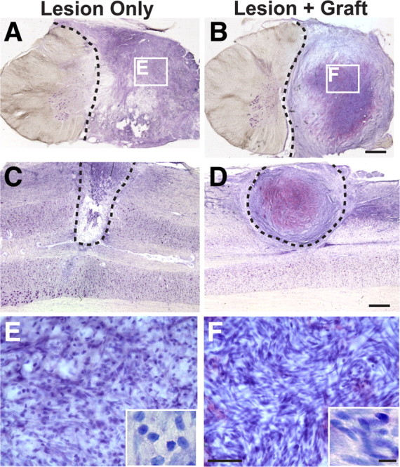
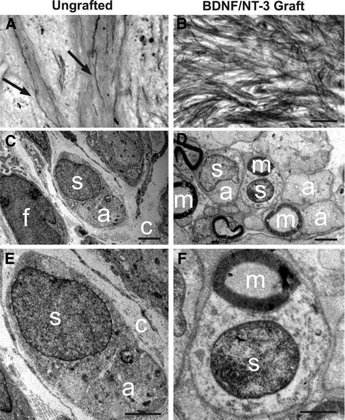
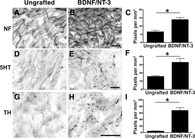
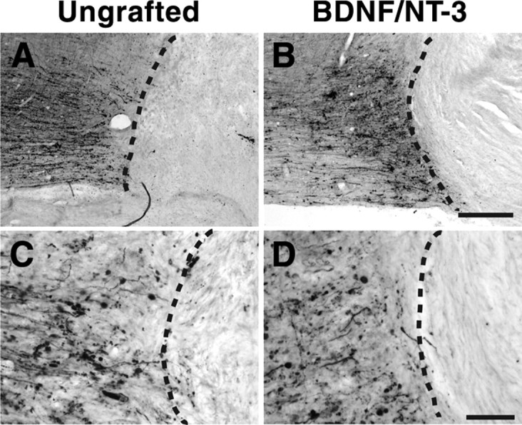
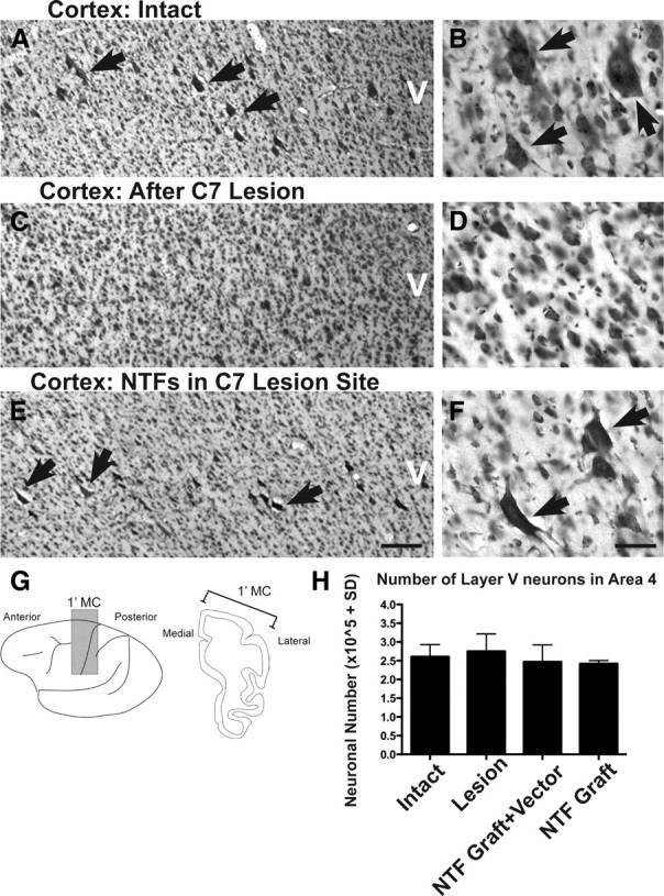
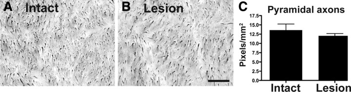
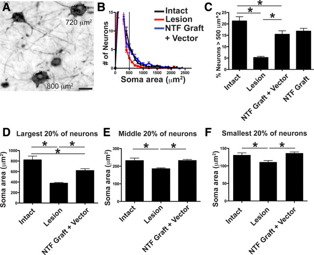
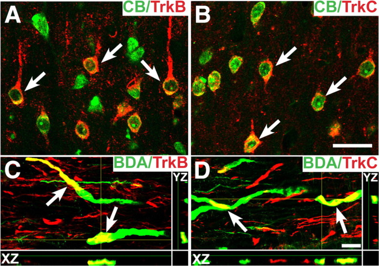
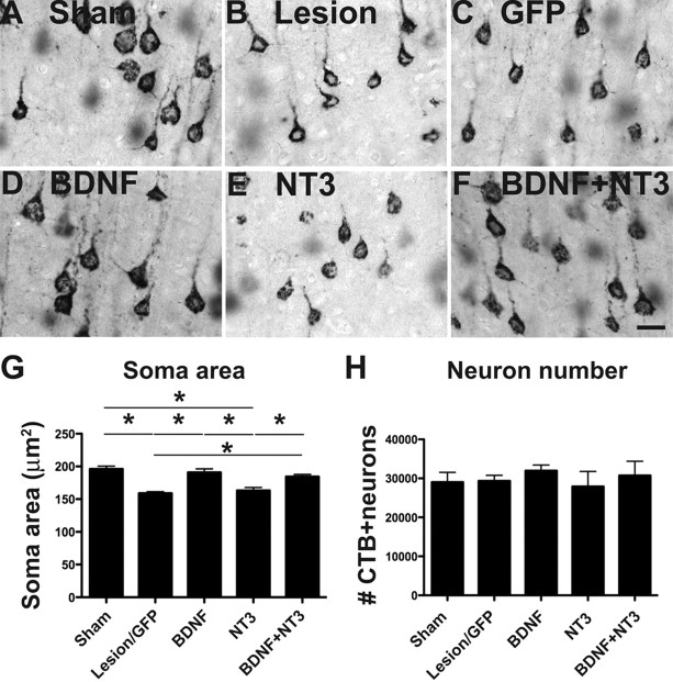
Primate models of spinal cord injury differ from rodent models in several respects, including the relative size and functional neuroanatomy of spinal projections. Fundamental differences in scale raise the possibility that retrograde injury signals, and treatments applied at the level of the spinal cord that exhibit efficacy in rodents, may fail to influence neurons at the far greater distances of primate systems. Thus, we examined both local and remote neuronal responses to neurotrophic factor-secreting cell grafts placed within sites of right C7 hemisection lesions in the rhesus macaque. Six months after gene delivery of brain-derived neurotrophic factor (BDNF) and neurotrophin-3 (NT-3) into C7 lesion sites, we found both local effects of growth factors on axonal growth, and remote effects of growth factors reflected in significant reductions in axotomy-induced atrophy of large pyramidal neurons within the primary motor cortex. Additional examination in a rodent model suggested that BDNF, rather than NT-3, mediated remote protection of corticospinal neurons in the brain. Thus, injured neural systems retain the ability to respond to growth signals over the extended distances of the primate CNS, promoting local axonal growth and preventing lesion-induced neuronal degeneration at a distance. Remote cortical effects of spinally administered growth factors could "prime" the neuron to respond to experimental therapies that promote axonal plasticity or regeneration.


Figures

Figure 1.
Figure 1.
Lesion/graft site after primate C7 hemisection. Thionin-stained transverse (A, B) and horizontal (C, D) sections through monkey spinal cord lesion site 8 months after C7 lateral hemisection (the dashed line indicates lesion border). In both nongrafted subjects (A, C) and subjects grafted with autologous fibroblasts genetically modified to secrete BDNF and NT-3 (B, D), cells fill the lesion site. Cells are both rounded and spindle-shaped, typical of fibroblast, Schwann cells, and leptomeningeal cell morphology (Fig. 2). Higher magnification insets in E and F reveal greater density cell within grafted subjects. Scale bars: B (for A, B), D (for C, D), 1 mm; F (for E, F), 50 μm; inset, 10 μm.
Figure 2.
Figure 2.
Myelin stain and ultrastructure of lesion/graft site. A, Myelin stain of the lesion site within ungrafted subject indicates occasional penetration of myelinated axons (arrows) into spontaneous cell matrix that forms in injury zone. B, In contrast, myelinated axons extensively penetrate BDNF/NT-3-secreting fibroblast graft placed in lesion site. C, Ultrastructure of lesion site in ungrafted subject demonstrates cellular influx consisting of both fibroblasts (f) and occasional ensheathing Schwann cells (s). Bundles of unmyelinated axons (a) are observed within cytoplasm of associated Schwann cell in ungrafted lesion cavity; nuclear–axonal juxtaposition is typical of Schwann cell morphology. Ungrafted subjects also exhibit abundant extracellular collagen fibrils (c), providing a substrate for axonal penetration into control lesion site. D, Subjects that received grafts of BDNF/NT-3-secreting fibroblasts into the lesion cavity generally exhibit a greater density of both cells and axons. Abundant Schwann cells are present and are associated with many myelinated axons (m). E, Higher magnification of nongrafted lesion site, demonstrating association of Schwann cell with unmyelinated axon, and extracellular collagen fibrils. F, Higher magnification of BDNF/NT-3 grafted subject within lesion site, demonstrating association of Schwann cell with myelinated axon. Scale bars: B (for A, B), 100 μm; C, D, 2 μm; E, F, 1 μm.
Figure 3.
Figure 3.
Local and supraspinal axons regenerate into neurotrophin-secreting cell grafts in the lesioned primate spinal cord. A, Neurofilament-labeled axons extend into the cellular matrix spontaneously filling the lesion in a nongrafted injury site, 8 months after injury. B, C, Significantly greater numbers of neurofilament-labeled axons penetrate the lesion site in subjects that received grafts of autologous fibroblasts genetically modified to secrete BDNF and NT-3 (B), quantified in C (*p < 0.05). D–F, Raphespinal axons grow into control lesion site (D) but exhibit a 2.5-fold increase in growth into BDNF/NT-3-secreting grafts (E), quantified in F. G–I, Cerulospinal axons labeled by TH exhibit little growth into control lesion site (G), but growth is significantly increased in the presence of growth factors (H), quantified in I. Error bars indicate SEM. Scale bars: B (for A, B), E (for D, E), H (for G, H), 25 μm.
Figure 4.
Figure 4.
Neurotrophin-secreting grafts do not promote CST regeneration. Shown are photomicrographs of horizontal spinal cord sections: rostral, left; caudal, right (dashed line indicates lesion interface). A–D, Transected CST axons anterogradely labeled with dextran-488 do not enter the cellular matrix of either ungrafted lesion site (A, C) or lesion site containing fibroblasts that secrete BDNF and NT-3 (B, D). Scale bars: B (for A, B), 250 μm; D (for C, D), 100 μm.
Figure 5.
Figure 5.
Cortical responses to injury and spinally administered neurotrophins. A, C, E, Representative thionin-stained coronal section from an intact monkey (A), lesion-only monkey (C), and lesion plus neurotrophin (NTF)-treated monkey (E). The arrows indicate large pyramidal neurons located within layer V. B, D, F, Higher magnification demonstrates a reduction in large layer V neurons after C7 hemisection that is prevented by growth factor administration to the spinal cord. G, H, Stereologic quantification of thionin-stained layer V total neuron number within the primary motor cortex (dark shading; G) indicates that total neuronal number in motor cortex is preserved after C7 lesions in all groups. Error bars indicate SEM. Scale bar: E (for A, C, E), 250 μm; F (for B, D, F), 25 μm.
Figure 6.
Figure 6.
Axons are retained within the medullary pyramids after SCI. A, B, To further confirm that CST neurons and their projections are retained 8 months after C7 hemisection, axon numbers were quantified in the medullary pyramids of intact (A) and lesioned (B) subjects (that did not receive BDNF/NT-3 grafts), using NF70 immunolabeling. C, Quantification of NF70 labeling density reveals no significant difference between intact or lesioned monkeys (p = 0.4). Error bars indicate SEM. Scale bar: B (for A, B), 25 μm.
Figure 7.
Figure 7.
Spinal growth factor treatment in primates ameliorates cortical neuronal atrophy: stereological assessment. A, Retrograde labeling with CTB indicates that a majority of identified layer V corticospinal neurons exhibit a somal area of >500 μm2 (see text). B, Frequency distribution of all thionin-stained neurons within layer V. The number of neurons within 100-μm2-sized bins is uniformly reduced after spinal cord injury (red line), and these numbers are restored by BDNF/NT-3 treatment (blue line) compared with intact animals (black line). C, C7 lesions induce a significant reduction in the proportion of large cortical motor neurons (>500 μm2), and treatment with BDNF/NT-3 substantially prevents this neuronal atrophy (NTF graft plus vector: BDNF/NT-3-secreting cell grafts in lesion cavity plus lenti-BDNF and lenti-NT-3 vector injections in host spinal cord parenchyma; NTF graft: BDNF/NT-3-secreting graft in lesion site; ANOVA, p < 0.001; post hoc Fisher's, *p < 0.001). Furthermore, neurotrophin treatment prevents atrophy of a range of layer V somal sizes. D–F, Somal sizes were divided into largest 20% (D), middle 20% (E), and smallest 20% (F). The size of layer V neurons after lesion only were significantly smaller compared with intact monkeys. BDNF/NT-3 treatment prevented the atrophy of both the middle 20% and the smallest 20% and partially prevented atrophy of the largest 20% of neurons. *p < 0.05, intact versus lesion and NTF graft plus vector. Error bars indicate SEM. Scale bar: A, 25 μm.
Figure 8.
Figure 8.
Primate TrkB and TrkC immunolabeling. A, TrkB and Cascade Blue (CB) double labeling showing expression of TrkB in intact corticospinal motor neuron cell somata. Corticospinal neurons are backlabeled after injection of CB into the spinal cord. The arrows indicate cells containing both TrkB and CB. B, TrkC and CB double labeling also shows expression of TrkC in retrogradely identified corticospinal motor neurons. C, Confocal z-stacks demonstrate that TrkB is also detected on corticospinal axons in the cervical spinal cord, double-labeled with BDA (arrows). D, TrkC is also present on corticospinal axons (arrows). The yellow crosshairs indicate the position of a single x–z and y–z plane to confirm colocalization. Scale bars: B (for A, B), 25 μm; D (for C, D), 5 μm.
Figure 9.
Figure 9.
Rodent study: spinally administered BDNF prevents cortical neuronal atrophy. A–F, Representative photomicrographs of corticospinal neurons retrogradely labeled with Cascade Blue within the rat motor cortex, after cervical corticospinal dorsal column lesion. Neuronal atrophy is evident after lesion-alone, lesion with GFP graft, or lesion with NT-3 graft. Atrophy is ameliorated by BDNF graft and BDNF plus NT-3 grafts to spinal cord. G, Stereological quantification confirms that corticospinal somal size exhibits a 20% reduction after C5 spinal cord dorsal column lesion; this atrophy is prevented by BDNF-secreting and BDNF/NT-3 grafts, but not NT-3 or control GFP grafts. H, Stereological quantification of CTB-labeled CST neuronal number indicates no cell loss after injury in any group, consistent with findings of primate study. *p < 0.05. Error bars indicate SEM. Scale bar: F (for A–F), 25 μm.

References

    1. Alto LT, Havton LA, Conner JM, Hollis Ii ER, Blesch A, Tuszynski MH. Chemotropic guidance facilitates axonal regeneration and synapse formation after spinal cord injury. Nat Neurosci. 2009;12:1106–1113. - PMC - PubMed
    1. Ben-Yaakov K, Fainzilber M. Retrograde Injury Signaling in Lesioned Axons. Results Probl Cell Differ. 2009;48:327–338. - PubMed
    1. Blesch A, Tuszynski MH. Spinal cord injury: plasticity, regeneration and the challenge of translational drug development. Trends Neurosci. 2009;32:41–47. - PubMed
    1. Chen Q, Zhou L, Shine HD. Expression of neurotrophin-3 promotes axonal plasticity in the acute but not chronic injured spinal cord. J Neurotrauma. 2006;23:1254–1260. - PubMed
    1. Conner JM, Varon S. Characterization of antibodies to nerve growth factor: assay-dependent variability in the cross-reactivity with other neurotrophins. J Neurosci Methods. 1996;65:93–99. - PubMed

Publication types

MeSH terms