. 2010 Jul 21;30(29):9910-8.
doi: 10.1523/JNEUROSCI.1111-10.2010.

The human basal ganglia modulate frontal-posterior connectivity during attention shifting

Affiliations

The human basal ganglia modulate frontal-posterior connectivity during attention shifting

Martine R van Schouwenburg et al. J Neurosci. .

Abstract

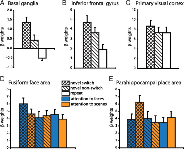
Current models of flexible cognitive control emphasize the role of the prefrontal cortex. This region has been shown to control attention by biasing information processing in favor of task-relevant representations. However, the prefrontal cortex does not act in isolation. We used functional magnetic resonance imaging combined with nonlinear dynamic causal modeling to demonstrate that the basal ganglia play a role in modulating the top-down influence of the prefrontal cortex on visual processing in humans. Specifically, our results reveal that connectivity between the prefrontal cortex and stimulus-specific visual association areas depends on activity in the ventral striatopallidum, elicited by salient events leading to shifts in attention. These data integrate disparate literatures on top-down control by the prefrontal cortex and selective gating by the basal ganglia and highlight the importance of the basal ganglia for high-level cognitive control.


Figures

Figure 1.
Figure 1.
The attention-switching paradigm used in this study required subjects to select one stimulus exemplar (left vs right) within one dimension (faces vs scenes) on every trial. A, Each trial consisted of two consecutive responses followed by feedback. Red boxes indicate a possible response sequence. B–D show two consecutive trials with responses defining the three different trial types. For clarification, the stimuli are displayed schematically (F1, face 1; S1, scene1; F2, face 2; S2, scene 2). B, In this example, the subject is attending to F1 on the first trial (attended stimuli are displayed in italic). On the next trial, no novel stimuli are introduced and the subject keeps attending to F1. The second trial is thus defined as a repeat trial. C, On a novel switch trial, novel stimuli of the unattended dimension, in this case scenes, are introduced (S3 and S4). The subject detects this change and switches attention to one of two novel stimuli (here S3). D, Alternatively the subject can fail to detect the novel stimuli and keep responding to the previously relevant stimulus exemplar, in this case F1. The subject will then receive negative feedback and the second trial is defined as a novel nonswitch trial.
Figure 2.
Figure 2.
Dynamic causal modeling was used to investigate the modulation of connections between the IFG and FFA/PPA by switch-related activity in the BG. A, The basic architecture of the model included connections from the IFG to the FFA and PPA (black) and the following inputs: novelty to the IFG, switch to the BG, attention to faces to the FFA, and attention to scenes to the PPA. B, We tested 16 alternative models that could include connections from IFG to BG (orange), from BG to IFG (blue), reciprocal connection between FFA and PPA (red), and modulation of IFG to FFA/PPA connectivity by the BG (green). Dark gray boxes indicate that this connection was included in the model. The best model (16) included all connections.
Figure 3.
Figure 3.
BOLD responses from a whole-brain analysis. Bars indicate t values, and figures are thresholded for a t value of 3.65, corresponding to a p-value of 0.001 uncorrected for multiple comparisons. A, Contrasting novel switch trials with repeat trials showed increased responses in the BG, anterior cingulate cortex, IFG, midbrain, parietal cortex, and posterior visual regions. B, When comparing novel nonswitch trials with repeat trials, the BG and frontoparietal regions also showed an increase in BOLD responses, but this effect was not observed in posterior visual regions. C, Contrasting novel switch trials with novel nonswitch trials showed increased responses in posterior visual regions and the BG.
Figure 4.
Figure 4.
Individual differences in behavior could be explained by BOLD signal in the BG. More specifically, the level of BOLD signal on novel switch trials in the BG correlated negatively with the switch likelihood (left BG, r18 = −0.54, p < 0.05; right BG, r18 = −0.48, p < 0.05). β weights were extracted from the group peak voxel from the novel switch versus repeat contrast in the right BG [MNI coordinates x, y, z (10, 10, 0)].
Figure 5.
Figure 5.
To illustrate the pattern of responses in our VOIs, we extracted the β weights for each subject from the group peak voxels [MNI coordinates (x, y, z)] from the novel switch versus repeat contrast. Here we display the mean ± SEM β weights across subjects. A, B, Novel stimuli increased BOLD responses in the BG [coordinates (10, 10, 0)] (A) and the IFG [coordinates (48, 10, 28)] (B) even when they were not detected. Supplementary repeated-measures ANOVA revealed a significant interaction between region (BG vs IFG) and novelty (novel switch + novel nonswitch vs repeat) (F(1,1) = 10.2, p < 0.01), suggesting that the IFG is particularly important for processing novel information. C, In contrast, in the primary visual cortex [coordinates (14, −80, 8)], BOLD responses increased only when the novel information elicited an attention switch. D, E, β weights for the FFA (D) and the PPA (E) were extracted from the individual localizer-defined VOIs using the supplementary GLM (model B). These areas showed stimulus-specific effects, such that the BOLD response in the FFA increased when an attention switch was elicited by a novel face but not a novel scene, whereas the reverse effect was found in the PPA.

References

    1. Alexander GE, DeLong MR, Strick PL. Parallel organization of functionally segregated circuits linking basal ganglia and cortex. Annu Rev Neurosci. 1986;9:357–381. - PubMed
    1. Braver TS, Cohen JD. On the control of control: the role of dopamine in regulating prefrontal function and working memory. In: Monsell S, Driver J, editors. Attention and performance XVIII; Control of cognitive processes. Cambridge, MA: MIT; 2000. pp. 713–737.
    1. Brett M, Anton JL, Valabregue R, Poline JB. Region of interest analysis using an SPM toolbox. Presented at the 8th International Conference on Functional Mapping of the Human Brain; June 2–6; Sendai, Japan. 2002.
    1. Bundesen C. A theory of visual-attention. Psychol Rev. 1990;97:523–547. - PubMed
    1. Bunzeck N, Düzel E. Absolute coding of stimulus novelty in the human substantia nigra/VTA. Neuron. 2006;51:369–379. - PubMed

Publication types

LinkOut - more resources