. 2010 Jul 21;30(29):9939-46.
doi: 10.1523/JNEUROSCI.1643-10.2010.

Histone deacetylase Rpd3 regulates olfactory projection neuron dendrite targeting via the transcription factor Prospero

Affiliations

Histone deacetylase Rpd3 regulates olfactory projection neuron dendrite targeting via the transcription factor Prospero

Joy S Tea et al. J Neurosci. .

Abstract

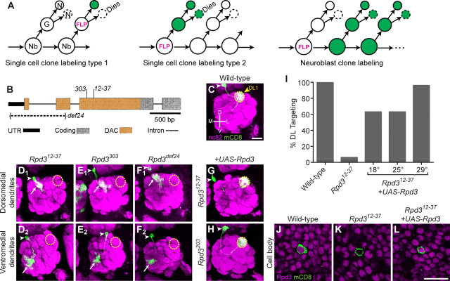
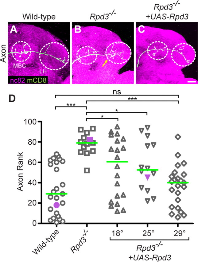
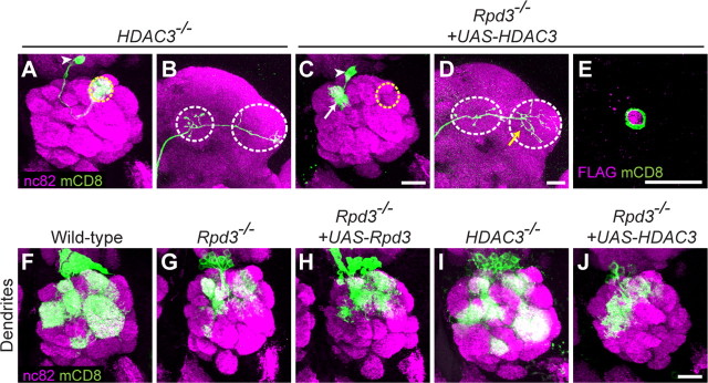
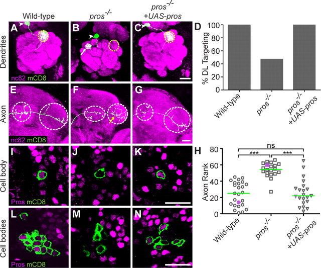
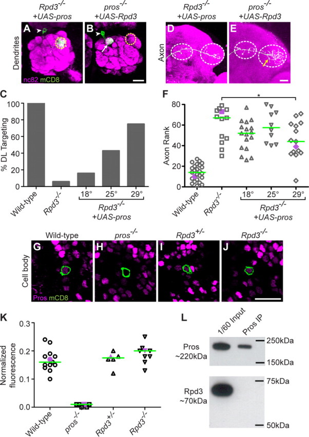
Compared to the mechanisms of axon guidance, relatively little is known about the transcriptional control of dendrite guidance. The Drosophila olfactory system with its stereotyped organization provides an excellent model to study the transcriptional control of dendrite wiring specificity. Each projection neuron (PN) targets its dendrites to a specific glomerulus in the antennal lobe and its axon stereotypically to higher brain centers. Using a forward genetic screen, we identified a mutation in Rpd3 that disrupts PN targeting specificity. Rpd3 encodes a class I histone deacetylase (HDAC) homologous to mammalian HDAC1 and HDAC2. Rpd3(-/-) PN dendrites that normally target to a dorsolateral glomerulus mistarget to medial glomeruli in the antennal lobe, and axons exhibit a severe overbranching phenotype. These phenotypes can be rescued by postmitotic expression of Rpd3 but not HDAC3, the only other class I HDAC in Drosophila. Furthermore, disruption of the atypical homeodomain transcription factor Prospero (Pros) yields similar phenotypes, which can be rescued by Pros expression in postmitotic neurons. Strikingly, overexpression of Pros can suppress Rpd3(-/-) phenotypes. Our study suggests a specific function for the general chromatin remodeling factor Rpd3 in regulating dendrite targeting in neurons, largely through the postmitotic action of the Pros transcription factor.


Figures

Figure 1.
Figure 1.
Rpd3 acts in postmitotic neurons to regulate projection neuron dendrite targeting. A, Schematic of MARCM single-cell or neuroblast clone generation. Using MARCM, single-cell or neuroblast clones can be positively labeled (shown in green) after FLP-mediated mitotic recombination in the ganglion mother cell or neuroblast, as indicated by the three models. Recent studies show that the sibling of the DL1 neuron dies during development (Lin et al., 2010; Potter et al., 2010). Nb, Neuroblast; G, ganglion mother cell; N, postmitotic neuron; FLP, Flippase. B, Schematic of the Rpd3 gene locus, with indicated mutations (modified after Mottus et al., 2000). Allele Rpd312–37 was generated in this study; Rpd3303 and Rpd3def24 have been previously described. Allele Rpd3303 is a cysteine to tyrosine missense mutation which retains anti-Rpd3 immunoreactivity (data not shown). Allele Rpd3def24 is a large deletion encompassing the transcriptional start and much of the N terminus which results in a loss of anti-Rpd3 immunoreactivity (data not shown). UTR, Untranslated region; DAC, deacetylase domain. C, WT DL1 PN dendrites target specifically to the glomerulus DL1 (yellow dashed circle). White arrowhead denotes cell body. D, Representative images of Rpd312–37 mutant single-cell clones. Mistargeting to the dorsomedial (DM) quadrant of the antennal lobe occurs in 47% of clones (D1). Mistargeting to ventromedial (VM) quadrant occurs in 44% of clones (D2). A total of 9% showed dorsolateral (DL) targeting (data not shown). n = 32. White arrowhead denotes cell body, yellow dashed circle indicates location of the DL1 glomerulus, white arrow denotes mistargeted dendrites. E, Representative images of Rpd3303 mutant single-cell clones: 82% mistargeting to DM (E1), 14% mistargeting to VM (E2). A total of 4% showed DL targeting (data not shown). n = 22. F, Representative images of Rpd3def24 mutant single-cell clones: 61% mistargeting to DM (F1), 25% mistargeting to VM (F2). A total of 14% showed DL targeting (data not shown). n = 28. G, Postmitotic expression of UAS-Rpd3 in a single DL1 PN rescues the Rpd312–37 dendrite phenotype. H, Postmitotic expression of UAS-Rpd3 in a single DL1 PN rescues the Rpd3303 dendrite phenotype. I, Quantification of DL targeting for genotypes in C, D, and G. Number of analyzed clones left to right is 35, 32, 22, 19, 24. J, At 24hAPF, Rpd3 is expressed ubiquitously in all nuclei, including PNs. K, Rpd312–37 single-cell MARCM clone (green) lacks Rpd3 immunoreactivity. L, MARCM expression of UAS-Rpd3 restores Rpd3 immunoreactivity in an Rpd312–37 single-cell clone. Green marks mCD8-GFP-labeled PN dendrites (C–H) and cell bodies (J–L) generated by MARCM and labeled using GH146-GAL4. C–H show confocal stacks, magenta is the presynaptic marker nc82. J–L show single confocal sections, magenta is anti-Rpd3. Scale bars: C (for C–H), L (for J–L), 20 μm.
Figure 2.
Figure 2.
Rpd3 limits PN axon terminal arborization. A, WT DL1 axons show an L-shaped pattern in the lateral horn (LH), with additional branches in the mushroom body calyx (MBC), both outlined with white dashed circle. B, Rpd3−/− DL1 PN axons exhibit an overbranching phenotype in the lateral horn (yellow arrow). C, Postmitotic expression of UAS-Rpd3 in a single DL1 PN rescues the Rpd3−/− overbranching phenotype. D, Rank order of axon overbranching for different genotypes as indicated. Pairwise Mann–Whitney U tests were performed to determine significance. ns, not significant. *p < 0.05. ***p < 0.0001. Purple symbols indicate the examples shown in A–C. Green line denotes median. Number of clones is 25, 13, 20, 14, 23. Green marks mCD8-GFP-labeled PN axons (A–C) generated by MARCM and labeled using GH146-GAL4. A–C show confocal stacks; magenta is the presynaptic marker nc82. Scale bar, 20 μm.
Figure 3.
Figure 3.
HDAC3 does not regulate PN targeting or arborization. A, Dendrites of HDAC3−/− DL1 PN target normally to the DL1 glomerulus. B, HDAC3−/− PN axon exhibits a normal branching pattern in the lateral horn. C, Postmitotic expression of UAS-HDAC3 does not suppress the Rpd3−/−-mistargeting phenotype (compare with Fig. 1D,G). D, Postmitotic expression of UAS-HDAC3 does not suppress the Rpd3−/− axon overbranching phenotype (compare with Fig. 2B,C). E, UAS-HDAC3 is expressed in a MARCM-labeled Rpd3−/− cell body at 24hAPF, as visualized by anti-FLAG staining of HDAC3–3xFLAG. F, Wild-type PN neuroblast clones target their dendrites to stereotyped glomeruli in the antennal lobe. G, Rpd3−/− neuroblast clones show a reduced dendritic mass due at least partially to a 50% decrease in cell number, and those dendrites do not target their correct glomeruli. H, The reduced cell number phenotype of Rpd3−/− neuroblast clones is not rescued by postmitotic expression of UAS-Rpd3. Because of the reduction of cell number and labeling of multiple neurons, it cannot be determined whether the dendrite-mistargeting phenotype is rescued by UAS-Rpd3. I, HDAC3−/− neuroblast clones show a 20% reduction in cell number, and their dendrites appear to target normally. J, The reduced cell number phenotype of Rpd3−/− neuroblast clones cannot be rescued by postmitotic expression of UAS-HDAC3. Because there are multiple neurons labeled, it cannot be determined whether the dendrite-mistargeting phenotype is rescued by UAS-HDAC3. Green marks mCD8-GFP-labeled PN dendrites (A, C, F–J) axons (B, D), and cell body (E) generated by MARCM and labeled using GH146-GAL4. A–D and F–J show confocal stacks; magenta is the presynaptic marker nc82. E, shows a single confocal section, magenta is anti-FLAG staining. Symbols are as in Figures 1 and 2. Scale bars: C (for A, C), D (for B, D), E, J (for F–J), 20 μm.
Figure 4.
Figure 4.
Rpd3 is the major histone deacetylase in PNs. A, A single confocal section in WT 24hAPF pupa showing the DL1 PN (green in A2) stained with an antibody against acetylated lysine (AcK; magenta). A low level of acetylated lysine is found throughout all cells. B, AcK is increased in an Rpd3−/− MARCM single-cell DL1 clone at 24hAPF. C, Postmitotic expression of UAS-Rpd3 restores wild-type acetylation levels in an Rpd3−/− MARCM single-cell DL1 clone. D, HDAC3−/− DL1 PN clone does not show an elevated level of acetylated lysine. E, Expression of HDAC3 cannot reduce the elevated acetylation in Rpd3−/− PNs. F, Single confocal section of WT 24hAPF pupa surrounding a PN neuroblast clone (green in F2) stained with an antibody against acetylated lysine (magenta). A low level of acetylated lysine is found throughout all cell bodies. G, A marked increase of acetylated lysine is evident in an Rpd3−/− MARCM neuroblast clone. H, Postmitotic expression of UAS-Rpd3 can restore wild-type acetylation levels in an Rpd3−/− neuroblast clone at 24hAPF. I, HDAC3−/− neuroblast clone does not show an elevated level of acetylated lysine. J, Expression of HDAC3 cannot reduce the elevated acetylation in Rpd3−/− PNs. Green marks mCD8-GFP-labeled PN cell body (A2–E2) or cell bodies (F2–J2) generated by MARCM and labeled using GH146-GAL4. A–J show single confocal sections; magenta is anti-acetylated lysine staining. *Increased acetylation in other cell bodies indicates additional unlabeled, GH146-negative, Rpd3−/− cells. Scale bars: E2 (for A1 to E2), J2 (for F1 to J2), 20 μm.
Figure 5.
Figure 5.
Prospero acts in postmitotic neurons to regulate PN dendrite targeting and axon terminal arborization. A, WT DL1 PN dendrites target specifically to the glomerulus DL1, same as Figure 1C. B, pros−/− DL1 PN dendrites mistarget to medial glomeruli (white arrow). C, Postmitotic expression of UAS-pros rescues the pros−/− DL1 PN dendrite phenotype. D, Quantification of DL targeting for the genotypes in A–C. Number of clones is 25, 45, 34. E, WT DL1 axon, same as Figure 2A. F, pros−/− DL1 PN axon exhibits an overbranching phenotype in the lateral horn (yellow arrow). G, Postmitotic expression of UAS-pros rescues the pros−/− overbranching phenotype. H, Rank order of axon overbranching phenotype of different genotypes as indicated. Pairwise Mann–Whitney U tests were performed to determine significance. Number of clones is 25, 20, 21. I, At 24hAPF, Pros is expressed at varying levels in different PN nuclei. The DL1 PN (green) expresses an intermediate level of Pros protein. J, pros−/− single-cell MARCM clone lacks Pros immunoreactivity. K, MARCM expression of UAS-pros restores Pros immunoreactivity in a single PN. L, At 24hAPF, Pros is expressed at varying levels in PN cell bodies. PNs can express high, medium, low, or undetectable levels of Pros. This variable expression persists throughout development and adulthood (data not shown). M, Cell bodies of pros−/− neuroblast MARCM clones (green) lack Pros immunoreactivity. N, MARCM expression of UAS-pros restores Pros immunoreactivity in neuroblast clone PNs at 24hAPF. The level of UAS-pros expression varies similarly to endogenous Pros expression, suggesting a posttranscriptional mechanism in regulating Pros protein level. Green marks mCD8-GFP-labeled PN dendrites (A–C), axons (E–G), and cell bodies (I–N) generated by MARCM and labeled using GH146-GAL4. A–C and E–G show confocal stacks; magenta is the presynaptic marker nc82. I–N show single confocal sections; magenta is anti-Prospero staining. Symbols are as in Figures 1 and 2. Scale bars: C (for A–C), G (for E–G), K (for I–K), N (for L–N), 20 μm.
Figure 6.
Figure 6.
Postmitotic expression of Prospero suppresses Rpd3−/− PN phenotypes. A, Postmitotic expression of UAS-pros suppresses the Rpd3−/− PN phenotype, restoring DL1 PN dendrite targeting to the DL1 glomerulus. B, Postmitotic expression of UAS-Rpd3 does not suppress the pros−/−-mistargeting phenotype. C, Quantification of UAS-pros suppression of the Rpd3−/− phenotype at different temperatures. Number of clones is 25, 13, 20, 23, 16. D, Postmitotic expression of UAS-pros partially suppresses the Rpd3−/− axon overbranching phenotype. E, Postmitotic expression of UAS-Rpd3 does not suppress the pros−/− axon overbranching phenotype. F, Rank order of axon overbranching for different genotypes as indicated. Pairwise Mann–Whitney U tests were performed to determine significance. *p < 0.05 at 29°C; no significant difference at 18°C or 25°C. Number of clones is 25, 13, 17, 10, 15. G, At 24hAPF, Pros is expressed at an intermediate level in the DL1 PN (green). H, pros−/− DL1 MARCM clone lacks Pros immunoreactivity. I, Heterozygous Rpd3+/− MARCM clone shows a normal level of Pros immunoreactivity. J, Homozygous Rpd3−/− MARCM clone shows a normal level of Pros immunoreactivity. K, Normalized fluorescence of DL1 PNs for different genotypes as indicated. Purple symbols indicate the examples shown in G–J. Green line denotes median. Number of clones is 13, 6, 6, 9. Graph shows the ratio of DL1 to highest intensity. L, Immunoprecipitation of Prospero does not result in coimmunoprecipitation of Rpd3. Top shows 1/60 input and final immunoprecipitation of Pros blotted with anti-Pros. Bottom shows 1/60 input and denatured immunoprecipitate blotted with anti-Rpd3. Green marks mCD8-GFP-labeled PN dendrites (A, B), axons (D, E), and cell bodies (G–J) generated by MARCM and labeled using GH146-GAL4. (A, B, D, E) show confocal stacks; magenta is the presynaptic marker nc82. G–J show single confocal sections; magenta is anti-Prospero staining. Symbols are as in Figures 1 and 2. Scale bars: B (for A, B), E (for D, E), J (for G–J), 20 μm.

References

    1. Bischof J, Maeda RK, Hediger M, Karch F, Basler K. An optimized transgenesis system for Drosophila using germ-line-specific phiC31 integrases. Proc Natl Acad Sci U S A. 2007;104:3312–3317. - PMC - PubMed
    1. Broadus J, Fuerstenberg S, Doe CQ. Staufen-dependent localization of prospero mRNA contributes to neuroblast daughter-cell fate. Nature. 1998;391:792–795. - PubMed
    1. Chihara T, Luginbuhl D, Luo L. Cytoplasmic and mitochondrial protein translation in axonal and dendritic terminal arborization. Nat Neurosci. 2007;10:828–837. - PubMed
    1. Cho Y, Griswold A, Campbell C, Min KT. Individual histone deacetylases in Drosophila modulate transcription of distinct genes. Genomics. 2005;86:606–617. - PubMed
    1. Choksi SP, Southall TD, Bossing T, Edoff K, de Wit E, Fischer BE, van Steensel B, Micklem G, Brand AH. Prospero acts as a binary switch between self-renewal and differentiation in Drosophila neural stem cells. Dev Cell. 2006;11:775–789. - PubMed

Publication types

MeSH terms

LinkOut - more resources