. 2010 Aug 4;102(15):1131-47.
doi: 10.1093/jnci/djq218. Epub 2010 Jul 21.

KLF6 Gene and early melanoma development in a collagen I-rich extracellular environment

Affiliations

KLF6 Gene and early melanoma development in a collagen I-rich extracellular environment

Sung Jin Huh et al. J Natl Cancer Inst. .

Abstract

Background: A putative tumor suppressor gene at chromosome 10p15, which contains KLF6 and other genes, is predicted to be lost during melanoma development, and its identity is unknown. In this study, we investigated the biological roles and identity of this tumor suppressor gene.

Methods: The human UACC 903 melanoma cell line containing introduced DNA fragments from the 10p15 region with (10E6/3, 10E6/11, and 10E6/18) and without (10ER4S.2/1) the tumor suppressor gene was used. Xenograft tumors were generated in a total of 40 mice with melanoma cell lines, and tumor size was measured. Cells were cultured on plastic or a gel of type I collagen. Viability, proliferation, and apoptosis were assessed. Expression of KLF6 protein was assessed by immunohistochemistry and immunoblot analysis. Expression of phosphorylated Erk1/2 and cyclin D1 was assessed by immunoblot analysis. Protein expression of KLF6 was inhibited with small interfering RNA (siRNA). KLF6 protein expression was assessed in 17 human nevi and human melanoma specimens from 29 patients. Statistical analyses were adjusted for multiple comparisons by use of Dunnett method. All statistical tests were two-sided.

Results: Melanoma cells containing KLF6 generated smaller subcutaneous xenograft tumors with fewer proliferating cells than control cells. When grown on collagen 1, viability of cells with ectopic KLF6 expression (72%) was lower than that of control cells (100%) (group difference = -28%, 95% confidence interval = -31.3% to -25.2%, P < .001). Viability of melanoma cells with or without the KLF6 tumor suppressor gene on plastic dishes was similar. When KLF6 expression was inhibited with KLF6 siRNA, viability of cells with the tumor suppressor gene on collagen I gel increased compared with that of control cells carrying scrambled siRNA. KLF6 protein was detected in all nevi examined but not in human metastatic melanoma tissue examined. Ectopic expression of KLF6 protein in melanoma cells grown on collagen I decreased levels of phosphorylated Erk1/2 and cyclin D1 in the mitogen-activated protein kinase signaling pathway.

Conclusions: In melanoma cells, the tumor suppressor gene at 10p15 appears to be KLF6. Signaling from the collagen I-rich extracellular matrix appears to be involved in the tumor suppressive activity of KLF6 protein.


Figures

Figure 1
Figure 1
Characterization of xenografted melanoma tumors carrying the 10p15 tumor suppressor gene. A) Tumor growth. Data are the mean value from eight tumors per group (four mice per group and two tumors per mouse) and are presented on a per mouse basis. The experiment was repeated twice. Open bars = control cells; shaded bars = cells containing the 10p15 tumor suppressor region. * = P < .001 (F4,34 = 41.77, two-sided one-way analysis of variance test followed by Dunnett multiple comparison test in which UACC 903 were compared with hybrid cells); error bars = 95% confidence interval. B) Electron micrographs of UACC 903 cell tumors and 10E6/11 tumors. Bar = 0.5 μm. C) Extensive extracellular matrix and basement membrane organization in xenograft tumors, as indicated. The extracellular matrix was stained with trichrome and collagen IV was stained with anti-collagen IV. Arrowheads = extracellular matrix (blue areas); solid arrows = collagen IV bundles; open arrows = randomly distributed collagen IV. Scale bars = 50 μm. BM = basement membrane; EM = extracellular matrix; V = blood vessels.
Figure 2
Figure 2
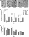
Cell proliferation and apoptosis in xenograft tumors containing or lacking the tumor suppressor fragment. A) Proliferating cells in xenograft tumors at day 16 after injection. Proliferative cells were stained by use of the bromodeoxyuridine (BrdUrd) method (37). Arrows = proliferating tumor cells. Scale bars = 50 μm. B) BrdUrd incorporation in tumors at days 16–18 after injection. Data are the mean percentage of BrdUrd-positive staining tumor cells of total tumor cells. For each tumor type, at least four different tumors and six fields per tumor were assessed (from minimum of 15 fields per group). * = P < .001 (F4,30 = 19.90 for day 16, 30.94 for day 17, and 18.30 for day 18) two-sided one-way analysis of variance test followed by Dunnett multiple comparison test for the comparisons of UACC 903 control tumor cells with 10E6/3, 10E6/11, and 10E6/18 hybrid tumor cells. C) Apoptosis rate of tumors containing or lacking the 10p15 suppressor gene at days 16–18. Data are the mean value from minimum of 15 fields per group. Bars are as described in panel (B). Data in panels (B and C) are presented as the mean per mouse. Error bars = upper 95% confidence intervals.
Figure 3
Figure 3
Viability of melanoma cells containing the 10p15 tumor suppressor gene in different culture systems. Cell viability was measured by use of a 3-(4,5-dimethylthiazon-2-yl)-5-(3-carboxymethoxyphenyl)-2-(4-sulfophenyl)-2H-tetrazolium salt (MTS) assay and is reported as the percentage of control UACC 903 cell cultures. UACC 903 and hybrid cells were cultured in 96-well dishes. A) Viability of cells cultured in plastic dishes. Cells were cultured in Dulbecco's modified Eagle medium (DMEM) containing either 10% or 1% fetal bovine serum (FBS). B) Viability of cells in anchorage-independent culture. Cells were cultured in polyHEMA-coated 96-well plates. C) Viability of cells in type I collagen culture. (AC) Data are the mean value from eight wells per group. Experiments were repeated three times. * = P < .001 (F4,35 = 129.31, two-sided one-way analysis of variance test followed by Dunnett multiple comparison test to compare UACC 903 control cells with 10E6/3, 10E6/11, and 10E6/18 hybrid cells that carry the tumor suppressor gene fragment); error bars = 95% confidence intervals. D) Mitogen-activated protein (MAP) kinase pathway signaling. Protein lysates were collected from cells growing in DMEM containing 10% FBS on plastic or type I collagen. Levels of phosphorylated extracellular-related kinase (pErk1/2), cyclin D1, and cyclin-dependent kinase 4 (CDK4) were assessed by immunoblot analysis. α-Enolase was the control for equal protein loading. Experiments were repeated three times.
Figure 4
Figure 4
KLF6 protein and mRNA expression in melanoma cell lines. A) Level of KLF6 protein in normal melanocytes (NHEM) and melanoma cell lines established from primary tumors at the radial (WM35 and WM3211), vertical (WM115, WM98.1, and WM278), and metastatic (UACC 903 and SK-MEL-24) growth phase of melanoma progression. Data are levels of KLF6 protein normalized to that of extracellular-related kinase 2 (the control for protein loading), which were averaged from three independent experiments. # = P = .014; & = P = .22, * = P < .001 (F7,16 = 14.15, two-sided one-way analysis of variance [ANOVA] test followed by Dunnett multiple comparison test to compare melanocyte control cells with WM35, WM3211, WM98, WM115, WM278, UACC 903, and SK-MEL-24 cells); arrows = decreased expression of KLF6 compared with normal melanocytes. B) KLF6 mRNA levels in control UACC 903 and 10ER4S.2/1 cells and in 10E6/3, 10E6/11, and 10E6/18 hybrid cells with the tumor suppressor gene fragment, as well as WM35 and SK-MEL-24 melanoma cells. Human GAPDH mRNA was the internal control, and level of KLF6 mRNA was normalized to that of GAPDH mRNA. Data are the mean of triplicate samples from three independent experiments. Square bracket = percentage of increased expression in hybrid cells; * = P < .001 (F4,40 = 27.33, two-sided one-way ANOVA test followed by Dunnett multiple comparison test to compare UACC 903 control with 10E6/3, 10E6/11, and 10E6/18 hybrid cells); error bars = 95% confidence intervals. Experiments were repeated three times.
Figure 5
Figure 5
Ectopic expression of KLF6 and viability of UACC 903 and SK-MEL-24 melanoma cells on type I collagen and on plastic. Expression of wild-type and mutant KLF6 protein in melanoma cells on type I collagen. A, Upper) Immunoblots of total lysates collected from UACC 903 or SK-MEL-24 cells ectopically expressing wild-type KLF6, hemagluttinin A (HA)-tagged KLF6 (HA3KLF6), and mutant forms of KLF6. Cells carrying empty puro vectors were the control. Blots were probed with antibodies against the HA tag or wild-type KLF6 protein. Intact = ectopical expression of wild-type HA-KLF6; truncated = mutant KLF6 form HA-W162X. Endogenous levels of KLF6 were probed with an antibody against KLF6 protein. α-Enolase was the control for equal protein loading. A, Lower) Relative levels of KLF6 protein. Levels of KLF6 protein in blots in panel (A) were normalized to that of α-enolase, and the relative expression was plotted. Endogenous levels of KLF6 in UACC 903 vector control were set as 1 (dash line). The normal physiological range of KLF6 protein is 1.5 as shown in WM35 cells, which has similar expression levels as normal melanocytes. Blots were repeated twice, and both showed similar results. B) Expression of KLF6 and viability of melanoma cells on collagen I. Viability of UACC 903 or SK-MEL-24 cells ectopically expressing wild-type or HA-tagged KLF6 (HA3KLF6) was assessed in type I collagen cell culture and compared with that of control cells expressing inactive (HA3K209R and HA3W162X) protein or carrying the empty puro vector, by use of 3-(4,5-dimethylthiazon-2-yl)-5-(3-carboxymethoxyphenyl)-2-(4-sulfophenyl)-2H-tetrazolium salt (MTS) assay. Data are the mean value from eight samples; the experiment was repeated three times. * = P < .001 (F4,35 = 641.17 for UACC 903 cells and F4,35 = 102.76 for SK-MEL-24 cells; two-sided one-way analysis of variance test followed by Dunnett multiple comparison test to compare UACC 903 or SK-MEL-24 puro vector control cells with KLF6 ectopically expressing cells); error bars = 95% confidence intervals.
Figure 6
Figure 6
KLF6 small interfering RNA (siRNA), melanoma cells containing chromosome 10p15 fragments, and viability on type I collagen of UACC 903, 10E6/3, 10E6/11, 10E6/18, and 10ER4S.2/1 cells. A) KLF6 siRNA and KLF6 protein expression. siRNA (100 pmol) of was introduced by nucleofection with an Amaxa Nucleofector, and 2 days later, protein lysates were prepared and KLF6 protein expression was assessed by immunoblot analysis with KLF6 antibody. Untransfected or cells nucleofected with scrambled siRNA were used as controls, and α-enolase was the control for equal protein loading. Arrow = reduction of KLF6 expression compared with control. Experiments were repeated three times. B) Cell viability at day 5 of cells cultured on type I collagen. Data are the mean value of eight samples; the experiment was repeated three times. * = P < .001 (F2,21 = 361.7 for 10E6/3 cells, 161.2 for 10E6/11 cells, and 260.6 for 10E6/18 cells; two-sided one-way analysis of variance test followed by Dunnett multiple comparison test to compare the effect of siRNA-mediated targeting KLF6 with that of untransfected and scrambled siRNA controls); error bars = 95% confidence intervals; arrow = increased cell viability compared with controls.
Figure 7
Figure 7
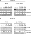
Mitogen-activated protein (MAP) kinase pathway signaling, KLF6 expression, and culture on type I collagen or plastic in UACC 903 or SK-MEL-24. A) Cell lines derived from UACC 903 melanoma cells. B) Cell lines derived from SK-MEL-24 melanoma cells. Levels of phosphorylated extracelluar-related kinase (pErk1/2) and cyclin D1 were assessed in lysates collected from UACC 903 and SK-MEL-24 cells ectopically expressing wild-type KLF6, HA-KLF6, or mutant forms of KLF6 by immunoblot analysis with anti-phosphorylated Erk1/2 and anti-cyclin D1 antibodies. Numerical values below each blot are the normalized averages of pErk1/2 and cyclin D1 expression from two independent experiments. Averages were normalized to the value for α-enolase (control for protein loading). Experiments were repeated two times.
Figure 8
Figure 8
KLF6 protein and mRNA expression in human melanomas and human nevi. A) Immunohistochemical analysis of KLF6 protein expression in melanoma tumors and nevi. Formalin-fixed paraffin-embedded sections of atypical nevi, primary melanomas of greater than 0.75 mm in depth, and metastatic melanomas were stained with hematoxylin–eosin (H&E) or for KLF6 protein expression by immunohistochemistry with anti-KLF6 and secondary avidin–biotin horseradish peroxidase anti-rabbit IgG antibody. Brown staining = KLF6 protein; arrowheads = KLF6 staining. Staining of KLF6 in epidermis served as the positive control. Bottom row is a higher magnification of the section in middle row. Scale bars = 50 μm. B) KLF6 protein expression in xenografted UACC 903 tumors at day 17. Arrowheads = KLF6 staining; arrows = exterior and interior of tumors at high magnification in the right panel. Scale bars = 50 μm. C) KLF6 protein expression in human metastatic melanomas. Protein lysates were prepared from of 29 human melanoma tumors (numbered 1–29) and analyzed for expression of KLF6, ITIH5 (a gene also located at 10p15), PTEN (a gene located at 10q23), and extracellular-related kinase2 (Erk2) proteins. PTEN protein status: + = present moderate expression; ++ = high expression; − = absent. Erk2 was the control for equal protein loading. WM35 and SK-MEL-24 cell lysates served as positive and negative controls for high and low KLF6 protein expression, respectively. Experiments were repeated three times. D) KLF6 mRNA expression in five human melanoma tumors that lacked KLF6 protein expression. KLF6 mRNA levels were measured with a quantitative real-time polymerase chain reaction in triplicate samples and in three experiments and were compared with that in WM35 cells, which have KLF6 protein levels similar to that in normal melanocytes. Human GAPDH sequences were amplified for use as an internal control and for normalization of KLF6 mRNA. The mRNA from WM35 cells served as a positive control for high KLF6 mRNA expression. Data are the mean value from triplicate sample, and each experiment was repeated three times. Error bars = 95% confidence intervals.

Comment in

References

    1. Newton JA. Genetics of melanoma. Br Med Bull. 1994;50(3):677–687. - PubMed
    1. DeClerck YA, Mercurio AM, Stack MS, et al. Proteases, extracellular matrix, and cancer: a workshop of the path B study section. Am J Pathol. 2004;164(4):1131–1139. - PMC - PubMed
    1. Robertson GP, Herbst RA, Nagane M, Huang HJ, Cavenee WK. The chromosome 10 monosomy common in human melanomas results from loss of two separate tumor suppressor loci. Cancer Res. 1999;59(15):3596–3601. - PubMed
    1. Isshiki K, Elder DE, Guerry D, Linnenbach AJ. Chromosome 10 allelic loss in malignant melanoma. Genes Chromosomes Cancer. 1993;8(3):178–184. - PubMed
    1. Bastian BC, LeBoit PE, Hamm H, Brocker EB, Pinkel D. Chromosomal gains and losses in primary cutaneous melanomas detected by comparative genomic hybridization. Cancer Res. 1998;58(10):2170–2175. - PubMed

Publication types

MeSH terms