. 2010 Sep 1;70(17):6824-36.
doi: 10.1158/0008-5472.CAN-10-1992. Epub 2010 Jul 21.

Inactivation of the orphan nuclear receptor TR3/Nur77 inhibits pancreatic cancer cell and tumor growth

Affiliations

Inactivation of the orphan nuclear receptor TR3/Nur77 inhibits pancreatic cancer cell and tumor growth

Syng-Ook Lee et al. Cancer Res. .

Abstract

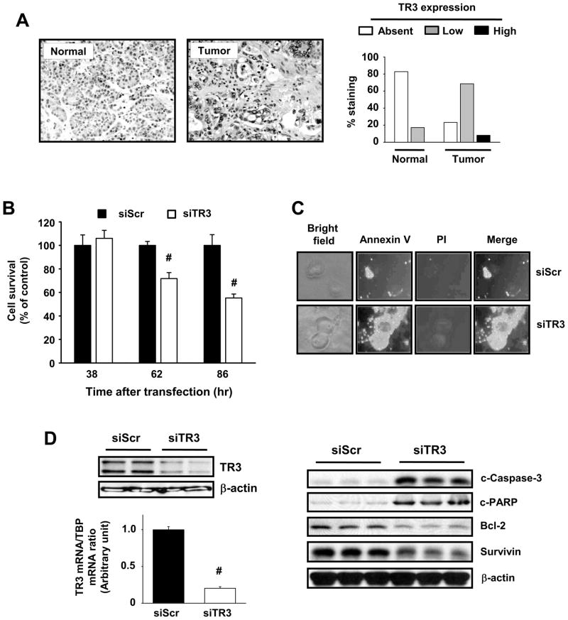
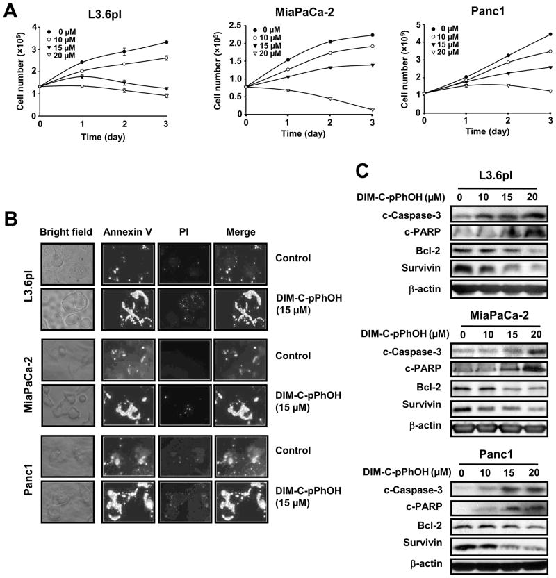
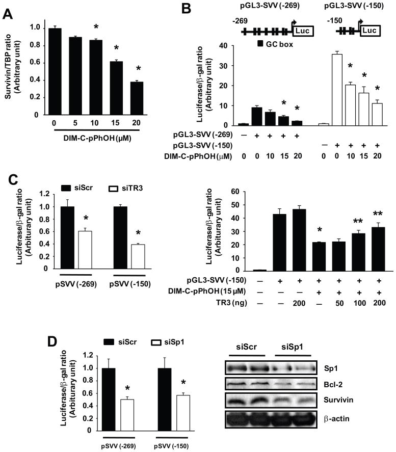
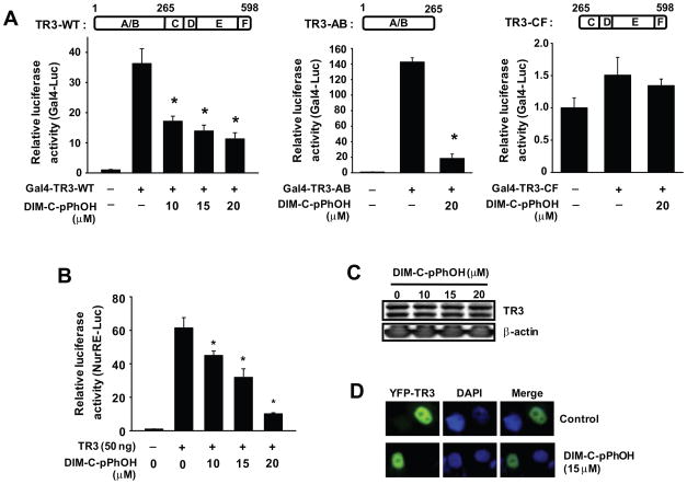
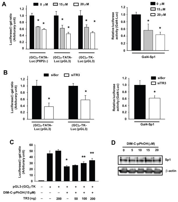
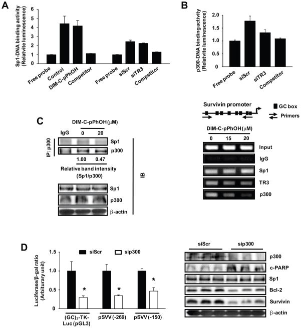
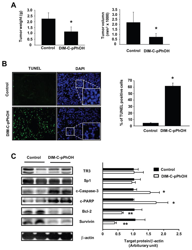
Activation of the orphan nuclear receptor TR3/Nur77 (NR4A1) promotes apoptosis and inhibits pancreatic tumor growth, but its endogenous function and the effects of its inactivation have yet to be determined. TR3 was overexpressed in human pancreatic tumors compared with nontumor tissue. Small interfering RNA-mediated knockdown of TR3 or cell treatment with the TR3 antagonist 1,1-bis(3'-indolyl)-1-(p-hydroxyphenyl)methane (DIM-C-pPhOH) decreased proliferation, induced apoptosis, and decreased expression of antiapoptotic genes including Bcl-2 and survivin in pancreatic cancer cells. Survivin suppression was mediated by formation of a TR3-Sp1-p300 DNA binding complex on the proximal GC-rich region of the survivin promoter. When administered in vivo, DIM-C-pPhOH induced apoptosis and inhibited tumor growth in an orthotopic model of pancreatic cancer, associated with inhibition of the same antiapoptotic markers observed in vitro. Our results offer preclinical validation of TR3 as a drug target for pancreatic cancer chemotherapy, based on the ability of TR3 inhibitors to block the growth of pancreatic tumors.


Conflict of interest statement

Conflict of Interest: All authors have no conflicts of interest to disclose.

Figures

Figure 1
Figure 1
TR3 expression in human pancreatic tumors and cells and effects of knockdown by RNAi. (A) TR3 protein staining in pancreatic tumor and non-tumor tissue. TR3 was immunostained in pancreatic tumor and non-tumor tissue from 89 patients, and histograms representing non-detectable, low and high staining were derived as outlined in the Materials and Methods. (B) Panc1 cell growth. After transfection with siScr or siTR3 for the indicated times, the number of cells in each well was counted (* significant growth inhibition; p<0.05). (C) Annexin V staining. Panc1 cells were transfected with siScr or siTR3 for 72 hr and stained for Annexin V and propidium iodide. Knockdown of TR3 induced the translocation of phosphatidylserine from the inner to outer leaflet of the plasma membrane, which in combination with negative propidium iodide staining, as seen in the merged images, indicates the induction of apoptosis. (D) siTR3 decreases TR3 and affects TR3-regulated gene products. Panc1 cells were transfected with either siScr or siTR3 for 72 hr and whole cell lysates were analyzed by Western blot analysis for TR3 or TR3-regulated genes and TR3 mRNA levels were determined (in triplicate) by real-time PCR (* significant decrease; p<0.05).
Figure 2
Figure 2
DIM-C-pPhOH inhibits cell growth and induces apoptosis in pancreatic cancer cells. (A) Cell growth inhibition. L3.6pL, MiaPaCa-2, and Panc1 cells were treated with either various concentrations of DIM-C-pPhOH or DMSO (control) for 3 days, and the number of cells in each well was counted on days 1, 2, and 3. (B) Annexin V staining. L3.6pL, MiaPaCa-2, and Panc1 cells were treated with either DMSO or 15 μM of DIM-C-pPhOH for 18 hr and stained for Annexin V and propidium iodide. (C) Western blot analysis. L3.6pL, MiaPaCa-2, and Panc1 cells were treated with either DMSO or various concentrations of DIM-C-pPhOH for 24 hr, and whole cell lysates were analyzed by western blots as described in the Materials and Methods.
Figure 3
Figure 3
DIM-C-pPhOH inhibits TR3 transactivation via its N-terminal A/B domain. (A) Deactivation of Gal4-TR3 chimeras. Gal4-Luc (0.1 μg) was cotransfected with 10 ng of each Gal4-TR3-WT, Gal4-TR3-AB, or Gal4-TR3-CF into Panc1 cells for 5 hr and treated with various concentrations of DIM-C-pPhOH for 18 hr. Luciferase activity was determined as described in the Materials and Methods (* significantly decreased activity; p<0.05). (B) NuRE-luc deactivation. Panc1 cells were cotransfected with NurRE-Luc (0.1 μg) and 50 ng of Flag-TR3 for 5 hr, and treated with various concentrations of DIM-C-pPhOH for 18 hr. Luciferase activity (relative to β-galactosidase activity) was determined as described in the Materials and Methods (* significantly decreased activity; p<0.05). (C) Effects of DIM-C-pPhOH on TR3 expression. Panc1 cells were treated with various concentrations of DIM-C-pPhOH for 24 hr, and whole cell lysates were analyzed by Western blot analysis as described in the Materials and Methods. (D) Subcellular localization of TR3. Panc1 cells were transfected with YFP-TR3 (0.5 μg) and treated with either DMSO or 15 μM of DIM-C-pPhOH for 12 hr. Images were obtained as described in the Materials and Methods.
Figure 4
Figure 4
DIM-C-pPhOH and TR3 knockdown inhibit survivin expression via down-regulation of its transcription in pancreatic cancer cells. (A) Decreased survivin mRNa. Panc1 cells were treated with various concentrations of DIM-C-pPhOH for 18 hr, and TR3 mRNA levels were determined by real-time PCR (* significantly decreased activity; p<0.05). (B) Decreased luciferase activity. Panc1 cells were transfected with 0.1 μg of either pGL3-SVV (−269) or pGL3-SVV (−150) and treated with various concentrations of DIM-C-pPhOH for 18 hr. Luciferase activity was determined as outlined in the Materials and Methods (* significantly decreased activity; p<0.05). (C) Inhibition by siTR3 or DIM-C-pPhOH. Panc1 cells were cotransfected with each siRNA and pSVV(−269) or pSVV(−150) as indicated and/or cotransfected with Flag-TR3 and treated with 15 μM DIM-C-pPhOH. Luciferase activity was determined as described in the Materials and Methods (* significantly decreased activity by siTR3 or DIM-C-pPhOH alone; ** significant reversal of the effects by TR3 overexpression; p<0.05). (D) Effects of Sp1 knockdown. Panc1 cells were cotransfected with each siRNA and pGL3-SVV as indicated. Luciferase activity or western blot analysis of whole cell lysates were determined as described in the Materials and Methods (* significantly decreased activity; p<0.05).
Figure 5
Figure 5
DIM-C-pPhOH and TR3 knockdown decrease Sp1-dependent transactivation in pancreatic cancer cells. (A) Transfection with GC-rich constructs and Gal4-Sp1. Panc1 cells were transfected with GC-rich constructs or Gal4-Sp1/Gal4-luc as indicated and treated with various concentrations of DIM-C-pPhOH for 18 hr. Luciferase activity was determined as described in the Materials and Methods (*significantly decreased activity; p<0.05). (B) Effects of TR3 knockdown. Panc1 cells were cotransfected with each siRNA and (GC)3-Luc or Gal4-Sp1/Gal4-luc. Luciferase activity was determined as described in the Materials and Methods (* significantly decreased activity; p<0.05). (C) TR3 rescue experiment. Panc1 cells were cotransfected with Flag-TR3 (or empty vector) and pGL3-(GC)3-TK-Luc, and treated with 15 μM of DIM-C-pPhOH for 18 hr. Luciferase activity was determined as described in the Materials and Methods (* significantly decreased activity; ** significant rescue by TR3 overexpression; p<0.05). (D) Sp1 expression, Panc1 cells were treated with DIM-C-pPhOH for 24 hr and nuclear extracts were analyzed for Sp1 protein by western immunoblots as described in the Materials and Methods.
Figure 6
Figure 6
TR3-dependent regulation of survivin through interactions with Sp1 are coregulated by p300. (A) Effect of DIM-C-pPhOH and siTR3 on Sp1 binding to GC-rich Sp1 consensus sequence. Panc1 cells were treated with 20 μM of DIM-C-pPhOH for 6 hr or transfected with either siScr or siTR3, and nuclear extracts were tested for Sp1-DNA binding activity as described in the Materials and Methods. (B) ChIP and DNA binding assays. Panc1 cells were treated with either DMSO or various concentrations of DIM-C-pPhOH for 6 hr, and interactions of Sp1, TR3, and p300 with the survivin promoter were determined as described in the Materials and Methods. Panc1 cells were transfected with either siScr or siTR3, and nuclear extracts were tested for p300-DNA binding activity as described in the Materials and Methods. (C) Coimmunoprecipitation of p300 and Sp1 proteins. Panc1 cells were treated with 20 μM of DIM-C-pPhOH for 6 hr, and nuclear extracts were prepared. Endogenous p300 was immunoprecipitated with anti-p300 antibodies and the immunoprecipitates were analyzed by Western blot analysis for Sp1 and p300 as described in the Materials and Methods. (D) Effects of p300 knockdown. Panc1 cells were cotransfected with GC-rich constructs and siRNAs (siScr or sip300), and luciferase activity or analysis of whole cell lysates were determined as described in the Materials and Methods (* significantly decreased activity; p<0.05).
Figure 7
Figure 7
Effects of DIM-C-pPhOH on growth and apoptosis in an orthotopic mouse model of human pancreatic cancer. (A) Effects on tumor weights and volume. L3.6pL human pancreatic cancer cells were orthotopically implanted into athymic nude mice, and each mouse was dosed by oral gavage with either corn oil (control) or DIM-C-pPhOH (30 mg/kg/day) for 4 weeks starting 7 days after implantation. Median tumor weights and volumes were calculated as described in the Materials and Methods (* significantly decreased effects; p<0.05). (B) TUNEL assay. The TUNEL assay was performed on tumor sections as described in the Materials and Methods, and the number of TUNEL positive cells was counted from randomly selected microscopic fields. DAPI stains both apoptotic and nonapoptotic cells blue and fluorescein-12-dUTP incorporation results in localized green fluorescence within the nucleus of apoptotic cells. Fluorescent images were collected at high (x400) magnification (* significantly increased TUNEL staining; p<0.05). (C) Protein expressions in tumor lysates. Tumor lysates from tumor samples were further analyzed by Western blot analysis as described in the Materials and Methods, and each lane represents a different tumor sample [* significantly increased caspase-3 and PARP cleavage; **significantly decreased bcl-2 and survivin protein levels (relative to β-actin loading control); p<0.05].

References

    1. Milbrandt J. Nerve growth factor induces a gene homologous to the glucocorticoid receptor gene. Neuron. 1988;1:183–8. - PubMed
    1. Giguere V. Orphan nuclear receptors: from gene to function. Endocr Rev. 1999;20:689–725. - PubMed
    1. McKenna NJ, O’Malley BW. Combinatorial control of gene expression by nuclear receptors and coregulators. Cell. 2002;108:465–74. - PubMed
    1. Winoto A. Genes involved in T-cell receptor-mediated apoptosis of thymocytes and T-cell hybridomas. Semin Immunol. 1997;9:51–8. - PubMed
    1. Mullican SE, Zhang S, Konopleva M, et al. Abrogation of nuclear receptors Nr4a3 and Nr4a1 leads to development of acute myeloid leukemia. Nat Med. 2007;13:730–5. - PubMed

Publication types

MeSH terms