Comparative Study
. 2010 Sep;104(3):1717-25.
doi: 10.1152/jn.00161.2010. Epub 2010 Jul 21.

Abnormal activity of primary somatosensory cortex in central pain syndrome

Affiliations
Comparative Study

Abnormal activity of primary somatosensory cortex in central pain syndrome

Raimi L Quiton et al. J Neurophysiol. 2010 Sep.

Abstract

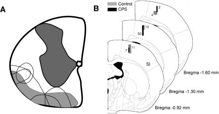
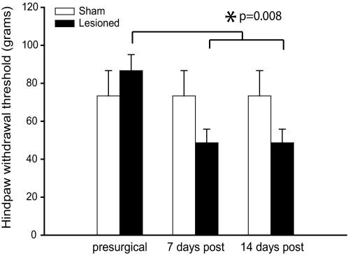
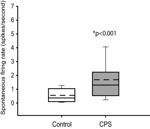
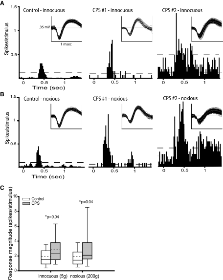
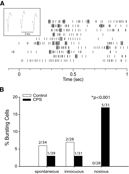
Central pain syndrome (CPS) is a debilitating and chronic pain condition that results from a lesion or dysfunction in the CNS. The pathophysiological mechanisms underlying CPS are poorly understood. We recently demonstrated that CPS is associated with suppressed inputs from the inhibitory nucleus zona incerta to the posterior thalamus (PO). As a consequence, activity in PO is abnormally increased in CPS. Because the perception of pain requires activity in the cerebral cortex, CPS must also involve abnormal cortical activity. Here we test the hypothesis that CPS is associated with increased activity in the primary somatosensory cortex (SI), a major projection target of PO that plays an important role in processing sensory-discriminative aspects of pain. We recorded activity of single units in SI in rats with CPS resulting from spinal cord lesions. Consistent with our hypothesis, SI neurons recorded from lesioned rats exhibited significantly higher spontaneous firing rates and greater responses evoked by innocuous and noxious mechanical stimulation of the hindpaw compared with control rats. Neurons from lesioned rats also showed a greater tendency than controls to fire bursts of action potentials in response to noxious stimuli. Thus, the excruciatingly painful symptoms of CPS may result, at least in part, from abnormally increased activity in SI.


Figures

Fig. 1.
Fig. 1.
A: drawing of coronal section through the cervical spinal cord, showing the location and size of lesions in animals with mechanical hyperalgesia (unfilled areas). Shaded areas represent the location of ascending spinothalamic tract axons, adapted from Fig. 5 in Giesler et al. (1981). B: coronal sections of the rat brain (Paxinos and Watson 1998) showing locations of all recorded neurons in this study. Numbers represent the number of neurons recorded in each shaded area of primary somatosensory (SI) cortex.
Fig. 2.
Fig. 2.
Hindpaw mechanical withdrawal thresholds decrease significantly over time after spinal lesions but not after sham surgery. All values represent means ± SE. Thresholds for the right and left hindpaw of each individual animal were identical; therefore only data for the hindpaw contralateral to the lesion are shown.
Fig. 3.
Fig. 3.
Group data showing that the spontaneous activity is significantly higher in SI neurons from animals with central pain syndrome (CPS, n = 39) than in control rats (n = 34). Boxes represent the 25th to 75th percentile of the distribution; whiskers show the 10th and 90th percentiles. - - -, mean values.
Fig. 4.
Fig. 4.
Neuronal activity in SI is enhanced in animals with CPS. A: peristimulus time histograms (PSTHs, 20 ms bins) showing the responses to innocuous mechanical hindpaw stimulation (5 g) of SI neurons from a sham-lesioned rat (left) and a spinal-lesioned rat with behaviorally confirmed CPS (middle and right). Sensory-evoked activity in SI neurons recorded from the CPS rat is markedly higher than in the neuron from the sham rat. Spontaneous activity is also higher in the second SI neuron from the CPS rat (right) than in the neuron from the sham rat. Dashed lines represent, the threshold at which the response significantly exceeded the spontaneous firing rate (99% confidence interval). Inset: representative spike waveforms. B: PSTHs showing the responses to noxious mechanical hindpaw stimulation (200 g) of SI neurons from a sham rat (left) and a CPS rat (middle and right). As in A, the sensory-evoked neuronal activity is higher in the CPS rats. C: group data showing that activity evoked by both innocuous and noxious mechanical stimulation of the hindpaw is significantly higher in SI neurons from animals with CPS (n = 30) than from control rats (n = 28). - - -, mean values.
Fig. 5.
Fig. 5.
A: raster plot of a bursting neuron's response to 10 applications of a noxious mechanical stimulus to the hindpaw at time = 0 s. Spikes enclosed in boxes represent bursts of action potentials (as defined in methods). Inset: spike waveforms of a burst from this neuron. B: percentage of cells in spinal and sham-lesioned animals that showed bursting activity either spontaneously or in response to innocuous and noxious hindpaw stimuli. The number of cells that showed bursting activity in each category is shown above the bars. The percentage of cells that burst in response to noxious stimuli was significantly higher in CPS rats than control rats (P < 0.001, χ2 test).

References

    1. Alden M, Besson JM, Bernard JF. Organization of the efferent projections from the pontine parabrachial area to the bed nucleus of the stria terminalis and neighboring regions: a PHA-L study in the rat. J Comp Neurol 341: 289– 314, 1994 - PubMed
    1. Alloway KD, Hoffer ZS, Hoover JE. Quantitative comparisons of corticothalamic topography within the ventrobasal complex and the posterior nucleus of the rodent thalamus. Brain Res 968: 54– 68, 2003 - PubMed
    1. Andersen G, Vestergaard K, Ingeman-Nielsen M, Jensen TS. Incidence of central post-stroke pain. Pain 61: 187– 193, 1995 - PubMed
    1. Andersson JL, Lilja A, Hartvig P, Langstrom B, Gordh T, Handwerker H, Torebjork E. Somatotopic organization along the central sulcus, for pain localization in humans as revealed by positron emission tomography. Exp Brain Res 117: 192– 199, 1997 - PubMed
    1. Apkarian AV, Bushnell MC, Treede RD, Zubieta JK. Human brain mechanisms of pain perception and regulation in health and disease. Eur J Pain 9: 463– 484, 2005 - PubMed

Publication types