Comparative Study
. 2010 Oct;104(4):2034-51.
doi: 10.1152/jn.00396.2010. Epub 2010 Jul 21.

Ion channels set spike timing regularity of mammalian vestibular afferent neurons

Affiliations
Comparative Study

Ion channels set spike timing regularity of mammalian vestibular afferent neurons

Radha Kalluri et al. J Neurophysiol. 2010 Oct.

Abstract

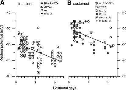
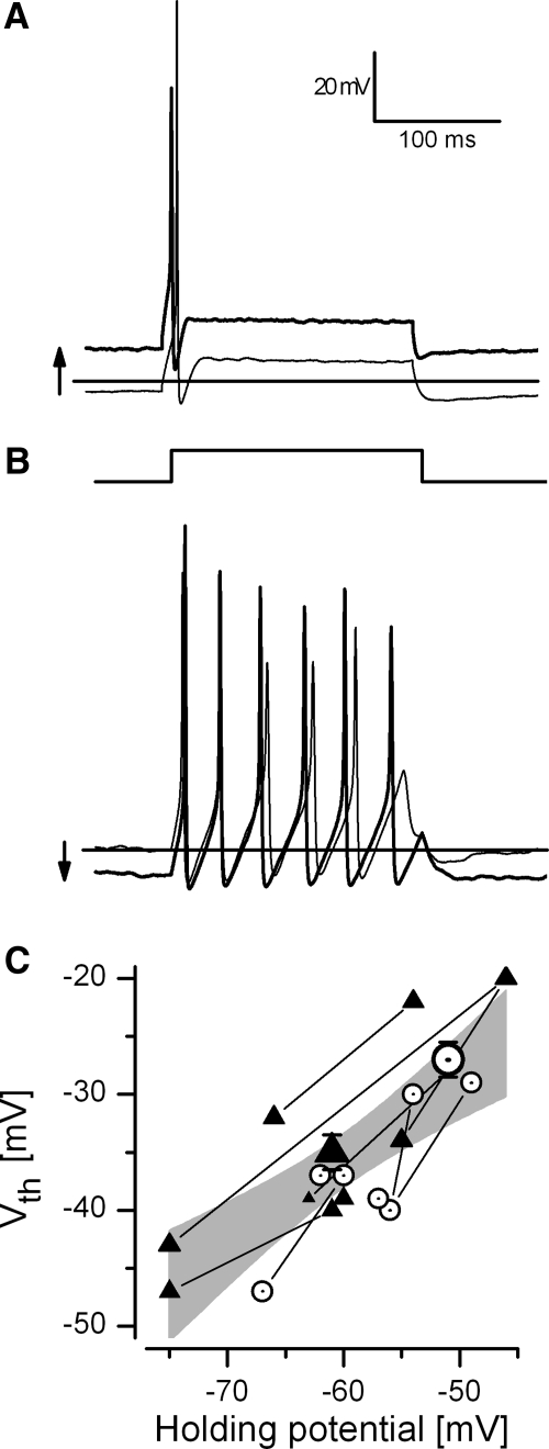
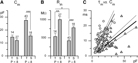
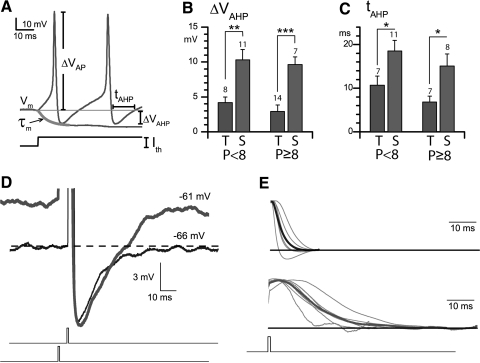
In the mammalian vestibular nerve, some afferents have highly irregular interspike intervals and others have highly regular intervals. To investigate whether spike timing is determined by the afferents' ion channels, we studied spiking activity in their cell bodies, isolated from the vestibular ganglia of young rats. Whole cell recordings were made with the perforated-patch method. As previously reported, depolarizing current steps revealed distinct firing patterns. Transient neurons fired one or two onset spikes, independent of current level. Sustained neurons were more heterogeneous, firing either trains of spikes or a spike followed by large voltage oscillations. We show that the firing pattern categories are robust, occurring at different temperatures and ages, both in mice and in rats. A difference in average resting potential did not cause the difference in firing patterns, but contributed to differences in afterhyperpolarizations. A low-voltage-activated potassium current (I(LV)) was previously implicated in the transient firing pattern. We show that I(LV) grew from the first to second postnatal week and by the second week comprised Kv1 and Kv7 (KCNQ) components. Blocking I(LV) converted step-evoked firing patterns from transient to sustained. Separated from their normal synaptic inputs, the neurons did not spike spontaneously. To test whether the firing-pattern categories might correspond to afferent populations of different regularity, we injected simulated excitatory postsynaptic currents at pseudorandom intervals. Sustained neurons responded to a given pattern of input with more regular firing than did transient neurons. Pharmacological block of I(LV) made firing more regular. Thus ion channel differences that produce transient and sustained firing patterns in response to depolarizing current steps can also produce irregular and regular spike timing.


Figures

Fig. 1.
Fig. 1.
Vestibular ganglion neurons produced 2 basic firing patterns, transient and sustained, in response to depolarizing current steps. For clarity, voltage traces are offset as current steps are iterated; current step levels are shown at right. Dashed line: −65 mV for the 0-pA current step. Scale bars (top left) apply to all traces. In this and all subsequent figures, recordings were made with the perforated-patch whole cell method. Data in A–E were from rat neurons, postnatal day 13 (P13) to P16. A and B: transient neurons fired one to 2 spikes at the onset of 200-ms depolarizing current steps above a threshold value. C–E: sustained neurons were either spikers (C and D) or resonators (E). Sustained-A spikers (C) fired long trains at spike threshold. In sustained-B spikers (D), spikes built up with increasing current amplitude. Sustained resonators (E) fired one or 2 spikes followed by prominent voltage oscillations. For hyperpolarizing steps, sustained spikers showed the hyperpolarization-activated cationic current (Ih)–mediated voltage sag also seen in transient neurons, but were more likely to spike at the step offset. F: current thresholds for spiking were much lower in sustained neurons in both the first postnatal week (P1–P7, left) and at later ages (P8–P16). In this and all figures: highly significant difference, ***p < 0.001; a very significant difference, **p < 0.01; and a significant difference, *p < 0.05.
Fig. 2.
Fig. 2.
Firing pattern categories were robust and stable with changes in age, temperature, and species. Vestibular ganglion somata were from rat (A–C) and mouse (D) at ages and temperatures indicated. Responses are shown for the smallest current step to produce a spike (resolution: 10 pA), except for the mouse sustained resonator (D, right), which never spiked. Dashed line: −55 mV. Scale bars (top left) apply to all traces.
Fig. 3.
Fig. 3.
Resting potential differed between firing pattern categories and became more negative with age. A: transient spikers. B: sustained spikers, divided into sustained-A (open circles) and sustained-B (filled circles). Most data (circles) are from rat at room temperature; some are from rat at warmer temperatures (inverted triangles), or mouse at room temperature (stars and crosses). After P7, sustained-A and sustained-B spikers diverged, such that sustained-B values were more negative than sustained-A values. Lines in A and B: linear regression fits (y = mx + b) to rat data at 22–25°C. Transient neurons (68): m = 0.7 ± 0.13 mV/d, b = −58.9 ± 0.96 mV, r2 = 0.32. Sustained neurons (45); m = 0.7 ± 0.17 mV/day, b = −49.9 ± 1.20 mV, r2 = 0.28.
Fig. 4.
Fig. 4.
Steady polarization did not change either the firing pattern or the voltage change required to evoke a spike. A: transient neuron (P7) driven by a 100-pA current step before (thin line) and during (thicker line) a steady approximately 12-mV depolarization. Horizontal line in A and B: −60 mV. B: sustained neuron (P8) driven by a 40-pA current step before (thin line) and during (thicker line) a steady approximately 10-mV hyperpolarization. C: dependence of the spike voltage threshold (Vth) on holding potential. Thin lines connect Vth values for single neurons held at different potentials. Triangles: transient neurons (n = 5); circles: sustained neurons (n = 4). Shaded area: 95% confidence interval (CI) for a linear regression of all data points (m = 0.7 ± 0.1 mV/mV, b = 7.5 ± 8.5 mV, r2 = 0.6). Large symbols: mean Vth ± SE (mean resting potential) for 10 transient neurons (triangle) and 10 sustained neurons (open circle); P < 8 for all neurons.
Fig. 5.
Fig. 5.
Passive membrane properties of transient and sustained neurons differed. A and B: membrane capacitance (Cm) and input resistance (Rin) for P1–P7 and P8–P16. Rin was significantly smaller for transient neurons over both age ranges. Cm of transient neurons increased from the first to the second week to be much larger than Cm of sustained neurons. C: membrane time constant (τm) as a function of Cm, for 45 sustained (circles) and 57 transient (triangles) neurons. Mean τm values are not significantly different but the linear regression slopes are: 340 ± 28 μs/pF (transient) vs. 800 ± 44 μs/pF (sustained). Thin lines, 95% CIs.
Fig. 6.
Fig. 6.
Differences between transient and sustained neurons in afterhyperpolarizations (AHPs) and subthreshold activity. A: how spike and AHP metrics were calculated. AHPs of sustained neurons were (B) larger and (C) longer than those of transient neurons at all ages tested. D: AHPs of a transient neuron (thin black line) and a sustained neuron (thicker gray line), each excited by a 1-ms current pulse of amplitude just above threshold. We aligned the AHPs on the trough to illustrate the similar recovery kinetics [alignment caused a leftward shift of the stimulus pulse (below) for the sustained neuron because of its broader spike]. E: subthreshold voltage responses of sustained neurons (n = 7, top) to 1-ms pulses were longer than those of transient neurons (n = 6, bottom). Traces were normalized to peak amplitude and averaged (thick traces).
Fig. 7.
Fig. 7.
Low-voltage-activated outward currents were larger in transient neurons than those in sustained neurons and grew with age. A and B: whole cell currents in a transient neuron (A, P13) and a sustained neuron (B, P13) in response to 3 voltage steps (top). See text. C: current–voltage (I–V) relationships taken 100 ms after the onset of the voltage step for transient (triangles) and sustained (circles) neurons, for the first (open symbols) and second (filled symbols) postnatal weeks. The outward current grew for transient neurons but not for sustained neurons (open circles are obscured by filled circles because of their similar values). By the second week, the I–V relation for transient neurons (filled triangles) was offset by close to −20 mV relative to that for sustained neurons (filled circles).
Fig. 8.
Fig. 8.
Low-voltage-activated potassium current (ILV) comprised α-DTX-sensitive and linopirdine-sensitive components. A–D: whole cell currents recorded in perforated-patch mode (27°C) from a transient neuron (P10, rat) in response to a family of voltage steps. A: family of currents evoked by a standard voltage protocol (bottom): prepulse to −125 mV followed by steps between −105 and −15 mV, control (Liebovitz-15 [L-15]) external solution. The cell had INa, IA, IHV, and ILV (as in Fig. 7A). B: currents at the onset of a depolarizing step in: L-15 (black), L-15 containing 100 nM a-dendrotoxin (α-DTX, red), and L-15 containing 100 nM α-DTX + 10 μM linopirdine (dark gray). C: I–V relations taken 100 ms after the onset of voltage steps, from the cell in B. Low-voltage (LV) currents (at potentials negative to −30 mV) were partly suppressed in α-DTX (red circles) and strongly suppressed in α-DTX + linopirdine (triangles). D and E: α-DTX-sensitive and linopirdine-sensitive components of ILV activated over different timescales. D: α-DTX blocked a fast component (bottom panel; obtained by subtraction from curves above). The slow remaining component was blocked by linopirdine (E). Activation was fit with single-exponential functions (red curves) with τ values of 4.3 ms for the α-DTX-sensitive component (D, bottom), 96 ms for the α-DTX-insensitive component (E, top), and 72 ms for the linopirdine-sensitive component (E, bottom). Currents in D and E are from 2 cells, different from the cell in A–C. F: conductance–voltage (g–V) relations for the α-DTX-sensitive current (circles) and linopirdine-sensitive current (triangles). Curves: Boltzmann fits (Eq. 1; methods). G: Boltzmann fits to g–V curves, normalized by maximum conductance for each curve (thin red lines, α-DTX-sensitive conductance; thin black lines, linopirdine-sensitive conductance). Thick red and thick black lines: average g–V curves for α-DTX-sensitive current (half-maximum activation voltage [V1/2] = −44 ± 0.2 mV, slope factor [S] = 7.1 ± 0.20 mV, n = 7) and linopirdine-sensitive current (V1/2 = −41 ± 0.3 mV, S = 7.4 ± 0.41 mV, n = 5), respectively. H: activation time constants were much faster for α-DTX-sensitive currents (red circles, n = 8 neurons, some at multiple voltages) than those for α-DTX-insensitive currents (gray triangles, n = 6) and linopirdine-sensitive currents (open down triangles, n = 3). All data in this figure were taken at room temperature, P9–P15.
Fig. 9.
Fig. 9.
Both Kv1 and KCNQ channels contributed to the transient firing pattern. A: spiking response of a P11 transient neuron to +160-pA current steps, in control solution (left), solution containing 100 nM α-DTX (middle), and solution containing 100 nM α-DTX + 10 μM linopirdine (right). α-DTX and linopirdine converted the transient response at spike threshold to a sustained-A response. For larger steps, α-DTX by itself increased spike number (see insets in control, α-DTX panels). B: as in A, but for a P15 transient neuron and with 10 μM linopirdine as the solo drug treatment (middle). Linopirdine had no effect at spike threshold, but increased spike number for much larger current steps (inset). Linopirdine blocked ILV at −45 mV by 40% in this neuron (not shown). C: sustained-B neurons also had ILV. At spike threshold in control conditions (+80 pA), the sustained-B firing pattern of a P10 neuron was modestly affected by α-DTX but was converted to the sustained-A pattern by α-DTX + linopirdine. This neuron fired spontaneously in the dual-blocker treatment (right, inset). D–F: LV channel blockers also made transient neurons more like sustained neurons in terms of resting potential (D), current threshold for spiking (E), and input resistance (F).
Fig. 10.
Fig. 10.
Pseudorandom trains of excitatory postsynaptic current (EPSC)–like currents evoked firing that was more regular in sustained neurons than in transient neurons. A: examples from a sustained-A, P4 neuron (top) and a transient P2 neuron (bottom), selected to have the same mean spike rate (∼13 spikes/s). Pseudo-EPSCs were delivered at the same mean rate (250/s) to each neuron but at different unitary amplitudes (40 pA for the sustained neuron vs. 200 pA for the transient neuron), as needed to drive them at similar rates. B and C: spike trains and pseudo-excitatory postsynaptic potentials (EPSPs) from the sustained and transient neurons are expanded in C for the interval in A marked by a black bar and are compared in B to intracellular, in vivo records from 2 lizard canal afferents, one regular and one irregular [adapted from Figs. 11 and 17 in Schessel et al. (1991), with permission from Elsevier and the authors]. Note similarities between the AHPs and EPSPs of the transient neuron (C, bottom) and irregular afferent (B, bottom) on the one hand and the sustained neuron (C, top) and regular afferent (B, top) on the other hand.
Fig. 11.
Fig. 11.
Spike regularity differences between transient and sustained neurons were robust over a range of mean spike intervals. A: spiking was driven at different mean intervals by varying the size of the unitary EPSC-like event from 30 to 220 pA. B: interspike interval histograms constructed from pseudo-EPSC-evoked spike trains 3.5 s in duration. Mean spike interval decreased as unitary EPSC amplitude increased. C: coefficient of variation (CV) as a function of mean interval in 22 neurons. Each symbol corresponds to one neuron, driven at multiple rates: 7 sustained-A neurons (red); 5 sustained-B neurons (green); 10 transient neurons (blue). Sustained neurons fired with shorter intervals and had lower CV values, with sustained-A neurons being the most regular. Lines: locally linear regression fits (loess algorithm, Matlab). Even at comparable rates, sustained neurons fired more regularly. D–F: step-evoked firing patterns of exemplar neurons from the set in C; the current step and the cell's symbol (C) are given at the far right.
Fig. 12.
Fig. 12.
Blocking ILV made the spike trains of transient neurons more regular. A, top: an irregular spike train (CV 0.8, mean interval 73 ms) from a P11 transient neuron, driven by pseudo-EPSCs with a unitary amplitude of 340 pA. Bottom: coapplication of α-DTX and linopirdine made spikes faster and more regular. (Spike rate increased in the blocker solution, so we reduced unitary amplitude to 170 pA, for a mean interval of 53 ms.) B–D: CV values as functions of mean interval are shown for 4 transient neurons before (black symbols) and after (gray symbols) ILV block: lines, average CV-interval curves (Fig. 11) for transient (thick black line) and sustained (thin gray line) neurons. Perforated-patch recordings. Data in B are from the neuron in A. E: patch rupture made a neuron more regular. Transient neuron, recorded in perforated-patch mode in control solution (black symbols; CV 0.6–0.8) and 100 μM α-DTX (black and gray symbols), which reduced CV to 0.3–0.5. The patch then ruptured and the neuron became extremely regular (gray symbols; CV 0.1–0.2).
Fig. 13.
Fig. 13.
Sustained neurons had longer integration times than did transient neurons. Uniformly spaced trains of pseudo-EPSCs were applied to a sustained neuron (A–C, P4, neuron from Fig. 10; step-evoked firing pattern shown in D) and a transient neuron (E–G, P5; step-evoked firing pattern shown in H). All spikes are truncated. Stimulus current train, shown below each response, delivered pseudo-EPSCs at intervals of 30 ms (A and E), 20 ms (B and F), or 10 ms (C and G). A–C: the sustained neuron integrated pseudo-EPSCs that were individually subthreshold (10 pA), to produce spiking for intervals <30 ms. At 30-ms intervals, the neuron did not fire (A); at 20-ms intervals, it began to fire (B); and at 10-ms intervals, it fired faster (C). E–G: the transient neuron integrated little, spiking only for pseudo-EPSCs that were individually suprathreshold; responses to 80-pA pseudo-EPSCs are shown. At 30-ms intervals (E), the neuron fired for every pseudo-EPSC. At 20-ms intervals (F), the neuron did not fire for every EPSC, but the timing of each spike was tightly coupled to the timing of a pseudo-EPSC. At 10-ms intervals (G) the neuron fired just one spike at the start of the pseudo-EPSC train.

References

    1. Adamson CL, Reid MA, Davis RL. Opposite actions of brain-derived neurotrophic factor and neurotrophin-3 on firing features and ion channel composition of murine spiral ganglion neurons. J Neurosci 22: 1385–1396, 2002b - PMC - PubMed
    1. Adamson CL, Reid MA, Mo ZL, Bowne-English J, Davis RL. Firing features and potassium channel content of murine spiral ganglion neurons vary with cochlear location. J Comp Neurol 447: 331–350, 2002a - PubMed
    1. Amir R, Devor M. Electrical excitability of the soma of sensory neurons is required for spike invasion of the soma, but not for through-conduction. Biophys J 84: 2181–2191, 2003 - PMC - PubMed
    1. Armstrong CE, Roberts WM. Electrical properties of frog saccular hair cells: distortion by enzymatic dissociation. J Neurosci 18: 2962–2973, 1998 - PMC - PubMed
    1. Baird RA, Desmadryl G, Fernández C, Goldberg JM. The vestibular nerve of the chinchilla. II. Relation between afferent response properties and peripheral innervation patterns in the semicircular canals. J Neurophysiol 60: 182–203, 1988 - PubMed

Publication types