. 2010 Nov;104(5):2624-33.
doi: 10.1152/jn.00752.2009. Epub 2010 Jul 21.

Representation of the ipsilateral visual field by neurons in the macaque lateral intraparietal cortex depends on the forebrain commissures

Affiliations

Representation of the ipsilateral visual field by neurons in the macaque lateral intraparietal cortex depends on the forebrain commissures

Catherine A Dunn et al. J Neurophysiol. 2010 Nov.

Abstract

Our eyes are constantly moving, allowing us to attend to different visual objects in the environment. With each eye movement, a given object activates an entirely new set of visual neurons, yet we perceive a stable scene. One neural mechanism that may contribute to visual stability is remapping. Neurons in several brain regions respond to visual stimuli presented outside the receptive field when an eye movement brings the stimulated location into the receptive field. The stored representation of a visual stimulus is remapped, or updated, in conjunction with the saccade. Remapping depends on neurons being able to receive visual information from outside the classic receptive field. In previous studies, we asked whether remapping across hemifields depends on the forebrain commissures. We found that, when the forebrain commissures are transected, behavior dependent on accurate spatial updating is initially impaired but recovers over time. Moreover, neurons in lateral intraparietal cortex (LIP) continue to remap information across hemifields in the absence of the forebrain commissures. One possible explanation for the preserved across-hemifield remapping in split-brain animals is that neurons in a single hemisphere could represent visual information from both visual fields. In the present study, we measured receptive fields of LIP neurons in split-brain monkeys and compared them with receptive fields in intact monkeys. We found a small number of neurons with bilateral receptive fields in the intact monkeys. In contrast, we found no such neurons in the split-brain animals. We conclude that bilateral representations in area LIP following forebrain commissures transection cannot account for remapping across hemifields.


Figures
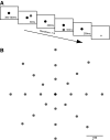
Fig. 1.
Fig. 1.
Receptive field (RF) mapping task. A: the monkey begins each trial fixating a central location (black dot). While the monkey fixates, a random number (1–9) of visual stimuli (gray dot) are presented for 50 ms with a 200 ms interstimulus interval. When the fixation point is extinguished the monkey is required to make an eye movement (arrow) to the last stimulus presented. B: the stimulus was placed at one of 8 locations in 3 rings with amplitudes of 7, 14, or 21°. Each gray dot represents a possible target location (24 total).
Fig. 2.
Fig. 2.
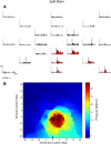
Responses from a single left-hemisphere neuron in a split-brain monkey. A: each histogram and raster represent activity for one particular stimulus location. This cell was recorded from the left hemisphere. Histograms are red when the activity during the visual epoch is significantly greater than the baseline epoch (see methods). The data are aligned on stimulus onset. The vertical line represents the neural latency for that location. If a neural latency could not be determined, then no line is present. B: contour plot of responses in A. Black asterisks indicate that the firing rate at the location was ≥75% of the peak firing rate. This neuron had a receptive that was down and slightly to the right (contralateral visual field).
Fig. 3.
Fig. 3.
Responses from a single left-hemisphere neuron in an intact monkey. Conventions as like those in Fig. 2. This neuron had a receptive that was down and to the left (ipsilateral visual field).
Fig. 4.
Fig. 4.
Number of lateral intraparietal cortex (LIP) neurons with responses to stimuli in ipsilateral, contralateral, or both hemifields. A: in the split-brain monkey, no neurons respond to stimuli is in the ipsilateral field. B: in the intact monkey, a small number of neurons have bilateral RFs, and even fewer are ipsilateral only.
Fig. 5.
Fig. 5.
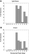
Location of peak response for LIP neurons in split-brain and intact monkeys. Positive values on the x-axis represent contralateral space; negative values represent ipsilateral space. A: in split-brain monkeys, the location of peak response varies from 0 to 20°. B: in intact monkeys, the location of peak firing varies from 16° in the ipsilateral field to 20° in the contralateral field.
Fig. 6.
Fig. 6.
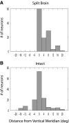
RF distance from the vertical meridian. On the x-axis, positive values indicate contralateral space and negative values indicate ipsilateral space. A: in the split-brain monkey, the inner edge of the RF ranges from 0 to 20°. B: in the intact monkey, the inner edge of the RF ranges from 16° ipsilateral to 20° contralateral.
Fig. 7.
Fig. 7.
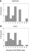
RF widths. Only neurons with clear boundaries are included. RF widths vary from a few degrees to as much as 20° in both split-brain and intact monkeys.
Fig. 8.
Fig. 8.
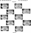
Coronal magnetic resonance images of monkeys EM and OP. Gray arrows indicate recording locations. A: images from split-brain monkey EM. The chamber was removed before the magnetic resonance imaging (MRI) scan. The location of the chamber was estimated based on the depression that remained after the chamber was removed. B: images from intact monkey OP. Recording locations in the split-brain monkey were aligned with recordings in the intact monkey.
Fig. 9.
Fig. 9.
Coronal MRIs of monkeys CH and FF. Conventions as in Fig. 8. Recordings in the split-brain monkey were more posterior.
Fig. 10.
Fig. 10.
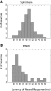
Distributions of visual response latency in split-brain and intact monkeys. On the x-axis, the latency of neural response. A: in split-brain monkeys, the mean latency is 106 ms. B: in intact monkeys, the mean latency is 86 ms. There is a significant difference in latency between the split-brain and intact populations.

References

    1. Andersen RA, Asanuma C, Essick G, Siegel RM. Corticocortical connections of anatomically and physiologically defined subdivisions within the inferior parietal lobule. J Comp Neurol 296: 65–113, 1990 - PubMed
    1. Antonetty CM, Webster KE. The organisation of the spinotectal projection. An experimental study in the rat. J Comp Neurol 163: 449–465, 1975 - PubMed
    1. Barash S, Bracewell RM, Fogassi L, Gnadt JW, Andersen RA. Saccade-related activity in the lateral intraparietal area. II. Spatial properties. J Neurophysiol 66: 1109–1124, 1991 - PubMed
    1. Basso MA, Liu P. Context-dependent effects of substantia nigra stimulation on eye movements. J Neurophysiol 97: 4129–4142, 2007 - PubMed
    1. Basso MA, Pokorny JJ, Liu P. Activity of substantia nigra pars reticulata neurons during smooth pursuit eye movements in monkeys. Eur J Neurosci 22: 448–464, 2005 - PubMed

Publication types

LinkOut - more resources