. 2010 Sep 24;285(39):30192-202.
doi: 10.1074/jbc.M110.103986. Epub 2010 Jul 26.

Impaired binding of the age-related macular degeneration-associated complement factor H 402H allotype to Bruch's membrane in human retina

Affiliations

Impaired binding of the age-related macular degeneration-associated complement factor H 402H allotype to Bruch's membrane in human retina

Simon J Clark et al. J Biol Chem. .

Abstract

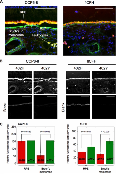
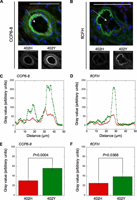
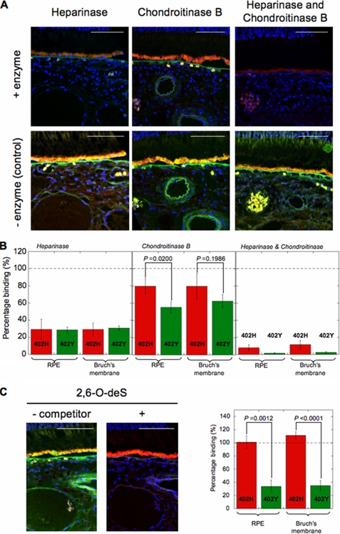
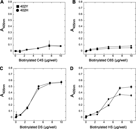
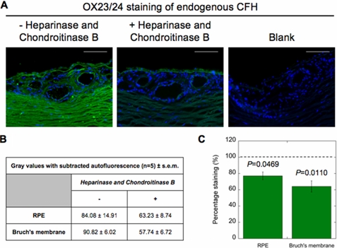
Age-related macular degeneration (AMD) is the predominant cause of blindness in the industrialized world where destruction of the macula, i.e. the central region of the retina, results in loss of vision. AMD is preceded by the formation of deposits in the macula, which accumulate between the Bruch's membrane and the retinal pigment epithelium (RPE). These deposits are associated with complement-mediated inflammation and perturb retinal function. Recent genetic association studies have demonstrated that a common allele (402H) of the complement factor H (CFH) gene is a major risk factor for the development of AMD; CFH suppresses complement activation on host tissues where it is believed to bind via its interaction with polyanionic structures. We have shown previously that this coding change (Y402H; from a tyrosine to histidine residue) alters the binding of the CFH protein to sulfated polysaccharides. Here we demonstrate that the AMD-associated polymorphism profoundly affects CFH binding to sites within human macula. Notably, the AMD-associated 402H variant binds less well to heparan sulfate and dermatan sulfate glycosaminoglycans within Bruch's membrane when compared with the 402Y form; both allotypes exhibit a similar level of binding to the RPE. We propose that the impaired binding of the 402H variant to Bruch's membrane results in an overactivation of the complement pathway leading to local chronic inflammation and thus contributes directly to the development and/or progression of AMD. These studies therefore provide a putative disease mechanism and add weight to the genetic association studies that implicate the 402H allele as an important risk factor in AMD.


Figures

FIGURE 1.
FIGURE 1.
Identification of 402H- and 402Y-binding sites in human macula. The 402H and 402Y variants (in the context of recombinant CCP6–8 or flCFH) were conjugated with Alexa Fluor 594 (red) and 488 (green), respectively, and used to label sections obtained from five human eyes (without AMD); nuclei were stained with DAPI (blue). Merged images (A) and corresponding grayscale data (B) are illustrated for one representative donor eye (a 402H/402Y heterozygote), where the scale bar corresponds to 100 μm (here and in all other figures); yellow/orange staining (e.g. on the RPE) is indicative of regions to which both 402H and 402Y variants bind equivalently. C, the relative fluorescent intensities of the RPE and Bruch's membrane were determined for the five different eyes, and the autofluorescence from blanks was subtracted; the autofluorescence observed in the red channel is due to the accumulation of lipofuscin granules within the RPE (7). Data are shown as mean values ± S.E., where statistical analysis of the 402H versus 402Y binding data was determined using the paired Student's t test. The five donors (donor group A) used here and throughout the rest of the study (unless indicated otherwise) corresponded to three heterozygous (402H/402Y) and two homozygous 402Y/402Y samples (Table 1).
FIGURE 2.
FIGURE 2.
Visualization of 402H- and 402Y-binding sites in choroidal blood vessels. CFH-binding sites were detected in large choroidal blood vessels using the 402H and 402Y variants in the context of either CCP6–8 (A) or flCFH (B). Merged images for 402H (red), 402Y (green), and DAPI (blue), illustrated for one donor eye (corresponding to the same 402H/402Y heterozygote section used in Fig. 1), are shown with the corresponding grayscale images for 402H and 402Y underneath. The white arrows denote 50-μm sections through representative blood vessels (from outside to inside) from which the grayscale values (fluorescent intensities) shown in C and D were determined. E and F, the relative fluorescent intensities were determined for five donor eyes, and the autofluorescence from blanks was subtracted; eight and seven vessels in total were analyzed for the CCP6–8 (E) and flCFH (F), respectively. Data, which are shown as mean values ± S.E., were compared using the paired Student's t test.
FIGURE 3.
FIGURE 3.
Heparan sulfate and dermatan sulfate represent major CFH-binding sites in human macula. Fluorescently labeled 402H and 402Y variants of CFH (in the context of the CCP6–8 construct) were used to stain eye sections essentially as in Fig. 2, except that sections were pretreated with GAG-digesting enzymes (A and B) or the labeled proteins were preincubated with a defined GAG preparation (C). A, merged images illustrated for one representative donor eye (a 402Y/402Y homozygote) with and without enzymatic pretreatment using Heparinase and/or chondroitinase B. B, the relative fluorescent intensities of the RPE and Bruch's membrane were determined for the five donor eyes where sections were treated as described in A, and the autofluorescence from blanks was subtracted (grayscale values are provided in supplemental Fig. S5). Data are shown as mean percentage of binding (n = 5; ± S.E.) relative to untreated sections (dotted lines). C, merged images for the same donor as in A where the 402H and 402Y proteins were preincubated with or without 2,6-O-desulfated heparin (2,6-O-deS) prior to staining and the corresponding mean percentage of binding (relative to the absence of competitor; dotted line) for the five donor eyes (± S.E.). Grayscale values are provided in supplemental Fig. S5. Statistical analysis between pairs of data (in B and C) was performed using the paired Student's t test.
FIGURE 4.
FIGURE 4.
402H and 402Y variants of CFH bind directly to dermatan sulfate and heparan sulfate. The 402H and 402Y variants of CFH (in the context of the CCP6–8 construct) were immobilized onto microtiter plates, and the binding of biotinylated GAGs was determined over a range of concentrations. A, chondroitin-4-sulfate (C4S); B, chondroitin-6-sulfate (C6S); C, DS; D, HS. Data (402H, circles; 402Y, squares) are plotted as mean absorbance (A405 nm) determined from two independent experiments (n = 8; ± S.E.).
FIGURE 5.
FIGURE 5.
Endogenous CFH associates with GAGs in Bruch's membrane and RPE within human macula. Endogenous CFH was detected (using OX23/OX24 monoclonal antibodies and the appropriate Alexa Fluor 488-labeled secondary antibody) in human eye sections from five heterozygous individuals (donor group C; Table 1) either untreated or following treatment with Heparinase and chondroitinase B. A, merged images for untreated, treated, and ‘blank‘ sections illustrated for a single donor eye (a 402Y/402H heterozygote). B, the relative fluorescent intensities of the RPE and Bruch's membrane (determined for the donor sections as described above and with the autofluorescence from blanks subtracted), where data are shown as gray values (n = 5; ± S.E.). C, data from B are shown as the mean percentage of binding (n = 5; ± S.E.) relative to untreated sections (dotted line). Statistical analyses of these data relative to untreated controls were done using the paired Student's t test.

Similar articles

Cited by

References

    1. Coleman H. R., Chan C. C., Ferris F. L., 3rd, Chew E. Y. (2008) Lancet 372, 1835–1845 - PMC - PubMed
    1. Hageman G. S., Luthert P. J., Victor Chong N. H., Johnson L. V., Anderson D. H., Mullins R. F. (2001) Prog. Retin. Eye Res. 20, 705–732 - PubMed
    1. Day A. J., Willis A. C., Ripoche J., Sim R. B. (1988) Immunogenetics 27, 211–214 - PubMed
    1. Edwards A. O., Ritter R., 3rd, Abel K. J., Manning A., Panhuysen C., Farrer L. A. (2005) Science 308, 421–424 - PubMed
    1. Klein R. J., Zeiss C., Chew E. Y., Tsai J. Y., Sackler R. S., Haynes C., Henning A. K., SanGiovanni J. P., Mane S. M., Mayne S. T., Bracken M. B., Ferris F. L., Ott J., Barnstable C., Hoh J. (2005) Science 308, 385–389 - PMC - PubMed

Publication types