. 2010 Aug 2;207(8):1713-26.
doi: 10.1084/jem.20100771. Epub 2010 Jul 26.

Exploitation of conserved eukaryotic host cell farnesylation machinery by an F-box effector of Legionella pneumophila

Affiliations

Exploitation of conserved eukaryotic host cell farnesylation machinery by an F-box effector of Legionella pneumophila

Christopher T D Price et al. J Exp Med. .

Abstract

Farnesylation involves covalent linkage of eukaryotic proteins to a lipid moiety to anchor them into membranes, which is essential for the biological function of Ras and other proteins. A large cadre of bacterial effectors is injected into host cells by intravacuolar pathogens through elaborate type III-VII translocation machineries, and many of these effectors are incorporated into the pathogen-containing vacuolar membrane by unknown mechanisms. The Dot/Icm type IV secretion system of Legionella pneumophila injects into host cells the F-box effector Ankyrin B (AnkB), which functions as platforms for the docking of polyubiquitinated proteins to the Legionella-containing vacuole (LCV) to enable intravacuolar proliferation in macrophages and amoeba. We show that farnesylation of AnkB is indispensable for its anchoring to the cytosolic face of the LCV membrane, for its biological function within macrophages and Dictyostelium discoideum, and for intrapulmonary proliferation in mice. Remarkably, the protein farnesyltransferase, RCE-1 (Ras-converting enzyme-1), and isoprenyl cysteine carboxyl methyltransferase host farnesylation enzymes are recruited to the LCV in a Dot/Icm-dependent manner and are essential for the biological function of AnkB. In conclusion, this study shows novel localized recruitment of the host farnesylation machinery and its anchoring of an F-box effector to the LCV membrane, and this is essential for biological function in vitro and in vivo.


Figures
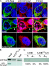
Figure 1.
Figure 1.
Ectopically expressed 3xFlag AnkB is farnesylated and is targeted to the plasma membrane. (A) Representative confocal microscopy images of cells ectopically expressing 3xFlag-tagged AnkB, AnkB169C/A, and BAP. Green indicates labeling with anti-Flag antibody, whereas red indicates labeling with antifarnesyl antibody, and the nucleus is stained blue (DAPI). The arrowheads indicate strong colocalization of AnkB with farnesylation at the plasma membrane. (B) Transfected HEK293 cells expressing 3xFlag-tagged proteins were lysed, immunoprecipitated with an anti-Flag antibody, and immunoblotted with antifarnesyl and anti-Flag antibodies. (C) Partitioning of 3xFlag AnkB in the detergent or aqueous phases of Triton X-114. Transfected cells were lysed with Triton X-114 and separated into detergent (D)-soluble and aqueous (Aq) phases. Samples were then analyzed by immunoblotting with anti-Flag antibodies. All experiments were performed three times, and representative examples are shown.
Figure 2.
Figure 2.
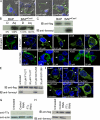
Host farnesyltransferase modifies ectopically expressed 3xFlag-tagged AnkB and BAP4CaaX through the addition of a farnesyl moiety. (A) Representative confocal microscopy images of cells transfected with 3xFlag-tagged BAP, BAP12CaaX, BAP8CaaX, and BAP4CaaX. The cells were labeled with anti-Flag antibody (green), whereas the nucleus is stained with DAPI (blue). Arrowheads indicate localization of the proteins to the plasma membrane. (B) Partitioning of 3xFlag-tagged BAP4CaaX in Triton X-114 detergent (D) or aqueous (Aq) phases. Transfected cells were lysed with Triton X-114 and separated into detergent-soluble and aqueous phases. Samples were then analyzed by immunoblotting with anti-Flag antibodies. (C) 3xFlag-tagged BAP and BAP4CaaX were immunoprecipitated from transfected HEK293 cells with anti-Flag resin and then analyzed by immunoblotting (IB) with antifarnesyl and anti-Flag antibodies. (D) Representative confocal images of HEK293 cells transfected with 3xFlag AnkB and treated with FTI-277 or GGTI-298 at concentrations of 0–20 µM. Anti-Flag labeling is represented in green, whereas the nucleus is stained blue (DAPI). Arrowheads indicate localization at the plasma membrane. All experiments were performed three times, and representative examples are shown. (E) Ectopically expressed 3xFlag AnkB in HEK293 cells treated with and without FTI-277 or GGTI-298 was immunoprecipitated with anti-Flag resin and then analyzed by immunoblotting with anti-Flag followed by antifarnesyl antibodies. (F) Knockdown expression of FT-α by RNAi blocks targeting of ectopically expressed 3xFlag-tagged AnkB and BAP4CaaX to the plasma membrane. Representative confocal microscopy images of transfected untreated HEK293 cells or treated with scrambled control or FT-α–specific RNAi. Anti-Flag labeling is represented in green, whereas the nucleus is stained blue (DAPI). Arrowheads indicate localization of Flag-tagged proteins at the plasma membrane. (G) HEK293 cell lysates were immunoblotted with an anti–FT-α antibody followed by antiactin antibody. (H) Lysates of RNAi-treated HEK293 cells expressing 3xFlag AnkB were immunoprecipitated by anti-Flag antibodies, followed by immunoblotting with anti-Flag followed by antifarnesyl antibodies. All experiments were performed three times, and representative examples are shown.
Figure 3.
Figure 3.
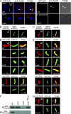
AnkB localizes to the LCV membrane through farnesylation of its CaaX motif. (A) The U937 cells and D. discoideum were infected and analyzed at 2 h by confocal microscopy using anti–L. pneumophila (Lpn) antibodies (green) and anti-AnkB antisera (red). Arrowheads indicate localization of AnkB with L. pneumophila. (B and C) Integrity of the membrane of purified LCVs from U937 cells was verified by labeling with mouse anti–L. pneumophila monoclonal antibody before permeabilization of the LCVs. After permeabilization, the LCVs were probed with rabbit anti–L. pneumophila antiserum to visualize the bacteria within the LCVs. (D and E) AnkB localizes to the cytosolic side of the LCV membrane. (D) The LCVs harboring the indicated strains were probed with anti-AnkB or anti-SidC antiserum before permeabilization (red) to determine whether the protein was localized to the cytosolic side of the LCV membrane. After permeabilization, the LCVs were probed with mouse anti–L. pneumophila monoclonal antibodies (green) to visualize the bacteria within the LCV. Quantitation is shown in the merged panels, where the numbers represent the percentage plus standard deviation of LCVs that bound anti-AnkB or anti-SidC antiserum before permeabilization. Analyses were based on the examination of 100 LCVs from triplicate samples. (E) The LCVs were permeabilized and then labeled with anti-AnkB or anti-SidC antiserum and anti–L. pneumophila monoclonal antibody, and quantitation of the LCVs that bound both antibodies is shown in the merged images and is based on analyses of 100 LCVs. (F) Farnesylation of AnkB anchors it to the LCV membrane. The AnkB proteins were immunoprecipitated using anti-AnkB antisera and then analyzed by immunoblotting (IB) with anti-AnkB followed by antifarnesyl antiserum. (G) The FTI-277 blocks localization of AnkB to the cytosolic side of the LCV membrane. The LCVs were isolated from untreated or 0.5 µM FTI-277–treated U937 cells at 1 h after infection and labeled with anti-AnkB antiserum before permeabilization (red). After permeabilization, the LCVs were labeled with anti–L. pneumophila monoclonal antibody (green) and analyzed by confocal microscopy. Quantification is shown in the merged panels, where the numbers represent the percentage plus standard deviation of LCVs that bound anti-AnkB before permeabilization. Analyses were based on examination of 100 LCVs from triplicate samples. All experiments were performed in triplicate, and representative examples are shown. All the results in this figure are representative of three independent experiments.
Figure 4.
Figure 4.
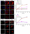
The CaaX motif of AnkB is essential for polyubiquitination of the LCV and for intracellular bacterial replication. (A and B) Representative confocal microscopy images of U937 cells (A) and D. discoideum cells (B) infected with L. pneumophila and examined at 2 h after infection for recruitment of polyubiquitinated proteins to the LCV. The U937 and D. discoideum cells were infected with the WT strain, the ankB mutant, and the ankB mutant complemented with the WT allele of ankB (ankB/c.ankB) or ankB169C/A (ankB/c.ankB169C/A) allele. Bacteria are labeled with anti-L. pneumophila (Lpn) antibody (green), polyubiquitinated (poly UB) proteins are labeled red, and the nucleus is stained blue (DAPI). Arrowheads indicate heavy colocalization of polyubiquitin with the LCVs. Numbers in the merged panels are quantitation of the percentage of LCVs positive for recruitment of polyubiquitinated proteins, based on the examination of 100 LCVs in triplicate samples. (C and D) Intracellular growth kinetics of L. pneumophila strains in U937 cells (C) or D. discoideum (D) infected with the WT strain, the ankB mutant, and the ankB mutant complemented with the WT copy of the ankB or the ankB169C/A allele. (E) Translocation of AnkB into U937 cells was determined at 1 h after infection. Strains harbored either empty vector (pCya) or Cya hybrids of RalF, AnkB, or AnkB169C/A. The translocation-defective dotA mutant harboring AnkB-Cya was used as a negative control. All experiments were performed three times, and representative examples are shown. (C–E) The data are the mean of triplicate samples, and the error bars are the standard deviations.
Figure 5.
Figure 5.
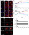
Chemical inhibition of host FT-α blocks recruitment of polyubiquitinated proteins to the LCV and abolishes intravacuolar bacterial replication. (A and B) Representative confocal microscopy images of cells untreated or treated with 0.5 µM FTI-277 for U937 cells (A) and D. discoideum (B). The host cells were infected with the WT strain or the ankB mutant, and recruitment of polyubiquitinated (poly UB) proteins to the LCV was examined at 2 h after infection. Bacteria are labeled by anti–L. pneumophila (Lpn) antibody (green), polyubiquitin is labeled red, and the nucleus is stained blue (DAPI). Arrowheads indicate heavy colocalization of polyubiquitinated proteins with the LCVs. Numbers in the merged panels are quantitation of the percentage of LCVs that colocalized with polyubiquitinated proteins at 2 h after infection, based on the examination of 100 LCVs from triplicate samples. (C and D) Intracellular growth kinetics of L. pneumophila strains in cells untreated or treated with and without 0.5 µM FTI-277 for U937 cells (C) or D. discoideum (D). The cells were infected with the WT strain, the ankB mutant, and the ankB mutant complemented with the WT copy of the ankB or the ankB169C/A allele. The results are representative of three independent experiments performed in triplicate, and error bars represent standard deviation.
Figure 6.
Figure 6.
Knockdown of expression of host FT-α by RNAi blocks recruitment of polyubiquitinated proteins to the LCV and abolishes intravacuolar bacterial replication. (A) Representative confocal images of untreated HEK293 cells or cells treated with FT-α–specific or scrambled RNAi control. The cells were infected with the WT strain or the ankB mutant bacteria, and recruitment of polyubiquitinated proteins to the LCV was examined at 2 h after infection. Bacteria are labeled with anti–L. pneumophila (Lpn) antibody (green), polyubiquitinated (polyUB) proteins are labeled red, and the nucleus is stained blue (DAPI). Arrowheads indicate heavy colocalization of the LCVs with polyubiquitinated proteins. Numbers in the merged panels are the percentage of LCVs positive for polyubiquitin plus standard deviation, based on the examination of 100 LCVs from triplicate samples. (B) Quantitation by single cell analysis of bacterial replicative phagosomes in HEK293 cells at 12 h after infection based on the analyses of 100 infected cells by confocal microscopy and presented as the number of bacteria/cell and the percentage of cells harboring a certain number of bacteria. The dotA mutant was used as a negative control. Infected cells from multiple coverslips were examined in each experiment. The results are representative of three independent experiments performed in triplicate, and error bars represent standard deviation.
Figure 7.
Figure 7.
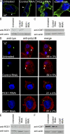
RNAi knockdown of ICMT and RCE-1 blocks targeting of AnkB to membranes and inhibits the biological function of the F-box protein in decorating the LCV by polyubiquitinated proteins. (A) Representative confocal microscopy images of untreated HEK293 cells or RNAi control–treated or ICMT- or RCE-1–specific RNAi–treated cells transfected with 3xFlag-tagged AnkB. The cells were labeled with anti-Flag antibody (green), whereas the nucleus is stained with DAPI (blue). Arrowheads indicate localization of the proteins to the plasma membrane. (B and C) RNAi-mediated knockdown expression of RCE-1 and ICMT, respectively. (D) Representative confocal images of untreated U937 cells or cells treated with the indicated RNAi. The cells were infected with the WT strain, and recruitment of polyubiquitinated proteins to the LCV was examined at 2 h after infection. Bacteria are labeled with anti–L. pneumophila (Lpn) antibody (green), polyubiquitinated (polyUB) proteins are labeled red, and the nucleus is stained blue (DAPI). Arrowheads indicate heavy colocalization of the LCVs with polyubiquitinated proteins. Numbers in the merged panels are the percentage of LCVs positive for polyubiquitin plus standard deviation, based on the examination of 100 LCVs from triplicate samples. (E and F) RNAi-mediated knockdown expression of RCE-1 and ICMT, respectively. All the results in this figure are representative of three independent experiments.
Figure 8.
Figure 8.
Dot/Icm-dependent colocalization of the LCV with the three enzymes FT-α, RCE-1, and ICMT. (A–C) The U937 cells were infected with WT, dotA, ankB, ankB/c.ankB, and ankB/c.ankB169C/A bacteria. At 2 h after infection, the cells were labeled with anti–L. pneumophila (Lpn) antibodies (green) and anti–FT-α (A), anti–RCE-1 (B), or anti-ICMT (C) antibodies (red). Nuclei were stained with DAPI (blue). The cells were analyzed by confocal microscopy. Arrowheads indicate colocalization of the respective marker with the LCVs. Quantification is shown in the merged images and represents a total percentage of LCVs colocalized with the maker plus standard deviation, based on the examination of 100 cells from triplicate samples. All experiments were performed three times, and representative examples are shown.
Figure 9.
Figure 9.
L. pneumophila expressing the ankB169C/A allele is defective in intrapulmonary proliferation in the A/J mice model of Legionnaires’ disease. Three A/J mice for each time point were infected with 106 CFUs of L. pneumophila WT strain, the ankB mutant, or the ankB mutant complemented with either the WT ankB allele (ankB/c.ankB) or with the ankB169C/A allele (ankB/c.ankB169C/A). At each time point, three mice were sacrificed, lungs were obtained and homogenized, and dilutions were plated on agar plated for CFU enumeration. The results are the mean of three mice/time point. These results are representative of two independent experiments. Error bars indicate standard deviation.

References

    1. Al-Khodor S., Price C.T., Habyarimana F., Kalia A., Abu Kwaik Y. 2008. A Dot/Icm-translocated ankyrin protein of Legionella pneumophila is required for intracellular proliferation within human macrophages and protozoa. Mol. Microbiol. 70:908–923 - PMC - PubMed
    1. Al-Khodor S., Al-Quadan T., Abu Kwaik Y. 2010a. Temporal and differential regulation of expression of the eukaryotic-like ankyrin effectors of L. pneumophila. Environ. Microbiol. In press 10.1111/j.1758-2229.2010.00159.x - DOI - PubMed
    1. Al-Khodor S., Price C.T.D., Kalia A., Abu Kwaik Y. 2010b. Functional diversity of ankyrin repeats in microbial proteins. Trends Microbiol. 18:132–139 10.1016/j.tim.2009.11.004 - DOI - PMC - PubMed
    1. Alli O.A., Zink S., von Lackum N.K., Abu-Kwaik Y. 2003. Comparative assessment of virulence traits in Legionella spp. Microbiology. 149:631–641 10.1099/mic.0.25980-0 - DOI - PubMed
    1. Asare R., Abu Kwaik Y. 2007. Early trafficking and intracellular replication of Legionella longbeachae within an ER-derived late endosome-like phagosome. Cell. Microbiol. 9:1571–1587 10.1111/j.1462-5822.2007.00894.x - DOI - PubMed

Publication types

MeSH terms