. 2010 Jul 26;190(2):197-207.
doi: 10.1083/jcb.200911156.

DNA damage signaling in response to double-strand breaks during mitosis

Affiliations

DNA damage signaling in response to double-strand breaks during mitosis

Simona Giunta et al. J Cell Biol. .

Abstract

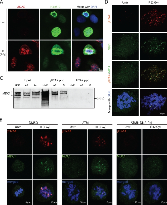
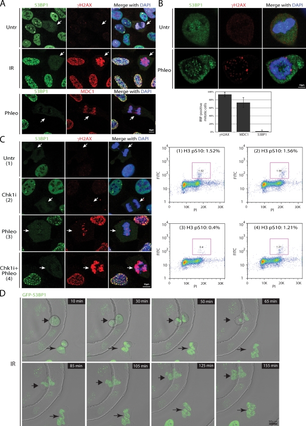
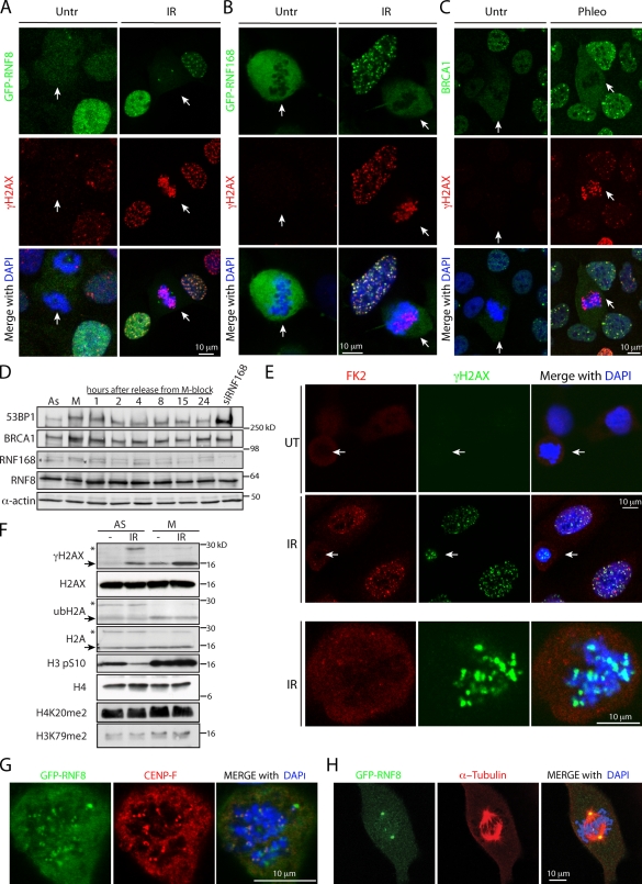
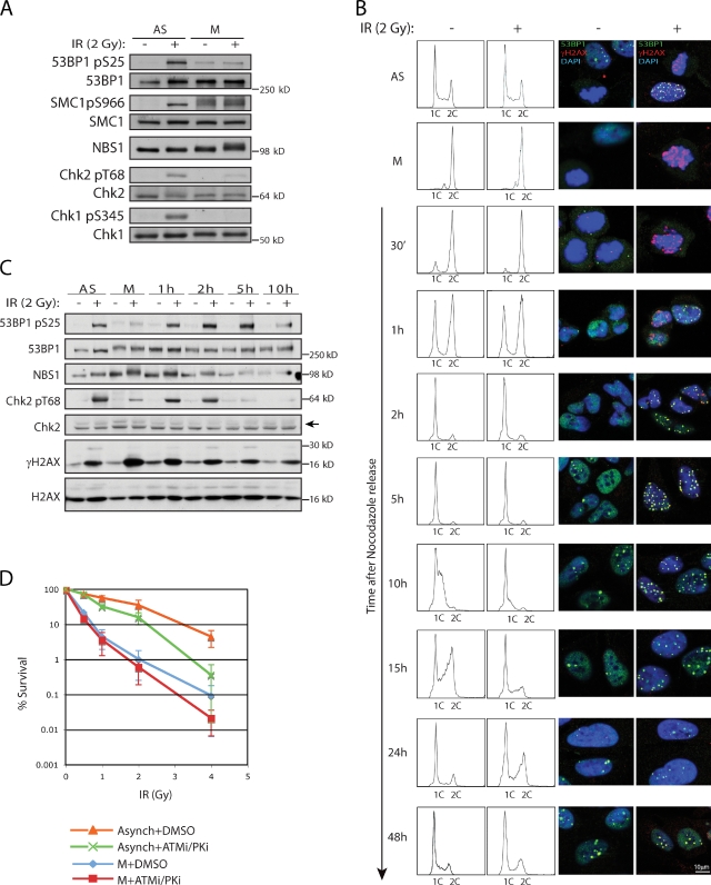
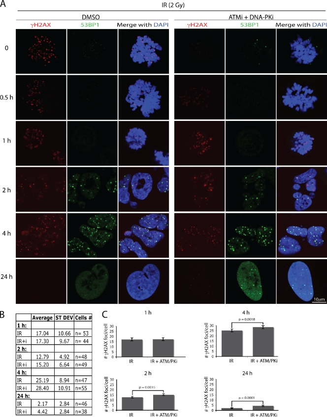
The signaling cascade initiated in response to DNA double-strand breaks (DSBs) has been extensively investigated in interphase cells. Here, we show that mitotic cells treated with DSB-inducing agents activate a "primary" DNA damage response (DDR) comprised of early signaling events, including activation of the protein kinases ataxia telangiectasia mutated (ATM) and DNA-dependent protein kinase (DNA-PK), histone H2AX phosphorylation together with recruitment of mediator of DNA damage checkpoint 1 (MDC1), and the Mre11-Rad50-Nbs1 (MRN) complex to damage sites. However, mitotic cells display no detectable recruitment of the E3 ubiquitin ligases RNF8 and RNF168, or accumulation of 53BP1 and BRCA1, at DSB sites. Accordingly, we found that DNA-damage signaling is attenuated in mitotic cells, with full DDR activation only ensuing when a DSB-containing mitotic cell enters G1. Finally, we present data suggesting that induction of a primary DDR in mitosis is important because transient inactivation of ATM and DNA-PK renders mitotic cells hypersensitive to DSB-inducing agents.


Figures

Figure 1.
Figure 1.
ATM and DNA-PK mediate IRIF formation in mitosis. All experiments were performed with U2OS cells. (A) IR induces γH2AX focus formation on mitotic chromosomes costained for histone H3pS10. (B) ATM inhibitor or a combination of ATM and DNA-PK inhibitors affect γH2AX and MDC1 IRIF formation in mitosis. (C) Immunoblot of MDC1 after peptide pull-downs with unmodified and phosphorylated H2AX C-terminal peptides. Inputs represent 10% of the total protein in the whole cell extracts prepared from asynchronous (AS) and mitotic (M) cells. HNE, HeLa nuclear extract. (D) NBS1 and γH2AX colocalize on mitotic chromosomes after IR.
Figure 2.
Figure 2.
53BP1 is excluded from IRIF during mitosis. White arrows point to mitotic cells. (A) Costaining of either mock- or IR-treated asynchronously growing U2OS cells with 53BP1 and γH2AX antibodies. Phleomycin-treated U2OS cells costained for 53BP1 and MDC1. (B) Enlarged images of mitotic cells show exclusion of 53BP1 from chromatin and a lack of colocalization between the punctate 53BP1 staining and γH2AX foci. The histogram represents a quantification of γH2AX, MDC1, and 53BP1 focus-positive mitotic cells after IR treatment from three independent experiments (n > 200). Error bars indicate standard deviation. (C) Active exclusion of 53BP1 from mitotic IRIF. After a 3-h incubation with 50 nM of Chk1 inhibitor (Chk1i), cells were treated with phleomycin for 2 h, fixed and costained for 53BP1 and γH2AX, or collected for FACS analyses to determine mitotic indices by H3 pS10 immunofluorescence (purple boxes). (D) Time-lapse frames of U2OS cells stably expressing EGFP-53BP1. After irradiation with 0.5 Gy, two mitotic cells marked with arrows were imaged for up to 3 h.
Figure 3.
Figure 3.
Lack of recruitment of the ubiquitin E3 ligases RNF8, RNF168, and BRCA1 to DSB sites in mitosis. (A–C) GFP-RNF8 (A), GFP-RNF168 (B), and BRCA1 (C) are excluded from chromatin and are not recruited to IRIF upon DSB induction during mitosis. White arrows indicate mitotic cells. (D) Levels of indicated proteins throughout the cell cycle. M-phase cells, obtained by a shake-off procedure after thymidine-nocodazole arrest, were released into fresh medium and collected at specified times. (E) FK2 antibody staining does not detect ubiquitin conjugates on mitotic chromosomes upon IR treatment. (F) Histones H2A and H2AX are deubiquitylated in mitosis. Arrows point to nonubiquitylated forms and asterisks mark ubiquitylated forms of the proteins. Note that histone H4K20me2 and H3K79me2 marks are present in mitosis. (G) GFP-RNF8 colocalizes with CENP-F at kinetochores. (H) GFP-RNF8 localizes to centrosomes.
Figure 4.
Figure 4.
Marking of DSBs in mitosis precedes full DDR activation in G1 and affects cell survival. Mitotic cells were obtained as in Fig. 3 D. (A) Diminished phosphorylation of ATM targets in IR-treated mitotic cells compared with asynchronous cells. (B) Untreated or irradiated mitotic cells were released from nocodazole and monitored for cell cycle progression and 53BP1 recruitment to IRIF (see text for details). (C) Chk2 T68 and 53BP1 S25 phosphorylation in cells released into G1 after IR treatment in mitosis. (D) Radiosensitivity of asynchronous and mitotic cells pretreated for 1 h with DMSO or a combination of ATM and DNA-PK inhibitors and then exposed to various doses of IR. 30 min after irradiation, inhibitors were washed away and cells were plated in fresh medium. Error bars represent standard deviations of the mean from six experiments. P-values were calculated at the standard 0.05 threshold. Treatment with PIKK inhibitors had statistically significant effects on radiosensitivity of both asynchronous (P = 0.00015) and mitotic (P = 0.041) cells.
Figure 5.
Figure 5.
Transient inhibition of ATM and DNA-PK during mitosis before irradiation results in the increase of persistent IRIF 24 h after treatment. U2OS cells were treated as in Fig. 4 D. (A) Representative images of the cells at different times after nocodazole and PIKK inhibitor release. The table (B) and the graph (C) summarize γH2AX focus quantification. Error bars represent standard errors (SE = SD/√n).

References

    1. Bakkenist C.J., Kastan M.B. 2003. DNA damage activates ATM through intermolecular autophosphorylation and dimer dissociation. Nature. 421:499–506 10.1038/nature01368 - DOI - PubMed
    1. Burma S., Chen B.P., Murphy M., Kurimasa A., Chen D.J. 2001. ATM phosphorylates histone H2AX in response to DNA double-strand breaks. J. Biol. Chem. 276:42462–42467 10.1074/jbc.C100466200 - DOI - PubMed
    1. Cai S.Y., Babbitt R.W., Marchesi V.T. 1999. A mutant deubiquitinating enzyme (Ubp-M) associates with mitotic chromosomes and blocks cell division. Proc. Natl. Acad. Sci. USA. 96:2828–2833 10.1073/pnas.96.6.2828 - DOI - PMC - PubMed
    1. Chin C.F., Yeong F.M. 2010. Safeguarding entry into mitosis: the antephase checkpoint. Mol. Cell. Biol. 30:22–32 10.1128/MCB.00687-09 - DOI - PMC - PubMed
    1. Deckbar D., Birraux J., Krempler A., Tchouandong L., Beucher A., Walker S., Stiff T., Jeggo P., Löbrich M. 2007. Chromosome breakage after G2 checkpoint release. J. Cell Biol. 176:749–755 10.1083/jcb.200612047 - DOI - PMC - PubMed

Publication types

MeSH terms

Substances