. 2010 Aug 10;107(32):14378-83.
doi: 10.1073/pnas.1009198107. Epub 2010 Jul 26.

Isolation of viable Plasmodium falciparum merozoites to define erythrocyte invasion events and advance vaccine and drug development

Affiliations

Isolation of viable Plasmodium falciparum merozoites to define erythrocyte invasion events and advance vaccine and drug development

Michelle J Boyle et al. Proc Natl Acad Sci U S A. .

Abstract

During blood-stage infection by Plasmodium falciparum, merozoites invade RBCs. Currently there is limited knowledge of cellular and molecular invasion events, and no established assays are available to readily measure and quantify invasion-inhibitory antibodies or compounds for vaccine and drug studies. We report the isolation of viable merozoites that retain their invasive capacity, at high purity and yield, purified by filtration of highly synchronous populations of schizonts. We show that the half-life of merozoite invasive capacity after rupture is 5 min at 37 degrees C, and 15 min at room temperature. Studying the kinetics of invasion revealed that 80% of invasion events occur within 10 min of mixing merozoites and RBCs. Invasion efficiency was maximum at low merozoite-to-RBC ratios and occurred efficiently in the absence of serum and with high concentrations of dialyzed nonimmune serum. We developed and optimized an invasion assay by using purified merozoites that enabled invasion-inhibitory activity of antibodies and compounds to be measured separately from other mechanisms of growth inhibition; the assay was more sensitive for detecting inhibitory activity than established growth-inhibition assays. Furthermore, with the use of purified merozoites it was possible to capture and fix merozoites at different stages of invasion for visualization by immunofluorescence microscopy and EM. We thereby demonstrate that processing of the major merozoite antigen merozoite surface protein-1 occurs at the time of RBC invasion. These findings have important implications for defining invasion events and molecular interactions, understanding immune interactions, and identifying and evaluating inhibitors to advance vaccine and drug development.


Conflict of interest statement

The authors declare no conflict of interest.

Figures
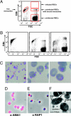
Fig. 1.
Fig. 1.
Purification of merozoites and invasion of RBCs. (A) Representative flow cytometry plot showing different cell populations of free merozoites, uninfected RBCs, RBCs with bound merozoites and infected RBCs. Note that this plot shows no infected RBCs (see B, Right and Fig. S1 for FACS plots containing infected RBCs). (B and C) Filtration effectively purifies viable merozoites from E64-treated schizonts. FACS plots (B) and Giemsa-stained smears (C) show merozoites of high purity after filtration of E64-treated schizonts (Left), and that purified merozoites were able to bind and invade RBCs (Center), resulting in a highly synchronous population of intraerythrocytic parasites (Right). (D) Surface labeling of purified merozoites with antibodies to AMA1 [red; counter-stained with DAPI (blue) to label the merozoites’ nucleus]. (E) Labeling of the rhoptry with antibodies to RAP1. (F) Transmission EM image of purified merozoites labeled to identify key structures.
Fig. 2.
Fig. 2.
Kinetics and requirements for merozoites invasion. (A) The proportion of merozoites that invade RBCs (invasion rate) is affected by the ratio of merozoites to RBCs. As the ratio of merozoites to RBCs increases, the invasion rate of merozoites decreases. Data are representative of four assays in duplicate. (B) The invasive potential of merozoites declines over time, and is affected by different temperatures. Merozoites were incubated at 37 °C, 22 °C, or on ice after purification before being mixed with RBCs to measure invasion. Data are mean ± SEM of seven assays in duplicate. (C) The rate of merozoite invasion over time is rapid following incubation with uninfected RBCs. The proportion of merozoites that have invaded with increasing time is shown as a percent of maximum invasion recorded in noninhibited samples. Data are means ± range of two assays in duplicate. (D) Merozoite invasion occurs in the presence and absence of serum components. Merozoites were tested for the ability to invade RBCs in the presence of serum at various concentrations. Serum was used with and without dialysis against RPMI-Hepes. Data are means ± SEM of three assays in duplicate and expressed as a percentage of invasion into RMPI-Hepes alone.
Fig. 3.
Fig. 3.
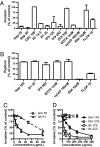
Development of an invasion inhibition assay using purified merozoites. (A) Various inhibitory and noninhibitory compounds and antibodies were tested for their ability to inhibit invasion of purified merozoites in IIAs. Invasion is expressed as a proportion of control. The concentration of inhibitors is in μg/mL unless otherwise indicated. Data are mean ± range of two assays in duplicate. (B) Compounds were tested for inhibition of schizont rupture by incubating with late stage parasites and measuring parasitemia and schizont rupture by flow cytometry over time. Rupture is expressed as a proportion of control. Data are mean ± range of two assays in duplicate. (C and D) Comparison of inhibitory activities in IIA versus conventional GIA of AMA1-binding peptide R1 (C) and the anti-AMA1 MAbs 1F9 and 2C5 (D).
Fig. 4.
Fig. 4.
Imaging of merozoite invasion in fixed cells. Purified merozoites were fixed in the process of invasion to visualize invasion events. Merozoites were examined at the point of initial binding to the RBC surface (Left), midway through RBC invasion (Center), and after invasion was complete (Right). Merozoites in the process of invasion were labeled with antibodies to MSP1-19 (A) or MSP1 block 2 (B) (green). Nucleus is stained with DAPI (blue). (C) EM images of fixed invading merozoites showed all stages of merozoite invasion.

References

    1. Snow RW, Guerra CA, Noor AM, Myint HY, Hay SI. The global distribution of clinical episodes of Plasmodium falciparum malaria. Nature. 2005;434:214–217. - PMC - PubMed
    1. Elliott SR, Beeson JG. Estimating the burden of global mortality in children aged <5 years by pathogen-specific causes. Clin Infect Dis. 2008;46:1794–1795. - PubMed
    1. Cowman AF, Crabb BS. Invasion of red blood cells by malaria parasites. Cell. 2006;124:755–766. - PubMed
    1. Dowse TJ, Koussis K, Blackman MJ, Soldati-Favre D. Roles of proteases during invasion and egress by Plasmodium and Toxoplasma. Subcell Biochem. 2008;47:121–139. - PubMed
    1. Richards JS, Beeson JG. The future for blood-stage vaccines against malaria. Immunol Cell Biol. 2009;87:377–390. - PubMed

Publication types