. 2010 Aug 10;107(32):14217-22.
doi: 10.1073/pnas.1008623107. Epub 2010 Jul 26.

Live imaging of Drosophila imaginal disc development

Affiliations

Live imaging of Drosophila imaginal disc development

Silvia Aldaz et al. Proc Natl Acad Sci U S A. .

Abstract

Live imaging has revolutionized the analysis of developmental biology over the last few years. The ability to track in real time the dynamic processes that occur at tissue and cellular levels gives a much clearer view of development, and allows greater temporal resolution, than is possible with fixed tissue. Drosophila imaginal discs are a particularly important model of many aspects of development, but their small size and location inside the larva and pupa has prevented live imaging techniques from extensively being used in their study. Here, we introduce the use of viscous culture medium to enable high resolution imaging of imaginal disc development. As a proof of principle, we have analyzed the transformation that occurs during metamorphosis of the wing imaginal disc into the mature wing and report several previously unobserved stages of this model of organogenesis. These imaging methods are especially useful to study the complex and dynamic changes that occur during morphogenesis, but we show that they can also be used to analyze other developmental and cellular events. Moreover, our viscous medium creates a platform for future adaptation of other tissue culture conditions to allow imaging of a wide range of developmental events and systems.


Conflict of interest statement

The authors declare no conflict of interest.

Figures

Fig. 1.
Fig. 1.
Wing imaginal disc and preparation of samples. (A) A late third instar imaginal disc. (Upper) A frontal view with the squamous peripodial epithelium (PE) in green. (Lower) An axial section showing a more detailed view of the three types of cells in the imaginal disc. (B) Custom observation chamber for use with an upright microscope. (C) Anterior half of a prepupa (3–4 h APF) cut along the axial plane. (D) Same pupa bisected along the sagittal plane. (E) One of the half-pupae showing a wing imaginal disc (arrow) close to the inner side of the pupal case. (F) Culture dish containing the sample embedded in agarose and immersed in liquid medium.
Fig. 2.
Fig. 2.
Ex vivo imaginal disc eversion. (A) Frames from Movie S1 showing a prepupal imaginal disc during the different steps of the eversion process. The disc is labeled with the Sqh-GFP transgene (green), and grunge-Gal4 driving UAS-mRFP (red); grunge-Gal4 is mainly expressed in peripodial cells but also in two bright spots in the region of the hinge of the columnar epithelium. This combination highlights the retraction process (white arrows mark the front of the retracting PE cells). The apoptotic bodies of the squamous cells accumulate at one side of the hinge region (orange arrow). The last images show the expansion of the wing pouch and the formation of the precursors of the wing margin sensory organs (yellow arrowheads). (B) Frames from Movie S2 showing a prepupal imaginal disc mounted on its side showing the distribution of Sqh-GFP (green) and His2A-RFP (red). The first two frames show the folding of the whole disc after the addition of the ecdysone. This lateral view allows observation of the opening of the hole in the stalk region and the start of retraction (white arrows).
Fig. 3.
Fig. 3.
Interaction between the wing disc and the pupal epidermis. (A) Frames from Movie S3 a prepupal imaginal disc attached to the pupal case. The disc expresses Sqh-GFP (green) and grunge-Gal4 >UAS-RFP (red). The red label also marks the close opposed pupal case and epidermis (white arrow). In the first frames, the PE cells are weakly stained, but when the contraction starts, intensity increases (orange arrows). At the same time the DP gets closer to the epidermis, folds, and protrudes its basal part (yellow arrows). (B) Frames from Movie S4, showing a His2A-mRFP (red), Sqh-GFP (green) wing disc. In this example, the separation between the disc proper and PE is very clear (orange arrows). The yellow arrows highlight the squamous epithelium retracting. The columnar epithelium protrudes from inside.
Fig. 4.
Fig. 4.
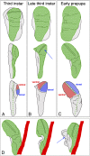
Schematic representation of disc eversion. (AC) Front view (Top in all panels) and side view (Middle in all panels) of a third instar wing disc. The squamous cells of the PE are represented in green and the columnar (and cuboidal) epithelium in gray. Note that in reality there is no sharp distinction between the squamous and columnar cells: a transition zone of cuboidal cells separates them. Here, we define the PE as the squamous cells alone. (Bottom) The image in each panel shows a side view of the disc, highlighting the dorsal and ventral compartments of the wing pouch of the imaginal disc and how they move during eversion. The green dotted line illustrates the bending between stages. (A) The third instar wing disc is flat, with ventral and dorsal compartments in the same plane. (B) By late third instar, the squamous epithelium has expanded, and the dorsal and ventral compartments of the wing pouch become apposed (arrow). (C) At the prepupal stage the disc starts to bend. The apposition of the two wing surfaces is complete. The peripodial cells cover almost the complete apical surface of the columnar epithelium. (D) Representation of the disc attached to the pupal case. The cuticle and the epidermis are represented in red. The three steps show how the disc approaches the epidermis, the PE contracts, folding the whole disc, and the basal part of the columnar epithelium protrudes (arrow).
Fig. 5.
Fig. 5.
Further applications of ex vivo culture. (A) Frames from Movie S7 showing a wing disc labeled with Sqh-GFP. The red arrows point to the nuclei of two peripodial cells. Using a 100× objective, intracellular structures like myosin II fibers can be tracked. (B) Frames from Movie S9 showing a prepupal imaginal disc labeled with Sqh-GFP (green) and annexin-V-Cy3 (red), which was added to the medium after dissection, as a reporter of apoptosis during retraction of the PE (indicated by arrows). (C) Two frames from Movie S10 showing numerous dividing cells (several marked by arrows) in a wing disc labeled with Arm-GFP. (D) Frames from Movie S11 showing a wing disc labeled with His2A-mRFP (red) and Sqh-GFP (green). Yellow arrows indicated dividing cells in which the different stages of mitosis can be tracked, both in the XY and XZ planes. (E) Initial (Upper) and final (Lower) frames, taken from Movie S12, showing the healing of a wound (red arrow) in the notum of a wing disc labeled with Sqh-GFP. (F) Frames from Movie S13 showing a wing disc labeled with Arm-GFP and the development of two wing vein sensillae in the wing pouch (green and red arrows).

References

    1. Bertet C, Sulak L, Lecuit T. Myosin-dependent junction remodelling controls planar cell intercalation and axis elongation. Nature. 2004;429:667–671. - PubMed
    1. Oda H, Tsukita S. Dynamic features of adherens junctions during Drosophila embryonic epithelial morphogenesis revealed by a Dalpha-catenin-GFP fusion protein. Dev Genes Evol. 1999;209:218–225. - PubMed
    1. Wood W, et al. Wound healing recapitulates morphogenesis in Drosophila embryos. Nat Cell Biol. 2002;4:907–912. - PubMed
    1. Richardson BE, Beckett K, Baylies MK. Live imaging of Drosophila myoblast fusion. Methods Mol Biol. 2008;475:263–274. - PMC - PubMed
    1. Ninov N, Martín-Blanco E. Live imaging of epidermal morphogenesis during the development of the adult abdominal epidermis of Drosophila. Nat Protoc. 2007;2:3074–3080. - PubMed

Publication types

LinkOut - more resources