. 2010 Jul 22;5(7):e11733.
doi: 10.1371/journal.pone.0011733.

HIV-1 inhibits autophagy in bystander macrophage/monocytic cells through Src-Akt and STAT3

Affiliations

HIV-1 inhibits autophagy in bystander macrophage/monocytic cells through Src-Akt and STAT3

Jennifer Van Grol et al. PLoS One. .

Abstract

Autophagy is a homeostatic mechanism of lysosomal degradation. Defective autophagy has been linked to various disorders such as impaired control of pathogens and neurodegeneration. Autophagy is regulated by a complex array of signaling pathways that act upstream of autophagy proteins. Little is known about the role of altered regulatory signaling in disorders associated with defective autophagy. In particular, it is not known if pathogens inhibit autophagy by modulation of upstream regulatory pathways. Cells infected with HIV-1 blocked rapamycin-induced autophagy and CD40-induced autophagic killing of Toxoplasma gondii in bystander (non-HIV-1 infected) macrophage/monocytic cells. Blockade of autophagy was dependent on Src-Akt and STAT3 triggered by HIV-1 Tat and IL-10. Neutralization of the upstream receptors VEGFR, beta-integrin or CXCR4, as well as of HIV-1 Tat or IL-10 restored autophagy in macrophage/monocytic cells exposed to HIV-1-infected cells. Defective autophagic killing of T. gondii was detected in monocyte-derived macrophages from a subset of HIV-1(+) patients. This defect was also reverted by neutralization of Tat or IL-10. These studies revealed that a pathogen can impair autophagy in non-infected cells by activating counter-regulatory pathways. The fact that pharmacologic manipulation of cell signaling restored autophagy in cells exposed to HIV-1-infected cells raises the possibility of therapeutic manipulation of cell signaling to restore autophagy in HIV-1 infection.


Conflict of interest statement

Competing Interests: The authors have declared that no competing interests exist.

Figures

Figure 1
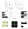
Figure 1. HIV-1 inhibits autophagy in bystander macrophages/monocytic cells.
A and B, MDM from healthy controls and HIV-1+ patients were incubated with or without CD154 (3 µg/ml) (A) or IFN-γ (200 U/ml) (B) followed by challenge with T. gondii tachyzoites. The number of parasites per 100 macrophages was assessed by light microscopy 24 h post-challenge. HIV-1+ patients were classified as non-responder when their macrophages exhibited a percentage decrease in parasite load that was less than the 10th percentile of the percentage decrease in parasite load observed in macrophages from healthy controls. CE, MDM from healthy controls were infected with pseudotyped HIV-1 (PV HIV) or pseudotyped control virus (PV Ctr) and were incubated with a monolayer of uninfected macrophages. In certain experiments macrophages were treated with or without zidovudine (AZT) 2 h after incubation with pseudotyped HIV-1 (E). Macrophage monolayers were treated with or without CD154 followed by challenge with T. gondii and assessment of parasite load at 24 h. F, Schematic representation of MonoMac6 cells infected with pseudotyped HIV-1 (PV HIV) or pseudotyped control virus (PV Ctr) incubated with uninfected MonoMac6 cells transfected with LC3-eGFP. Cells were treated with or without rapamycin (1 µM) and assessed for autophagy by expression of large (≥1 µm) LC3+ structures. G, Flourescent images of LC3-eGFP+ cells as treated in panel (F). H, Quantification of autophagic cells as treated in panel (F). Data are representative of 4 independent experiments presented as means ± SEM; ***p≤0.001; **p≤0.01; *≤0.05; ∧p≥0.05.
Figure 2
Figure 2. HIV-1 Tat inhibits autophagy.
A, MonoMac6 cells infected with pseudotyped HIV-1 (PV HIV) or pseudotyped control virus (PV Ctr) were incubated with uninfected MonoMac6 cells transfected with LC3-eGFP overnight in the presence or absence of either a neutralizing anti-Tat or control IgG mAb. Cells were treated with or without rapamycin and assessed for autophagy by expression of large LC3+ structures. B, MonoMac6 cells transfected with LC3-eGFP were incubated overnight with HIV-1 Tat (100 pg/ml) plus either neutralizing anti-Tat, a neutralizing anti-TNF-α, or control IgG mAb. Cells were then stimulated with rapamycin. Autophagy was assessed by examining expression of large LC3+ structures. C, MDM were treated with or without HIV-1 Tat followed by rapamycin. Cell lysates were obtained at 2 h and used for immunoblot for LC3 II and actin. D, MDM were treated with or without HIV-1 Tat followed by rapamycin in the presence of bafilomycin A1 (Baf A1; 100 nM). Cell lysates were obtained at 2 h and used for immunoblot for LC3 II and actin. E, and F, MDM were treated with increasing concentrations of HIV-1 Tat (E) or HIV-1 Tat (60 pg/ml) plus IFN- γ (100 U/ml) (F) with or without CD154. Macrophages were challenged with tachyzoites of T. gondii and assessed for parasite load at 24 h. G, MDM infected with pseudotyped HIV-1 were incubated with uninfected macrophages. Cells were cultured in the presence of neutralizing anti-Tat or control mAb. After stimulation with CD154, monolayers were challenged with T. gondii and parasite load was assessed at 24 h. H, MDM from CD40 non-responder HIV-1+ patients were incubated with neutralizing anti-Tat or control mAb followed by stimulation with CD154. Macrophages were challenged with T. gondii and assessed for parasite load at 24 h. Each symbol represents a different patient. The number of parasites per 100 macrophages was examined at 24 h by light microscopy. Data are representative of 3 to 4 independent experiments presented as means ± SEM; ***p≤0.001, ** p≤0.01, ∧p≥0.05.
Figure 3
Figure 3. HIV-1 inhibits autophagy in bystander macrophages/monocytic cells through Akt.
A, MDM incubated with or without HIV-1 Tat (1 ng/ml) were examined for expression of total Akt and phospho-Akt by immunoblot. B, MonoMac6 cells were transfected with siRNA against Akt1 or control siRNA. Expression of Akt and actin were examined 72 h post-transfection. C, MonoMac6 cells transfected with control or siRNA directed against Akt1 were transfected with LC3-eGFP. Cells were incubated with or without HIV-1 Tat (100 pg/ml) overnight followed by stimulation with rapamycin. Autophagy was assessed by examining expression of large LC3+ structures. D, MonoMac6 cells were transfected with control siRNA or siRNA directed against Akt. Cells were transfected with LC3-eGFP and incubated with MonoMac6 cells infected with pseudotyped control virus (PV Ctr) or pseudotyped HIV-1 (PV HIV). Cultures were treated with or without rapamycin. Autophagy was assessed by examining expression of large LC3+ structures. E, MDM were treated with Akt inhibitor IV (1.25 µM) or vehicle followed by incubation with HIV-1 Tat. Macrophages were incubated with or without CD154, challenged with T. gondii and assessed for parasite load at 24 h. Data are representative of 3 independent experiments presented as means ± SEM; *p≤0.05, **p≤0.01, ∧p≥0.05.
Figure 4
Figure 4. HIV-1 inhibits autophagy in bystander macrophages/monocytic cells through Src and Akt.
A, MonoMac6 cells were cultured with MonoMac6 cells infected with pseudotyped control virus (PV Ctr) or pseudotyped HIV-1 (PV HIV) or treated with HIV-1 Tat (100 pg/ml). Culture lysates were examined by immunoblot for expression of PTEN and actin. B, MDM were incubated with or without HIV-1 Tat and examined for expression of Src, phospho-Src, FAK and phospho-FAK by immunoblot. C, MDM were incubated with PP2 (20 µM) or vehicle followed by incubation with HIV-1 Tat. Expression of total Akt and phospho-Akt were examined by immunoblot. D, MonoMac6 cells were transfected with control siRNA or siRNA directed against Src and examined for expression of Src and actin. E, MonoMac6 cells transfected with control siRNA or siRNA directed against Src were transfected with LC3-eGFP and incubated with pseudotyped control virus (PV Ctr) or pseudotyped HIV-1 (PV HIV)-infected MonoMac6 cells overnight. Cultures were treated with or without rapamycin. Autophagy was assessed by examining expression of large LC3+ structures. Data are representative of 3 independent experiments presented as means ± SEM; ***p≤0.001, ∧p≥0.05.
Figure 5
Figure 5. HIV-1 inhibits autophagy in bystander macrophages/monocytic cells through CXCR4, VEGFR and β-integrin.
A, MonoMa6 cells were transfected with LC3-eGFP and incubated with cells infected with pseudotyped control virus (PV Ctr) or pseudotyped HIV-1 (PV HIV). Cultures were treated overnight with control mAb, control peptide cRAD, anti-CXCR4 mAb, anti-VEGFR1 mAb, VEGFR1 tyrosine kinase inhibitor, or β-integrin blocking peptide cRGD. MonoMac6 cells were treated with or with out rapamycin and assessed for large LC3+ structures. B, MonoMac6 cells transfected with LC3-eGFP were treated overnight with or without Tat (100 pg/ml) and either control mAb, control peptide cRAD, anti-CXCR4 mAb, anti-VEGFR1 mAb or β-integrin blocking peptide, cRGD. MonoMac6 cells were treated with or without rapamycin and assessed for large LC3+ structures. C, Schematic representation of MonoMac6 cells transfected with LC3-eGFP cultured overnight with MonoMac6 cells transfected with a control plasmid or a plasmid encoding Tat. Cultures were treated with rapamycin and assessed for large LC3+ structures. D, MonoMac6 cells transfected with LC3-eGFP were cultured overnight with MonoMac6 cells transfected with a control plasmid or a plasmid encoding either wild-type Tat, Tat CD26 mutant, Tat chemokine-like domain mutant (Tat31–101), Tat basic domain mutant, or Tat RGD mutant as depicted in panel (C). Cultures were treated with rapamycin and assessed for large LC3+ structures. Data are representative of 4 independent experiments presented as means ± SEM; *p≤0.05, **p≤0.01, ∧p≥0.05.
Figure 6
Figure 6. IL-10 is required for HIV-1 to inhibit autophagy in bystander macrophages/monocytic cells.
A, MonoMac6 cells transfected with LC3-eGFP were incubated with HIV-1 Tat (100 pg/ml) or IL-10 (100 pg/ml) overnight. Cells were then stimulated with rapamycin. Autophagy was assessed by examining expression of large LC3+ structures. B, MonoMac6 cells transfected with LC3-eGFP were incubated with MonoMac6 cells infected with pseudotyped control virus (PV Ctr) or pseudotyped HIV-1 (PV HIV). Cultures were treated with either neutralizing anti-IL-10 or control mAb overnight. Cells were then stimulated with rapamycin. Autophagy was assessed by examining expression of large LC3+ structures. C, MonoMac6 cells transfected with LC3-eGFP were incubated with HIV-1 Tat (100 pg/ml) plus either neutralizing anti-IL-10 or control mAb overnight. Cells were then stimulated with rapamycin. Autophagy was assessed by examining expression of large LC3+ structures. D, MDM infected with pseudotyped HIV-1 were incubated with uninfected MDM. Cells were cultured in the presence of either neutralizing anti-Tat, neutralizing anti-IL-10 or control mAb. After stimulation with CD154, monolayers were challenged with T. gondii. The number of parasite per 100 macrophages was examined at 24 h by light microscopy. E, MDM taken from a representative CD40 non-responder HIV-1+ infected patient were cultured in the presence of neutralizing anti-Tat, anti-IL-10 or control mAb. After stimulation with CD154, monolayers were challenged with T. gondii. The number of parasite per 100 macrophages was examined at 24 h by light microscopy. Data are representative of 3 independent experiments presented as means ± SEM; *p≤0.05, **p≤0.01, ***p≤0.001, ∧p≥0.05.
Figure 7
Figure 7. STAT3 and Akt are required for HIV-1 and IL-10 to inhibit autophagy.
A, MonoMac6 cells were transfected with siRNA against STAT3 or control siRNA. Expression of STAT3 and actin were examined 72 h post-transfection. B, MonoMac6 cells transfected with control siRNA or siRNA directed against STAT3 or Akt were transfected with LC3-eGFP. Cells were incubated with or without HIV-1 Tat or IL-10 (both at 100 pg/ml) overnight followed by stimulation with rapamycin. Autophagy was assessed by examining expression of large LC3+ structures. Data are representative of 3 independent experiments presented as means ± SEM; ***p≤0.001, ∧p≥0.05.
Figure 8
Figure 8. A schematic representation of the mechanism of HIV-1-induced inhibition of autophagy.
HIV-1 Tat interaction with the cell surface receptors CXCR4, VEGFR, and β-Integrin induces signaling through Src, it's binding partner FAK, and Akt. Concomitant upregulation of IL-10 by HIV-1 results in STAT3 activity, which functions in cooperation with Akt to initiate a robust inhibition of autophagy.

Similar articles

Cited by

References

    1. Mizushima N, Ohsumi Y, Yoshimori T. Autophagosome formation in mammalian cells. Cell Struct Funct. 2002;27:421–429. - PubMed
    1. Yoshimori T. Autophagy: a regulated bulk degradation process inside cells. Biochem Biophys Res Comm. 2004;313:453–458. - PubMed
    1. Levine B, Kroemer G. Autophagy in the pathogenesis of disease. Cell. 2008;132:27–42. - PMC - PubMed
    1. Gutierrez MG, Master SS, Singh SB, Taylor GA, Colombo MC, et al. Autophagy is defense mechanism inhibiting BCG and Mycobacterium tuberculosis survival in infected macrophages. Cell. 2004;119:753–766. - PubMed
    1. Nakagawa I, Amano A, Mizushima N, Yamamoto A, Yamaguchi H, et al. Autophagy defends cells against invading Group A Streptococcus. Science. 2004;306:1037–1040. - PubMed

Publication types