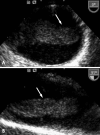
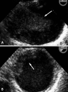
A case of huge thrombus in the aortic arch with cerebrovascular embolization
- PMID: 20661342
- PMCID: PMC2889393
- DOI: 10.4250/jcu.2009.17.4.148
A case of huge thrombus in the aortic arch with cerebrovascular embolization
Abstract
Pedunculated thrombus in the aortic arch that is associated with cerebral infarction is very rare requires prompt diagnosis and treatment to prevent occurrence of another devastating complication. Transesophageal echocardiography is useful for detecting source of embolism including aortic thrombi. The treatment options of aortic thrombi involves anticoagulation, thrombolysis, thromboaspiration, and thrombectomy. Here we report a case of huge thrombus in the aortic arch, resulting in acute multifocal cerebellar embolic infarct in patient without any risk factors for vascular thrombosis. Thrombi in the aortic arch were diagnosed by transesophageal echocardiography and treated with anticoagulants successfully.
Keywords: Aortic thrombus; Echocardiography.
Figures


References
-
- Panetta T, Thompson JE, Talkington CM, Garrett WV, Smith BL. Arterial embolectomy: a 34-year experience with 400 cases. Surg Clin North Am. 1986;66:339–353. - PubMed
-
- Gagliardi JM, BattM, Khodja RH, Le bas P. Mural thrombus of the aorta. Ann Vasc Surg. 1988;2:201–204. - PubMed
-
- Laperche T, Laurian C, Roudaut R, Steg PG. Mobile thromboses of the aortic arch without aortic debris. A transesophageal echocardiographic finding associated with unexplained arterial embolism. The Filiale Echocardiographie de la Société Française de Cardiologie. Circulation. 1997;96:288–294. - PubMed
-
- Sadony V, Walz M, Löhr E, Rimpel J, Richter HJ. Unusual cause of recurrent arterial embolism: floating thrombus in the aortic arch surgically removed under hypothermic cardiocirculatory arrest. Eur J Car-diothorac Surg. 1988;2:468–471. - PubMed
-
- Tunick PA, Kronzon I. Protruding atherosclerotic plaque in the aortic arch of patients with systemic embolization: a new finding seen by transesophageal echocardiography. Am Heart J. 1990;120:658–660. - PubMed
Publication types
LinkOut - more resources
Full Text Sources

