. 2010 Jun 23:4:27.
doi: 10.3389/fnana.2010.00027. eCollection 2010.

A comparative study of age-related hearing loss in wild type and insulin-like growth factor I deficient mice

Affiliations

A comparative study of age-related hearing loss in wild type and insulin-like growth factor I deficient mice

Raquel Riquelme et al. Front Neuroanat. .

Abstract

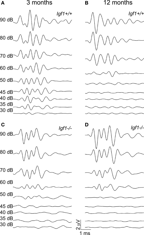
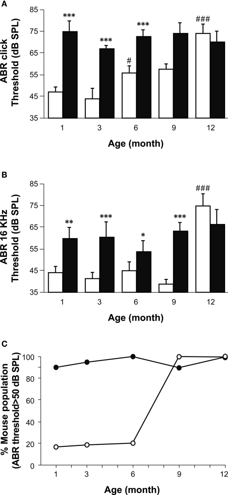
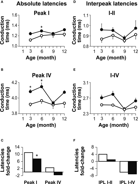
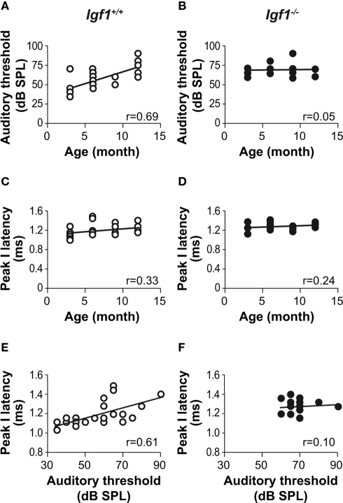
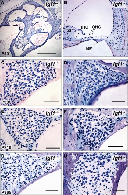
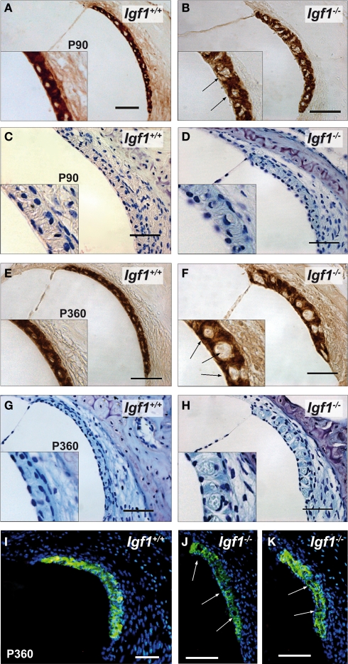
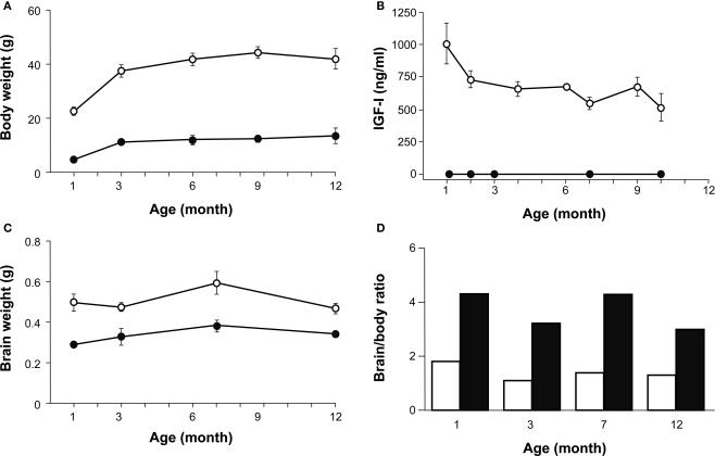
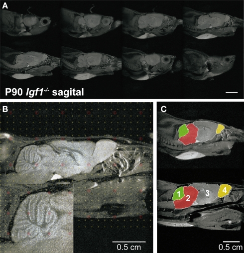
Insulin-like growth factor-I (IGF-I) belongs to the family of insulin-related peptides that fulfils a key role during the late development of the nervous system. Human IGF1 mutations cause profound deafness, poor growth and mental retardation. Accordingly, Igf1(-/-) null mice are dwarfs that have low survival rates, cochlear alterations and severe sensorineural deafness. Presbycusis (age-related hearing loss) is a common disorder associated with aging that causes social and cognitive problems. Aging is also associated with a decrease in circulating IGF-I levels and this reduction has been related to cognitive and brain alterations, although there is no information as yet regarding the relationship between presbycusis and IGF-I biodisponibility. Here we present a longitudinal study of wild type Igf1(+/+) and null Igf1(-/-) mice from 2 to 12 months of age comparing the temporal progression of several parameters: hearing, brain morphology, cochlear cytoarchitecture, insulin-related factors and IGF gene expression and IGF-I serum levels. Complementary invasive and non-invasive techniques were used, including auditory brainstem-evoked response (ABR) recordings and in vivo MRI brain imaging. Igf1(-/-) null mice presented profound deafness at all the ages studied, without any obvious worsening of hearing parameters with aging. Igf1(+/+) wild type mice suffered significant age-related hearing loss, their auditory thresholds and peak I latencies augmenting as they aged, in parallel with a decrease in the circulating levels of IGF-I. Accordingly, there was an age-related spiral ganglion degeneration in wild type mice that was not evident in the Igf1 null mice. However, the Igf1(-/-) null mice in turn developed a prematurely aged stria vascularis reminiscent of the diabetic strial phenotype. Our data indicate that IGF-I is required for the correct development and maintenance of hearing, supporting the idea that IGF-I-based therapies could contribute to prevent or ameliorate age-related hearing loss.

Keywords: Igf1−/− null mouse; aging; auditory brainstem responses; deafness; in vivo brain imaging; insulin-like factors; presbycusis; sensorineural deafness.


Figures

Figure 1
Figure 1
ABR wave recordings from representative 3- and 12-month-old Igf1+/+ wild type and Igf1−/− null mice. The waves represent the ABR in response to intensities of click stimuli decreasing from 90 dB SPL to 30 dB SPL of a representative mouse for each condition. (A,B) ABR waves from 3- (A) and 12- (B) month-old Igf1+/+ mice. (C,D) ABR waves from 3- (C) and 12- (D) month-old Igf1−/− mice. In (A) and (C) the intensity of the stimulus for each response is indicated, and the range of intensities is the same in (B) and (D).
Figure 2
Figure 2
A comparison of age-related hearing loss in Igf1+/+ and Igf1−/− mice. (A) ABR thresholds in response to click stimulus in Igf1+/+ (open bars) and Igf1−/− mice (closed bars) at different ages (1, 3, 6, 9 and 12 months). Wild type mice show an age-related increase in ABR thresholds, whereas the null mutant mice were deaf from the youngest age studied. (B) ABR responses to a 16 kHz stimuli were obtained at the same ages and showed a similar trait. (C) Percentage of individuals for each genotype showing ABR thresholds above normal hearing (≥50 dB SPL) at each of the ages studied. Wild type and null mice are represented as open and closed circles, respectively. Statistical analysis was performed with ANOVA and post hoc Bonferroni and Dunnet tests. Data are presented as the mean ± s.e.m; *indicates comparison between genotypes ***P < 0.001, **P < 0.01 and *P < 0.05; #indicates comparison between consecutive ages ###P < 0.001. The number (n) of mice studied were 15, 13, 9, 8 and 7 wild type mice of 1, 3, 6, 9 and 12 month old, respectively; and 10, 11, 10, 6 and 5 null mice of the same ages.
Figure 3
Figure 3
Age-related changes in absolute and interpeak latencies in both genotypes. (A,B) Absolute Peak I (A) and peak IV (B) latencies obtained at 80 dB SPL. Absolute latencies are plotted for Igf1+/+ (open circles) and Igf1−/− mice (closed circles) aged from 3 to 12 month old. (C) Percentage delay in latency with aging. Open and closed bars indicate wild type and null mice, respectively. The bars represent the percentage latency increase from 3 to 12 months of age for peaks I (bars to the left) and IV (bars to the right). The asterisk indicates that the 11% difference in peak I latency between 3- and 12-month-old wild type mice is significant (P < 0.05). Data are presented as the mean ± s.e.m. (D,E) Interpeak latencies (IPL) I–II (D) and I–IV (E) obtained at 80 dB SPL for Igf1+/+ (open circles) and Igf1−/− mice (closed circles) aged from 3 to 12 months old. (F) Percentage IPL increases with aging. As in (C), for wild type (open bars) and null mice (closed bars), the IPL I–II is shown by the bars on the left and IPL I–IV in the bars on the right. The number (n) of mice studied was: 7, 9, 4 and 5 wild type and 6, 6, 6 and 3 null mice of 3, 6, 9 and 12 month old, respectively.
Figure 4
Figure 4
Correlations between auditory thresholds and peak response latencies with aging and genotype. Product moment Pearson correlation coefficients were calculated for the pairs of parameters indicated. A clear correlation was evident between ABR thresholds and age in the Igf1+/+ mice [open circles; (A) P < 0.05; n = 24; r = 0.69] but not in the Igf1−/− mice [closed circles; (B) ns; n = 19; r = 0.05]. The absolute peak I latency was weakly correlated with age in both Igf1+/+ [(C) P < 0.1; n = 24; r = 0.33] and Igf1−/− [(D)P < 0.25; n = 19; r = 0.24] mice. The Pearson product moment correlation coefficient between peak I absolute latencies and auditory thresholds at different ages in Igf1+/+ mice was statistically significant (P < 0.05) [(E) P < 0.05; n = 24; r = 0.61]. The same correlation in Igf1−/− mice was not statistically significant. [(F) ns; n = 19; r = 0.09].
Figure 5
Figure 5
Comparative study of cochlear morphology with aging in Igf1+/+ wild type and Igf1−/− null mice. (A,B) Cochlear cross-section and close up of the cochlear basal turn in a representative 3-month-old null mice. The disposition of the cells in the organ of Corti is apparently normal. IHC: inner hair cell; OHC: outer hair cells; BM: basilar membrane. (C–H) Comparison in both genotypes of the spiral ganglion morphology and of the neuronal density in mice at the ages studied. The neuronal density decreased in the aged spiral ganglion, such that at the older ages studied the cochlear ganglia was morphologically similar in both genotypes. Some specimens displayed heavy hair cell loss at this age, which was correlated with the increase in hearing thresholds (data not shown). Scale bar 50 μm [0.5 mm in (A)].
Figure 6
Figure 6
Comparative study of the stria vascularis with the aging of Igf1+/+ wild type and Igf1−/− null mice. Na-K-ATPase (A,B,E,F) and cresyl violet staining (C,D,G,H) expression in the stria vascularis of 3- (A–D) and 12-month-old mice (E–H) showing the morphological differences associated with the aging of each genotype. The stria vascularis in the Igf1−/− mice had an abnormal morphology, it was shorter and thicker, and with evident dilation of the vascular spaces (arrows). Kir4.1 (KCNJ10) expression (I–K) in the stria vascularis of aged null mice was altered and there was a relative loss of expression in the stria and sacular dilations. Four to six mice per condition were analyzed. Scale bar 50 μm.
Figure 7
Figure 7
A comparison of age-related changes in IGF-I levels, and body and brain weight in both genotypes. (A) Body weight of Igf1+/+ (open circles) and Igf1−/− null (closed circles) mice was measured at 1, 3, 6 and 12 months of age. Statistical analysis in (A) and (C) was performed with ANOVA and post hoc Bonferroni and Dunnet tests. Data are presented as the mean ± s.e.m. Both genotypes presented a significant difference at all the ages studied (P < 0.001) and an age-related increase in body weight. (B) The serum IGF-I levels were followed during aging in Igf1+/+ and Igf1−/− mice in parallel. An age-related decline in serum IGF-I was observed in wild type mice (open circles) whereas no IGF-I could be detected at any time in the null mice (closed circles). Data are given in ng/ml as median ± s.e.m. At least six to eight mice were studied per group. (C) Brain weight measurements did not change significantly as the mice aged, although there was a significant difference between the two genotypes at the ages studied (P < 0.05). (D) The brain/body weight ratio was calculated for each age group of Igf1+/+ wild type (black bars) and Igf1−/− null mice (white bars). This ratio did not vary with age in any genotype. The number (n) of mice studied in (A), (C) and (D) were 19, 7, 9, 8 and 5 wild type and 8, 6, 7, 6 and 5 null mice of 1, 3, 6, 9 and 12 month old, respectively.
Figure 8
Figure 8
In vivo brain images of Igf1+/+ wild type and Igf1−/− null mice. (A) Complete series of brain slices in the sagittal plane of a representative 3-month-old Igf1−/− null mouse. (B) Sagittal MRI T2 weighted image of a representative 3-month-old wild type mouse showing the stereological grid that was randomly overlaid on each slide to estimate the volume. The inset shows the specific grid used to estimate the volume of specific brain areas, such as the cerebellum. (C) Comparison of the selected brain areas of Igf1−/− and Igf1+/+ mice: cerebellum (1), brainstem (2), diencephalon-telencephalon (3) and olfactory bulb (4). Note the reduced olfactory bulb in the deficient mice (yellow).
Figure 9
Figure 9
Brain distribution and expression of Igf2 and Igf1r. Igf1r (A) and Igf2 (B) mRNA levels were measured by quantitative RT-PCR of RNA from the indicated mouse brain regions and normalized to the levels of the 18S ribosomal RNA 3–5 mice per genotype were used. The results represent the mean ± s.d. Brainstem (BS), olfactory bulb (OB), cerebellum (CBL) and the remainder of the telencephalon-diencephalon (TD). Open and closed bars indicate Igf1+/+ and Igf1−/− mice, respectively.

Similar articles

Cited by

References

    1. Aberg D. (2010). Role of the growth hormone/insulin-like growth factor 1 axis in neurogenesis. Endocr. Dev. 17, 63–7610.1210/en.2006-1556 - DOI - PubMed
    1. Aleman A., Torres-Aleman I. (2009). Circulating insulin-like growth factor I and cognitive function: neuromodulation throughout the lifespan. Prog. Neurobiol. 89, 256–26510.1016/j.pneurobio.2009.07.008 - DOI - PubMed
    1. Barrenas M., Landin-Wilhelmsen K., Hanson C. (2000). Ear and hearing in relation to genotype and growth in Turner syndrome. Hear. Res. 144, 21–2810.1023/B:JARG.0000040239.40913.c3 - DOI - PubMed
    1. Barrenas M. L., Bratthall A., Dahlgren J. (2005). The association between short stature and sensorineural hearing loss. Hear. Res. 205, 123–13010.1016/j.heares.2005.03.019 - DOI - PubMed
    1. Bartke A. (2005). Minireview: role of the growth hormone/insulin-like growth factor system in mammalian aging. Endocrinology 146, 3718–372310.1210/en.2005-0411 - DOI - PubMed