. 2010 Jul 15;6(7):e1001025.
doi: 10.1371/journal.pgen.1001025.

The Caenorhabditis elegans homolog of Gen1/Yen1 resolvases links DNA damage signaling to DNA double-strand break repair

Affiliations

The Caenorhabditis elegans homolog of Gen1/Yen1 resolvases links DNA damage signaling to DNA double-strand break repair

Aymeric P Bailly et al. PLoS Genet. .

Abstract

DNA double-strand breaks (DSBs) can be repaired by homologous recombination (HR), which can involve Holliday junction (HJ) intermediates that are ultimately resolved by nucleolytic enzymes. An N-terminal fragment of human GEN1 has recently been shown to act as a Holliday junction resolvase, but little is known about the role of GEN-1 in vivo. Holliday junction resolution signifies the completion of DNA repair, a step that may be coupled to signaling proteins that regulate cell cycle progression in response to DNA damage. Using forward genetic approaches, we identified a Caenorhabditis elegans dual function DNA double-strand break repair and DNA damage signaling protein orthologous to the human GEN1 Holliday junction resolving enzyme. GEN-1 has biochemical activities related to the human enzyme and facilitates repair of DNA double-strand breaks, but is not essential for DNA double-strand break repair during meiotic recombination. Mutational analysis reveals that the DNA damage-signaling function of GEN-1 is separable from its role in DNA repair. GEN-1 promotes germ cell cycle arrest and apoptosis via a pathway that acts in parallel to the canonical DNA damage response pathway mediated by RPA loading, CHK1 activation, and CEP-1/p53-mediated apoptosis induction. Furthermore, GEN-1 acts redundantly with the 9-1-1 complex to ensure genome stability. Our study suggests that GEN-1 might act as a dual function Holliday junction resolvase that may coordinate DNA damage signaling with a late step in DNA double-strand break repair.


Conflict of interest statement

The authors have declared that no competing interests exist.

Figures
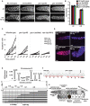
Figure 1
Figure 1. gen-1 mutants are defective in DNA damage–dependent cell cycle arrest and apoptosis and in positional cloning of GEN-1.
(A) Representative pictures of N2-wild type and gen-1 mitotic germ lines with and without IR treatment (60 Gy). Germ lines were dissected and stained with DAPI. Scale bar 10 µm (B) Statistical analysis of cell cycle arrest. All mitotic germ cells within 50 µm from the distal tip cell were counted (n = 7, error bars represent s.e.m.). (C) DNA damage-induced germ cell apoptosis is defective in gen-1 mutant worms (n = 15, error bars represent s.e.m.). Germ cell apoptosis was assayed as described . (D) gen-1 mutant germ cells fail to arrest in G2. Dissected germlines were stained with the human Cdk-1 phosphotyrosine 15 antibody as a G2 marker (red). (E) Mapping of gen-1(yp30). Linkage to the centre of chromosome III is shown in the upper panel. The ratio of yp30/versus “Hawaiian” DNA measured at various single nucleotide polymorphisms. Single recombination events placing yp30 map position between SNPs located at map position -1,43 and -1,38 are indicated in the lower panel. (F) Chromatograms showing the C to T transition found in the gen-1 (yp30) mutant. (G) Diagram of an adult hermaphrodite germ line adapted from .
Figure 2
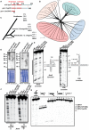
Figure 2. Phylogenetic analysis of GEN-1 and in vitro nuclease activity.
(A) Diagram showing the GEN-1 domain structure. (B) Unrooted phylogenetic tree of XPG-superfamily members. Bacterial Taq. Pol1 serves as an outgroup. Protein sequences were aligned using Jalview 2.4 employing the MAFT algorithm , and the Splitstree program was used to generate the tree shown using the Neighbour Joining Method. The length of the scale bar indicates the lengths of the branches of the phylogenetic trees corresponding to a 10% chance (p 0.1) of replacing an amino acid/site. An, Aspergillus nidulans, Bm, Brugia malayi, Ca, Candida albicans, Ci, Ciona intestinalis, Dm, Drosophila melanogaster, Gg, Gallus gallus, Hs, Homo sapiens, Nv, Nematostella vectensis, Pp, Pichia pasteuris, Sc, Saccharomyces cerevisiae, Sp, Schizosaccharomyces pombe. (C) Four distinct XPG-1 family members occur in all animals examined. The current view of phylogenetic relationships between vertebrates, insects and nematodes is indicated. (D) Affinity purified GEN-1 antibodies (guinea pig) detect a specific band corresponding to the predicted size of GEN-1 and GEN-1 (yp30) (for details Figure S7B, S7C). Equal loading is demonstrated by the Coomassie staining of the membrane. (E) Jbm5 (left panel) and X26 (right panel) junctions, radioactively [5′-32P]-labeled on the b and a strand respectively, were incubated at 37°C with the indicated protein (Figure S4A), and the cleavage products analyzed by denaturing gel electrophoresis in polyacrylamide. Lane 1 is a sequence marker for the b stand (left panel). The sequence of the junction is shown, with the homologous section shaded, and cleavage sites are indicated. (F) Symmetry of Holliday junction cleavage by recombinant GEN1. Holliday junction Jbm5, radioactively [5′-32P]-labeled on either the b or the d strand was incubated at 37°C with the indicated protein, and the cleavage products were analysed by electrophoresis in polyacrylamide gels under denaturing conditions. M indicates the sequence marker for each respective strand. (G) GEN-1 is specific for Holliday Junctions. Single-stranded, duplex, 3′ overhang and 5′ flap structures were subjected to GEN-1 nucleolytic activity as shown in E. For each reaction, 5 nM of gel purified substrates has been used. For more details see Materials and Methods.
Figure 3
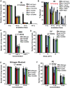
Figure 3. Sensitivity of gen-1 (tm2940) and gen-1 (yp30) in response to DNA damaging agents.
(A) L1 stage worms sensitivity assay to IR, (B) L4 stage worms sensitivity assay to IR. (C) MMS, (D) UV irradiation, (E) nitrogen mustard, and (F) HU exposure of L1 stage worms sensitivity assay. Assays were performed as described in Materials and Methods. Error bars represent s.e.m.
Figure 4
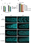
Figure 4. Persistent RAD-51 foci in gen-1(tm2940) and gen-1(yp30) worms.
Rad-51 foci were scored 48 h post IR with 30 Gy as described . A layer of only five z-stacks is displayed for clarity. DAPI and RAD-51 correspond to blue and red staining respectively. A quantification of foci per mitotic germ line is shown in the right panel. n = 5, error bars represent s.e.m. Foci were counted by projecting all the z-stacks using SoftWorks. Scale bar is 10 µm.
Figure 5
Figure 5. Chromosome fragmentation assay.
(A) L4 worms were irradiated with 90 Gy of IR and dissected 48 h later. Scale bar is 5 µm. (B) A quantification of the extent of chromosome fragmentation (n = 12). (C) No chromosome fragmentation is observed 20 h (top panel) and 8 h after irradiation (lower panel).
Figure 6
Figure 6. gen-1 acts in a non-canonical DNA damage checkpoint pathway.
(A) cep-1 mediated transcriptional induction of egl-1 and ced-13. Quantitative RT-PCRs were performed as described , induction compared to unirradiated N2 wild type is shown (B), gen-1 RNAi leads to synthetic lethality in conjunction with hus-1(op241) and mrt-2(e2663) mutations. Percent (%) survival indicates the number of eggs hatched and grown to adulthood. RNAi against GFP was used as negative RNAi feeding control. (C) Phospho-Chk-1 antibody staining 6 h after IR (60 Gy) treatment of the indicated genotypes. Scale bar is 10 µm.
Figure 7
Figure 7. L1 stage worms IR sensitivity assay with gen-1 and gen-1 atm-1 double mutants.
Assays were performed as described in Figure 3. Error bars represent s.e.m.
Figure 8
Figure 8. Model.
A processed DSB with a single stranded tail coated by RPA1 and ATR is indicated at the top of the panel. Signaling via the 9-1-1 complex, Chk-1 and CEP-1/p53 is indicated in the right. A Holliday junction is depicted on the left and the predicted symmetrical cleavage activity of GEN-1 is indicated by two arrows.

References

    1. Mimitou EP, Symington LS. DNA end resection: Many nucleases make light work. DNA Repair (Amst) 2009 - PMC - PubMed
    1. Zou L, Elledge SJ. Sensing DNA damage through ATRIP recognition of RPA-ssDNA complexes. Science. 2003;300:1542–1548. - PubMed
    1. Zhou BB, Elledge SJ. The DNA damage response: putting checkpoints in perspective. Nature. 2000;408:433–439. - PubMed
    1. Green CM, Erdjument-Bromage H, Tempst P, Lowndes NF. A novel Rad24 checkpoint protein complex closely related to replication factor C. Curr Biol. 2000;10:39–42. - PubMed
    1. Majka J, Burgers PM. Yeast Rad17/Mec3/Ddc1: a sliding clamp for the DNA damage checkpoint. Proc Natl Acad Sci U S A. 2003;100:2249–2254. - PMC - PubMed

Publication types

Substances