. 2010 Jul 22;5(7):e11469.
doi: 10.1371/journal.pone.0011469.

Tumor-derived microvesicles induce, expand and up-regulate biological activities of human regulatory T cells (Treg)

Affiliations

Tumor-derived microvesicles induce, expand and up-regulate biological activities of human regulatory T cells (Treg)

Marta Szajnik et al. PLoS One. .

Abstract

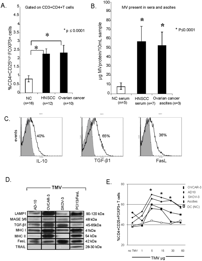
Background: Tumor-derived microvesicles (TMV) or exosomes are present in body fluids of patients with cancer and might be involved in tumor progression. The frequency and suppressor functions of peripheral blood CD4(+)CD25(high)FOXP3(+) Treg are higher in patients with cancer than normal controls. The hypothesis is tested that TMV contribute to induction/expansion/and activation of human Treg.

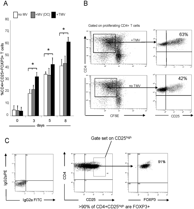
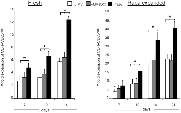
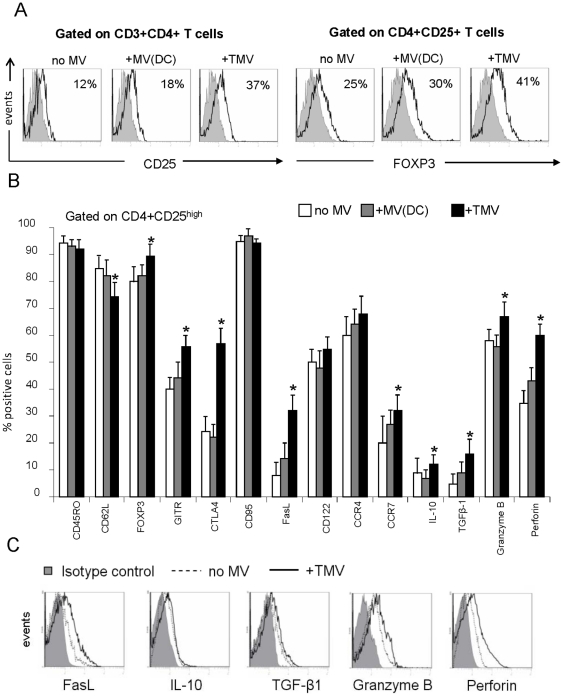
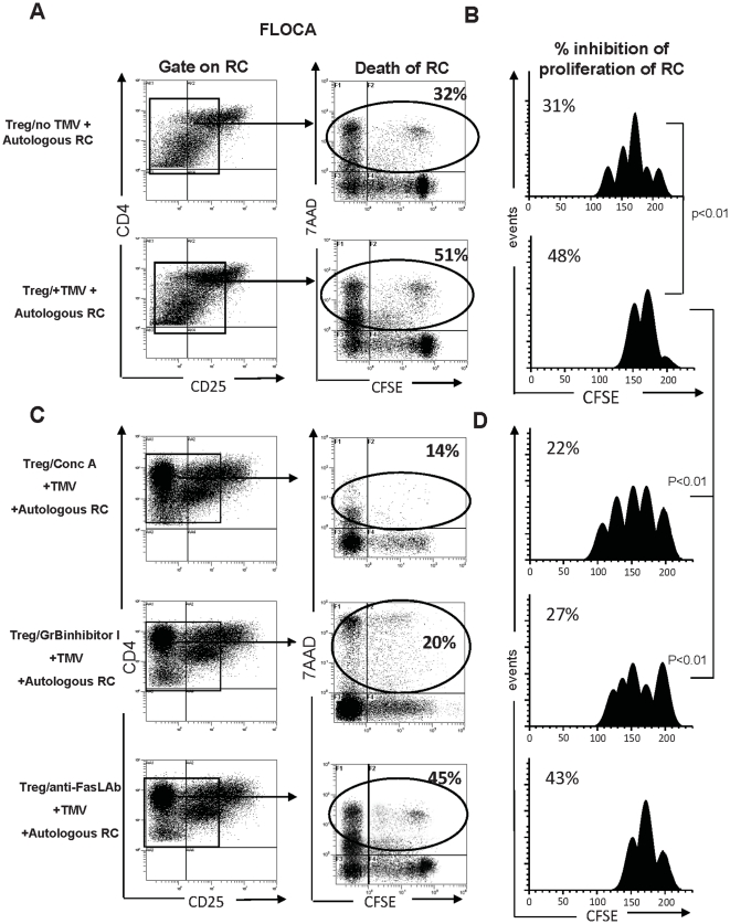
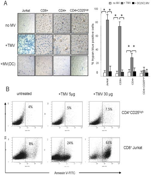
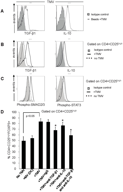
Methodology/principal findings: TMV isolated from supernatants of tumor cells but not normal cells induced the generation and enhanced expansion of human Treg. TMV also mediated conversion of CD4(+)CD25(neg) T cells into CD4(+)CD25(high)FOXP3(+) Treg. Upon co-incubation with TMV, Treg showed an increased FasL, IL-10, TGF-beta1, CTLA-4, granzyme B and perforin expression (p<0.05) and mediated stronger suppression of responder cell (RC) proliferation (p<0.01). Purified Treg were resistant to TMV-mediated apoptosis relative to other T cells. TMV also increased phospho-SMAD2/3 and phospho-STAT3 expression in Treg. Neutralizing Abs specific for TGF-beta1 and/or IL-10 significantly inhibited TMV ability to expand Treg.

Conclusions/significance: This study suggests that TMV have immunoregulatory properties. They induce Treg, promote Treg expansion, up-regulate Treg suppressor function and enhance Treg resistance to apoptosis. Interactions of TMV with Treg represent a newly-defined mechanism that might be involved in regulating peripheral tolerance by tumors and in supporting immune evasion of human cancers.


Conflict of interest statement

Competing Interests: The authors have declared that no competing interests exist.

Figures

Figure 1
Figure 1. CD4+CD25highFOXP3+ T cells and microvesicles (MV) in cancer patients and normal controls (NC).
(A) Percentages of CD4+CD25highFOXP3+ Treg in PBMC of cancer patients and NC. (B) the protein content/10 mL of serum or ascites in cancer patients and NC. The data in A and B are mean values ± SD. (C) Flow analyses of IL-10, TGF-β1 and FasL expression in MV purified from the ascites of OvCa patients and coated on latex beads. (D) Western blots of TMV isolated from OvCa SN. Molecular weights of the detected proteins are indicated. (E) Percentages of CD4+CD25+FOXP3+ cells in 8-day co-cultures of CD4+CD25neg T cells with TMV obtained from various sources and used at increasing concentrations. The asterisks indicate a significant increase at p<0.05.
Figure 2
Figure 2. TMV promote differentiation of human Treg in culture.
(A) Purified CD3+CD4+ T cells were labeled with CFSE and cultured as described in Materials and Methods ± TMV or DC-derived MV (5 µg/mL). On days 3, 5 and 8, the frequency of CD4+CD25+FOXP3+ Treg among proliferating T cells was determined by flow cytometry. The data (means ± SD) represent three independent experiments (*p<0.01). (B) Proliferating CD3+CD4+ T cells (squares) were tested for co-expression of CD25 in a representative co-culture ± TMV. A higher proportion of proliferating CD4+ T cells expressed CD25 in the co-culture with TMV than without TMV. (C) The proliferating CD4+CD25+ T cells in the co-cultures with TMV were evaluated for the frequency of FOXP3+ T cells upon gating on the CD4+CD25high subset (see box). Over 90% of these cells also expressed intracellular FOXP3. Data are representative for one out of 6 cultures tested.
Figure 3
Figure 3. TMV promote expansion of human Treg in culture.
The fold expansion of fresh (left panel) or rapamycin-expanded (right panel) CD4+CD25high T cells to which TMV or DC-derived MV were added on day 0. Cells were stimulated with OKT3, anti-CD28 Abs and IL-2 (500 IU/mL) and cultured for 14–21 d. The data are means ± SD of six independent co-cultures. Asterisks indicate significant differences (p<0.05) between the cultures ± TMV.
Figure 4
Figure 4. TMV Convert CD25neg T cells to Treg.
(A) Flow cytometry histograms of cultured (d5) CD4+CD25neg T cells showing conversion of CD25neg T cells into CD25+ T cells ± TMV or DC-derived MV (left panel) and expression of FOXP3 in the converted CD4+CD25+ T cells (right panel) in the same cultures. (B) A phenotypic profile of CD4+CD25high T cells present in 7 day cultures of CD4+CD25+ T cells ± TMV or DC-derived MV. T cells were stained with various mAbs and evaluated by multiparameter flow cytometry. The gate is set on CD4+CD25high T cells. The data are mean percentages ± SD of positive cells from three independent experiments. (C) MFI for FasL, IL-10, TGF-β1, granzyme B and perforin expression in CD4+CD25high T cells cultured as described in (B) ± TMV. The data are representative of three independent experiments.
Figure 5
Figure 5. TMV increase suppressor activity of Treg.
The FLOCA was used to simultaneously measure suppression proliferation of CFSE-labeled autologous CD4+CD25neg RC and their apoptosis upon co-incubation with CFSE-negative Treg. RC cells stimulated with OKT3, anti-CD28 mAb and IL-2 (150 IU/mL) were co-cultured for 5 d with Treg pre-incubated or not with TMV. At harvest, cells were stained with 7-AAD and examined by flow cytometry. The suppressor assays were performed at the S:RC ratio of 1∶1. Treg pre-incubated with TMV induced higher levels of apoptosis (A) and greater inhibition of RC proliferation (B). The data are from one experiment of five performed. When Treg were pretreated with Concanamycin A or GrB inhibitor I and then incubated with TMV, the frequency of 7-AAD+ RC was lower (C) as was RC proliferation inhibition (D). Treg pretreated with FasL Ab and then incubated with TMV induced RC death (C) and inhibited RC proliferation (D).
Figure 6
Figure 6. CD4+CD25high Treg are resistant to TMV-induced death.
(A) Trypan blue positive cells after 6 h incubation ± TMV or DC-derived MV in primary T-cell subsets and CD8+ Jurkat cells (mag ×200) *p<0.001. (B) Percentages of ANXV binding to fresh CD4+CD25high T cells or CD8+ Jurkat cells incubated ± TMV for 6 h. The data are representative dot plots from one of five independent experiments.
Figure 7
Figure 7. TMV-associated TGF-β1 and IL-10 promote Treg expansion.
(A) Flow cytometry analysis of TGF-β1 and IL-10 expression on TMV purified from OVCAR-3 SN and coated onto latex beads. (B) CD4+CD25highFOXP3+ T cells were cultured with OKT3, anti-CD28 and IL-2 (150 IU/mL) +/− TMV for 72 h at 37°C in the presence of Golgistop and then stained for CD4, CD3, CD25 and intracellular TGF-β1 and IL-10. Expression of both cytokines was up-regulated in the presence of TMV (p<0.05). (C) SMAD2/3 and STAT3 phosphorylation in Treg before and after exposure to TMV. Representative results are from one of three independent experiments for A, B and C. (D) The percentage of CD4+CD25highFOXP3+ T cells increased among CD4+CD25+ T cells cultured in the presence of TMV but not DC-derived MV. Neutralizing anti-TGF-β1 and/or anti-IL-10 Abs inhibited the induction of Treg by TMV. Non-blocking IgG isotype control Abs were used as controls. Asterisks indicate decreases (p<0.05) in Treg percentages in the presence of neutralizing Abs. Results are means ± SD of three independent experiments.

References

    1. Whiteside TL. The tumor microenvironment and its role in promoting tumor growth. Oncogene. 2008;27:5904–5912. - PMC - PubMed
    1. Wolf D, Wolf AM, Rumpold H, Fiegl H, Zeimet AG, et al. The expression of the regulatory T cell-specific forkhead box transcription factor FoxP3 is associated with poor prognosis in ovarian cancer. Clin Cancer Res. 2005;11:8326–8331. - PubMed
    1. Liyanage UK, Moore TT, Joo HG, Tanaka Y, Herrmann V, et al. Prevalence of regulatory T cells is increased in peripheral blood and tumor microenvironment of patients with pancreas or breast adenocarcinoma. J Immunol. 2002;169:2756–2761. - PubMed
    1. Curiel TJ, Coukos G, Zou L, Alvarez X, Cheng P, et al. Specific recruitment of regulatory T cells in ovarian carcinoma fosters immune privilege and predicts reduced survival. Nat Med. 2004;10:942–949. - PubMed
    1. Wieckowski E, Visus C, Szajnik M, Szczepanski MJ, Storkus WJ, et al. Tumor-derived microvesicles promote regulatory T-cell expansion and induce apoptosis in tumor-reactive activated CD8+ T lymphocytes. J Immunol. 2009;183:3720–3730. - PMC - PubMed

Publication types

Substances