. 2010 Jul 22;6(7):e1001010.
doi: 10.1371/journal.ppat.1001010.

Uropathogenic Escherichia coli modulates immune responses and its curli fimbriae interact with the antimicrobial peptide LL-37

Affiliations

Uropathogenic Escherichia coli modulates immune responses and its curli fimbriae interact with the antimicrobial peptide LL-37

Ylva Kai-Larsen et al. PLoS Pathog. .

Abstract

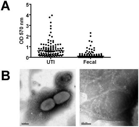
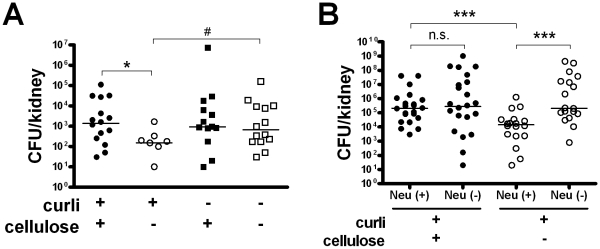
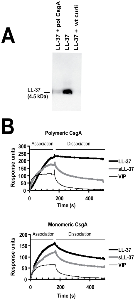
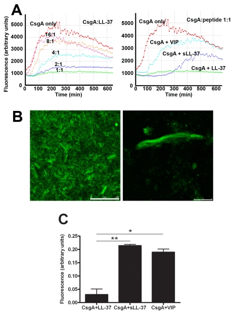
Bacterial growth in multicellular communities, or biofilms, offers many potential advantages over single-cell growth, including resistance to antimicrobial factors. Here we describe the interaction between the biofilm-promoting components curli fimbriae and cellulose of uropathogenic E. coli and the endogenous antimicrobial defense in the urinary tract. We also demonstrate the impact of this interplay on the pathogenesis of urinary tract infections. Our results suggest that curli and cellulose exhibit differential and complementary functions. Both of these biofilm components were expressed by a high proportion of clinical E. coli isolates. Curli promoted adherence to epithelial cells and resistance against the human antimicrobial peptide LL-37, but also increased the induction of the proinflammatory cytokine IL-8. Cellulose production, on the other hand, reduced immune induction and hence delayed bacterial elimination from the kidneys. Interestingly, LL-37 inhibited curli formation by preventing the polymerization of the major curli subunit, CsgA. Thus, even relatively low concentrations of LL-37 inhibited curli-mediated biofilm formation in vitro. Taken together, our data demonstrate that biofilm components are involved in the pathogenesis of urinary tract infections by E. coli and can be a target of local immune defense mechanisms.


Conflict of interest statement

The authors have declared that no competing interests exist.

Figures

Figure 1
Figure 1. Biofilm expression by uropathogenic and fecal E. coli isolates.
(A) Adhesion capacity and thickness of biofilm produced by E. coli isolates collected from urine of patients with urinary tract infections (UTI, n = 99) and from fecal samples of healthy individuals (Fecal, n = 77) was measured. Individual values and medians are presented, depicted as optical density (OD) at 570 nm after dissolution of crystal violet. The difference is significant (P<0.0001, Mann-Whitney U test). (B) Isolates from urine samples were investigated by electron microscopy. The left image shows an overview, the right image is a magnification showing immunogold-labelled curli. The scale bars show 0.5 µm (left) and 100 nm (right), respectively.
Figure 2
Figure 2. Adhesion and immune induction by E. coli expressing or lacking curli and/or cellulose.
(A) Adhesion to renal epithelial cells A498 was measured after 30 min. Curliated strains adhered significantly better to renal epithelial cells than strains lacking curli, independent of the expression of cellulose (### P<0.0001, t-test). Cellulose expression decreased the number of cell-associated bacteria in curliated (*** P<0.0001, t-test) and non-curliated strains (** P = 0.001, t-test). Results from three independent experiments in quadruplicates are shown as mean and standard deviation. Similar results were obtained for bladder epithelial cells (data not shown). (B) Induction of IL-8 was measured in culture supernatants of renal epithelial cells A498 stimulated with E. coli for 24 h. Curliated bacteria induced a significantly stronger IL-8 response than the mutants lacking curli in the presence (## P = 0.001, t-test) and absence (### P<0.0001, t-test) of cellulose. In curliated bacteria, the expression of cellulose reduced IL-8 induction (** P = 0.001, t-test). Results from three independent experiments in quadruplicate are shown as mean and standard deviation. Similar results were obtained for bladder epithelial cells (data not shown). (C+D) The phenotype of E. coli No. 12 could be restored by complementation of its mutants. Cellulose expression in strain B23 is inducible by aTc (left panels) and reduces adherence and IL-8 induction (*** P<0.0001 and ** P = 0.007, respectively, t-test). The curli subunits CsgA and CsgB are expressed from pWSK29-csgBA in strain WE11_1 (right panels) and increases adherence and IL-8 induction compared to WE11_1 carrying the vector pWSK29 only (* P = 0.048 and ** P = 0.003, respectively, t-test). Results in A498 cells are shown as mean and standard deviation. Data from three experiments in quadruplicate for adherence and from two experiments in triplicate for IL-8 induction are presented. (E) Mice were infected with the isogenic E. coli strains for 1 h. The curliated mutant were isolated from kidneys in significantly higher numbers than the double knockout (# P = 0.026, Mann-Whitney U test). Individual values from n = 8–10 mice/group and medians are shown. (F) Levels of MIP-2 were measured in kidney tissue of infected mice after 16 h. In the absence of cellulose, the curliated mutant strain induced higher levels of MIP-2 compared to the non-curliated strain (## P = 0.001, Mann-Whitney U test). Expression of cellulose reduced the induction of MIP-2 in the presence (*** P<0.0001, Mann-Whitney U test) and absence of curli (** P = 0.001, Mann-Whitney U test). Individual values from n = 5–10 mice/group and medians are shown.
Figure 3
Figure 3. Cellulose delays bacterial elimination in vivo.
(A) The expression of cellulose in curliated strains increased the number of bacteria in kidneys determined at 48 h p.i. (* P = 0.011, Mann-Whitney U test). The expression of curli in the absence of cellulose, on the other hand, mediated a more rapid elimination (# P = 0.011, Mann-Whitney U test). Individual values from n = 5–8 mice/group and medians are shown. (B) Cellulose expression reduces bacterial clearance by neutrophils. Control mice (Neu +) and mice with induced neutropenia (Neu −) were infected with curliated E. coli strains, and the bacterial load in the kidneys was determined 48 h p.i. A difference between bacteria with or without cellulose was only seen in the presence of neutrophils (P = 0.001, Mann-Whitney U test). Individual values from n = 9–12 mice/group and medians are shown.
Figure 4
Figure 4. Curli increase the resistance to the antimicrobial peptide LL-37.
(A) Bladder epithelial cells T24 were infected with bacteria for 30 min and adherent bacteria were subjected to LIVE/DEAD staining. Curli and cellulose expression enhanced bacterial resistance to antimicrobial properties (### P<0.001 for curliated strains versus non-curliated strains, * P = 0.023 and ** P = 0.003 for cellulose expressing strains with or without curli, t-test). Combined data from four experiments are shown. (B+C) Bacteria were exposed to conditioned medium of bladder epithelial cells T24 stimulated with phenylbutyrate to enhance LL-37 production. Curli expression enhanced bacterial survival over 30 min (## P = 0.006, t-test) (B). Results from three experiments in triplicate are shown. Conditioned medium was incubated with neutralizing anti-LL-37-antibodies (nAb) or isotype control antibodies (Co) prior to bacterial inoculation. Neutralizing of LL-37 had no effect on viability of the curliated strain (left) but enhanced viability of the double knockout (right, * P = 0.047, t-test) (C). Results from four experiments in triplicate are shown. (D–G) The susceptibility to LL-37 and mCRAMP of E. coli strains expressing or lacking curli or cellulose was tested by the broth dilution method. The expression of curli increased the resistance to both LL-37 (D+E) and mCRAMP (F+G). A significant difference of bacterial growth was observed at 10 µM LL-37 between curliated and non-curliated strains (### P<0.001, t-test). The curliated strains were also significantly more resistant to 5 µM mCRAMP than bacteria not producing curli (# P<0.05, t-test). An increased resistance to both cathelicidins was not observed for cellulose. Mean and standard deviation from data of two separate experiments in triplicates are shown. The IC50 is indicated by a broken line.
Figure 5
Figure 5. LL-37 binds to recombinant polymerized CsgA and isolated wild-type curli.
(A) Western blot analysis of supernatants after precipitation of LL-37 with curli. By adding polymeric CsgA (pol CsgA) or wild-type curli (wt curli) to a solution of 0.1 µM LL-37, the levels of LL-37 decreased in the supernatants after centrifugation. (B) Surface plasmon resonance. LL-37 exhibits a stronger association and lower dissociation rates to both polymeric (upper panel) and monomeric CsgA (lower panel) compared to the control peptides sLL-37 and VIP.
Figure 6
Figure 6. LL-37 prevents formation of biofilm by E. coli in vitro.
(A) Different concentrations of LL-37 and the control peptides sLL-37 and VIP were added to the curli-expressing mutant. (B) At 2.5 µM, LL-37 caused more than 80% reduction of biofilm production, whereas the same concentration of the control peptides gave a reduction of only ∼10%. Mean and standard deviation from data of two separate experiments in triplicates are shown. The difference between LL-37 versus sLL-37 or VIP at 2.5 µM was statistically significant (*** P = 0.001, t-test). Similar results were obtained for the wild-type strain expressing both curli and cellulose (data not shown).
Figure 7
Figure 7. LL-37 inhibits CsgA polymerization.
(A) Monomeric CsgA was incubated without or with different concentrations of LL-37 (left). The CsgA monomers formed fibers that could be detected by the fiber-specific fluorescent dye Thioflavin (ThT). When bound to fibers, ThT gave rise to fluorescence that was detected by a Tecan plate reader. The fiber formation was inhibited by LL-37 in a dose-dependent manner and was completely inhibited at a molar ratio of 1∶1 (CsgA∶LL-37). As control peptides, VIP and sLL-37 were included (right). (B+C) Confocal images of polymerized CsgA stained with ThT. (B) The left image shows an overview, the right image a magnification of an aggregate of fibers. Scale bars are 250 µm (left) and 25 µm (right), respectively. (C) Fluorescent signals from CsgA incubated with LL-37, sLL-37, or VIP were quantified. Values were corrected for the contribution of the peptides themselves and are expressed in relation to the signal from CsgA alone. The inhibitory effect of LL-37 is significantly stronger than the effect of sLL-37 (** P = 0.007) or VIP (* P = 0.011). Combined results from two experiments are presented.
Figure 8
Figure 8. The monomeric form of CsgA remains stable in the presence of LL-37.
(A) Monomeric CsgA was incubated for 20 h at 37°C without or with LL-37. When CsgA is incubated together with LL-37, the CsgA monomer is visible after SDS-PAGE, whereas the monomer is not detected in the absence of LL-37. When polymeric CsgA is treated with formic acid (FA), the CsgA monomer is detectable, excluding degradation of CsgA. The bands of ∼30 and ∼60 kDa are most likely the dimer and tetramer of CsgA, respectively. (B) The stability of the monomeric form of CsgA in the presence of LL-37 was also confirmed with CD spectroscopy after 60 h incubation. The CD spectrum reveals that CsgA alone exhibits a fiber-like structure with weak beta-sheet conformation and a decreased solubility. In contrast, CsgA incubated together with LL-37 displays an unstructured, random coil structure. The spectrum of LL-37 alone was subtracted from the spectrum of CsgA + LL-37.

Similar articles

Cited by

References

    1. Roberts RC, Mohr CD, Shapiro L. Developmental programs in bacteria. Curr Top Dev Biol. 1996;34:207–257. - PubMed
    1. Costerton JW, Lewandowski Z, Caldwell DE, Korber DR, Lappin-Scott HM. Microbial biofilms. Annu Rev Microbiol. 1995;49:711–745. - PubMed
    1. Folkesson A, Haagensen JAJ, Zampaloni C, Sternberg C, Molin S. Biofilm induced tolerance towards antimicrobial peptides. PLoS ONE. 2008;3:e1891. - PMC - PubMed
    1. Hall-Stoodley L, Costerton JW, Stoodley P. Bacterial biofilms: from the natural environment to infectious diseases. Nat Rev Micro. 2004;2:95–108. - PubMed
    1. Shapiro JA. Thinking about bacterial populations as multicellular organisms. Ann Rev Microbiol. 1998;52:81–104. - PubMed

Publication types