

Microcirculatory abnormalities in patients with severe influenza A (H1N1) infection
- PMID: 20661679
- PMCID: PMC7101965
- DOI: 10.1007/s12630-010-9365-6
Microcirculatory abnormalities in patients with severe influenza A (H1N1) infection
Abstract
Purpose: This study was designed to evaluate the degree of microcirculatory abnormalities in patients with severe influenza A (H1N1) infection.
Methods: We assessed the sublingual microcirculation in seven consecutive patients with acute lung injury related to influenza A (H1N1) infection. The evaluation was carried out using sidestream dark field (SDF) imaging within the first 96 hr after the patients were admitted to the intensive care unit. Thenar oxygen saturation (StO(2)) was also measured with near-infrared spectroscopy (NIRS) during a vascular occlusion test. In addition, the Lung Injury Score (LIS) and the APACHE II and SOFA scores were recorded.
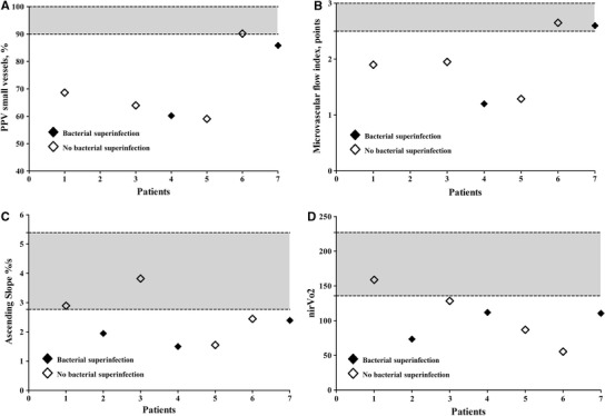
Results: All patients received invasive mechanical ventilation and at least one of the following adjuvant therapies: inhaled nitric oxide (n = 4), extracorporeal membrane oxygenation (n = 1), prone position (n = 4), recruitment maneuver (n = 3), and hydrocortisone 50 mg·hr(-6) (n = 6). The median time from admission to microcirculatory assessment was 21 hr. Three patients had bacterial superinfection. The median LIS and PaO(2)/F(i)O(2) were 2.5 (2.25-3.25) and 178 (158-212), respectively. Three subjects were treated with norepinephrine. During a vascular occlusion test, the microcirculation was moderately to severely compromised with a NIRS ascending slope of 2.39%·sec(-1) (1.75-2.67%·sec(-1)), 66% (60-86%) of perfused small vessels in the sublingual microcirculation, and a microvascular flow index of 1.9 (1.3-2.6). The degree of microcirculatory abnormalities detected by the NIRS and SDF imaging techniques was correlated with the severity of the disease, as reflected by the SOFA and APACHE II scores.
Conclusions: The microcirculation as assessed by SDF imaging and NIRS techniques was compromised in patients with acute respiratory distress syndrome (ARDS) and influenza A (H1N1) infection.
Objectif: Cette étude a été conçue afin d’évaluer le degré d’anomalies microcirculatoires chez des patients souffrant d’une infection grave à la grippe A (H1N1).
Méthode: Nous avons examiné la microcirculation sublinguale chez sept patients consécutifs souffrant d’une lésion pulmonaire aiguë liée à une infection à la grippe A (H1N1). L’évaluation a été réalisée à l’aide d’imagerie en champ noir à épi-illumination latérale (sidestream dark field – SDF) au cours des premières 96 h suivant l’admission des patients à l’unité des soins intensifs. La saturation thénarienne en oxygène (StO2) a également été mesurée par spectroscopie proche infrarouge (NIRS) pendant un test d’occlusion vasculaire. De plus, un Score de lésion pulmonaire (LIS) et les scores APACHE II et SOFA ont été enregistrés.
Résultats:
Tous les patients ont subi une ventilation mécanique invasive et au moins l’une des thérapies adjuvantes suivantes: monoxyde d’azote inhalé (n = 4), oxygénation par membrane extracorporelle (n = 1), position ventrale (n = 4), manœuvre de recrutement (n = 3), et hydrocortisone 50 mg·h−6 (n = 6). Le temps moyen entre l’admission et l’évaluation microcirculatoire était de 21 h. Trois patients souffraient de surinfection bactérienne. Les LIS et PaO2/F
Conclusion: La microcirculation, telle qu’analysée par des techniques d’imagerie SDF et de NIRS, a été compromise chez les patients souffrant de syndrome de détresse respiratoire aiguë (SDRA) et d’infection à la grippe A (H1N1).
Conflict of interest statement
None declared.
Figures
References
-
- Centers for Disease Control and Prevention (CDC). Swine influenza A (H1N1) infection in two children–Southern California, March-April 2009. MMWR Morb Mortal Wkly Rep 2009; 58: 400-2. - PubMed
-
- World Health Organization. WHO Pandemic (H1N1) 2009 - update 86. http://www.who int/csr/don/2010_02_5/en/index.html (accessed July 2010).
Publication types
MeSH terms
Substances
LinkOut - more resources
Full Text Sources
Medical
