. 2010 Nov;62(11):3237-48.
doi: 10.1002/art.27667.

Engagement of fatty acids with Toll-like receptor 2 drives interleukin-1β production via the ASC/caspase 1 pathway in monosodium urate monohydrate crystal-induced gouty arthritis

Affiliations

Engagement of fatty acids with Toll-like receptor 2 drives interleukin-1β production via the ASC/caspase 1 pathway in monosodium urate monohydrate crystal-induced gouty arthritis

Leo A B Joosten et al. Arthritis Rheum. 2010 Nov.

Abstract

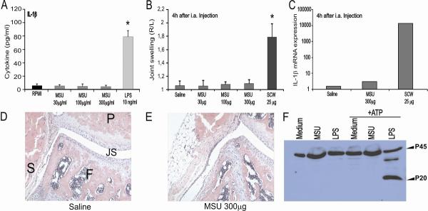
Objective: The concept that intraarticular crystals of uric acid by themselves trigger episodes of painful gouty arthritis is inconsistent with the clinical reality. Patients with large deposits of monosodium urate monohydrate (MSU) crystals (tophi) do not necessarily experience gouty attacks. In fact, it is the excessive consumption of food or alcohol that elicits the inflammation of the acute gout attack. The aim of this study was to identify the precise mechanism that initiates flares of gouty arthritis.

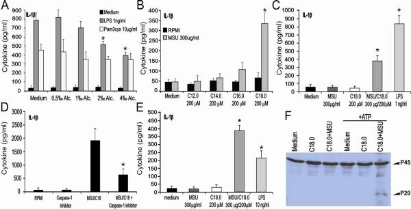
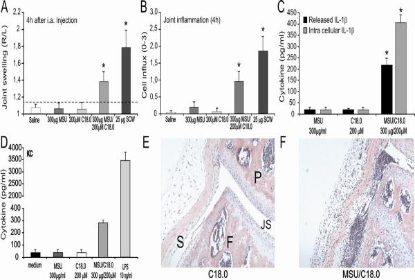
Methods: Human peripheral blood mononuclear cells (PBMCs) and murine macrophages were stimulated in vitro with MSU, free fatty acids (FFAs), or both in combination. Thereafter, production of interleukin-1β (IL-1β) and activation of caspase 1 were determined. Gouty arthritis was induced in mice with deficiencies in the genes for caspase 1, ASC, NALP3, or IL-1β, and the lack of inflammasome activity during joint swelling or other joint pathologic features was investigated in these mice.

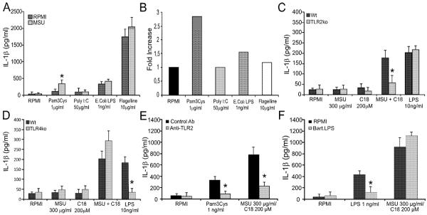
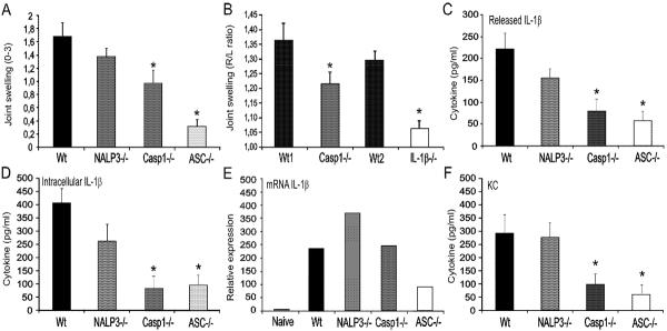
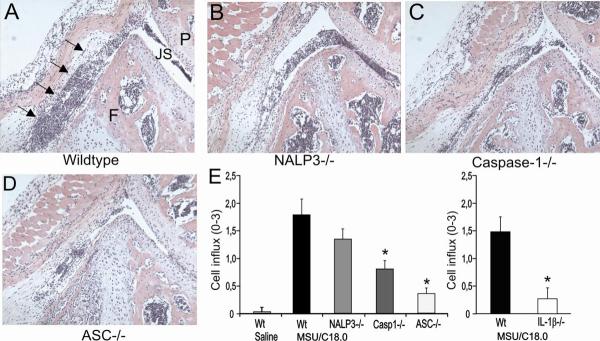
Results: MSU crystals had no biologic effects on PBMCs from healthy subjects, whereas the FFA C18:0 in the presence of MSU crystals induced the release of large amounts of IL-1β following engagement of Toll-like receptor 2 (TLR-2). Interaction of FFAs, but not alcohol, with TLR-2 synergized with MSU crystals to induce an inflammatory reaction. An important event of MSU/FFA-induced acute joint inflammation is the activation of the inflammasome. MSU/FFA-induced release of IL-1β was dependent on activation of caspase 1 and ASC, but surprisingly, not NALP3.

Conclusion: The synergistic effect between FFAs and MSU crystals leads to ASC/caspase 1-driven IL-1β release. This mechanism could explain how constitutionally derived metabolic events initiate attacks of gout via the induction of IL-1β-mediated joint inflammation.


Figures

Figure 1
Figure 1. MSU crystals do not induce IL-1β production or joint inflammation
A. Human PBMC's were exposed to a dose-range of MSU for 24h, and IL-1β was determined in the supernatants using ELISA. LPS (1ng/ml) was used as control. Data are expressed as mean±SEM of 8 healthy donors. B. MSU crystals fails to induce joint inflammation. Joint swelling at 4h after intraarticular injection of a dose-range MSU crystal or 25μg SCW fragments (positive control), measured by radioactive 99mTc-uptake method. The swelling is expressed as a right-left ratio and a ratio > 1.15 is indicated as inflammation. Data are expressed as mean±SEM of 7 mice per group. C. Synovial tissue was isolated 4h after MSU i.a. injection. Subsequently, RNA was isolated and used for determining IL-1β mRNA expression by q-PCR. Values are expressed as fold increase compared to saline injected group (synovial biopsies of 7 mice were pooled). E/F Histology confirmed no joint inflammation after injection of MSU. P= patella, F= femur, JS = joint space and S = synovium. 200× magnification. G. No caspase-1 activation by MSU. BMDM were exposed to medium, MSU or LPS for 24h. Thereafter cells were left untreated of treated with ATP for 30 minutes. Experiment was repeated twice with similar results. *P<0.01, Mann-Whitney U-test.
Figure 2
Figure 2. MSU synergizes with FFA for cytokine prodcution
A. Human PBMC's were exposed to a dose-range of ethanol in combination with TLR2 or TLR4 agonists for 24h. Data are expressed as mean±SEM of 4 healthy donors. B. Several saturated FA (200μM) were tested in combination with MSU using human PBMCs. Data are expressed as mean±SEM of 4 healthy donors. C. Strong synergy between MSU and C18.0 for IL-1β production in human PBMCs. Data are expressed as mean±SEM of 4 healthy donors. D. MSU/C18.0 IL-1β production is reduced by a caspase-1 inhibitor. PBMCs were incubated with YVAD (10mM) and/or MSU/C18.0 (300μg/ml and 200μM concentration, respectively) for 24h. Data are expressed as mean±SEM of 4 healthy donors. E. Murine peritoneal macrophages were exposed to MSU, C18.0 or both for 24h. LPS (10ng/ml) served as positive control. Data are expressed as mean±SEM of 5 mice per group. F. MSU/C18.0 activates caspase-1 in maturated murine BMDCs. BMDCs were exposed to medium, C18.0 MSU/C18.0 for 24h. Thereafter cells were stimulated either with medium (RPMI) or with ATP for 30 minutes. Experiment was repeated twice with similar results. *P<0.01, Mann-Whitney U-test.
Figure 3
Figure 3. MSU/C18.0 synergy is TLR2 dependent
A. Human PBMCs were stimulated with several TLR ligands and MSU (300μg/ml) for 24h. Data are expressed as mean±SEM of 4 healthy donors. B. Ratio of IL-1β production by MSU/TLR ligand versus medium/TLR ligand. C/D. Peritoneal macrophages of wild type, TLR2ko or TLR4ko mice were stimulated with MSU, C18.0 or MSU/C18.0 for 24h. LPS (10ng/ml) served as positive control. Data are expressed as mean±SEM of 5 mice. Experiment was twice repeated with similar results. E/F. Blockade of TLR2 or TLR4 in combination with MSU/C18.0 stimulation. Human PBMCs were pretreated for 30 min with anti-TLR2 (eBioscience, Clone T2.5) or TLR4 antagonist (B.LPS) before exposure to MSU/C18.0. Data are expressed as mean±SEM of 4 healthy donors. *P<0.01, Mann-Whitney U-test.
Figure 4
Figure 4. MSU/C18.0 induced joint inflammation
A. MSU, C18.0, MSU/C18.0 or SCW fragments were injected i.a. and 4h later joint swelling was determined using 99mTC-uptake method. The swelling is expressed as a right-left ratio and a ratio > 1.15 is indicated as inflammation. Data are expressed as mean±SEM of 7 mice per group. B. Cell influx into the joint cavity at 4h was determined by histology. The amount of cells, predominantly PMNs infiltrating in the synovial lining and the joint cavity was scored from 0–3. C. Both intracellular and released IL-1β was measured in synovial tissue explants at 4h after injection of MSU/C18.0. Data are expressed as mean±SEM of 5 mice per group. D. KC production of peritoneal macrophages after exposure to MSU/C18.0 for 24h. Data are expressed as mean±SEM of 5 mice per group. E/F/Histology 4h after i.a. injection of C18.0 or MSU/C18.0. For details and MSU injection alone see figure 1. Note the cell influx in the joint cavity after MSU/C18.0 injection. *P<0.01, Mann-Whitney U-test.
Figure 5
Figure 5. Role of inflammasome components in MSU/C18.0 gouty arthritis
A. Joint swelling at 4h after injection of MSU/C18.0 in wild type, NALP3ko, caspase-1ko or ASCko mice. Joint swelling was scored macroscopically. B. Joint swelling determined by 99mTC uptake method. Note that MSU/C18.0 induced joint inflammation is highly IL-1β dependent. C/D. Released and intracellular IL-1β concentration 4h after injection of MSU/C18.0 in wild type, NALP3ko, caspase-1ko or ASCko mice. E. Expression of IL-1β mRNA in synovial tissue biopsies at 4h time point. F. Production of KC at 4h after induction of gouty arthritis by MSU/C18.0. For all panels, data are expressed as mean±SEM of 5 mice per group. Experiment was repeated once with similar results.
Figure 6
Figure 6. Joint inflammation after injection of MSU/C18.0 in wild type, NALP3ko, caspase-1ko or ASCko mice
A. Joint inflammation in wild type mice was induced by intraarticular injection of a combination of MSU and C18.0 FFA. For details see Figure 1. Note cell influx in the joint cavity as indicated by arrows. B. Knee joint of a NALP3ko mouse injected with MSU/C18.0. C. Caspase-1 mouse. D. ASCko mouse. E. Cell influx into the joint cavity at 4h. The amount of cells, predominantly PMNs infiltrating the synovial lining and the joint cavity (scored from 0–3). Note that ASCko and IL-1βko mice showed strong reduced influx of cells. *P<0.01, Mann-Whitney U-test.

Comment in

References

    1. Terkeltaub R. Update on gout: new therapeutic strategies and options. Nat Rev Rheumatol. 2010;6:30–8. - PubMed
    1. Pope RM, Tschopp J. The role of interleukin-1 and the inflammasome in gout: implications for therapy. Arthritis Rheum. 2007;56:3183–8. - PubMed
    1. Tramontini N, Huber C, Liu-Bryan R, Terkeltaub RA, Kilgore KS. Central role of complement membrane attack complex in monosodium urate crystal-induced neutrophilic rabbit knee synovitis. Arthritis Rheum. 2004;50:2633–9. - PubMed
    1. Abramson S, Hoffstein ST, Weissmann G. Superoxide anion generation by human neutrophils exposed to monosodium urate. Arthritis Rheum. 1982;25:174–80. - PubMed
    1. Di Giovine FS, Malawista SE, Thornton E, Duff GW. Urate crystals stimulate production of tumor necrosis factor alpha from human blood monocytes and synovial cells. Cytokine mRNA and protein kinetics, and cellular distribution. J Clin Invest. 1991;87:1375–81. - PMC - PubMed

Publication types

MeSH terms