Proteomic analysis identifies highly antigenic proteins in exosomes from M. tuberculosis-infected and culture filtrate protein-treated macrophages
- PMID: 20662102
- PMCID: PMC3664454
- DOI: 10.1002/pmic.200900840
Proteomic analysis identifies highly antigenic proteins in exosomes from M. tuberculosis-infected and culture filtrate protein-treated macrophages
Abstract
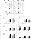
Exosomes are small 30-100 nm membrane vesicles released from hematopoietic and nonhematopoietic cells and function to promote intercellular communication. They are generated through fusion of multivesicular bodies with the plasma membrane and release of interluminal vesicles. Previous studies from our laboratory demonstrated that macrophages infected with Mycobacterium release exosomes that promote activation of both innate and acquired immune responses; however, the components present in exosomes inducing these host responses were not defined. This study used LC-MS/MS to identify 41 mycobacterial proteins present in exosomes released from M. tuberculosis-infected J774 cells. Many of these proteins have been characterized as highly immunogenic. Further, since most of the mycobacterial proteins identified are actively secreted, we hypothesized that macrophages treated with M. tuberculosis culture filtrate proteins (CFPs) would release exosomes containing mycobacterial proteins. We found 29 M. tuberculosis proteins in exosomes released from CFP-treated J774 cells, the majority of which were also present in exosomes isolated from M. tuberculosis-infected cells. The exosomes from CFP-treated J774 cells could promote macrophage and dendritic cell activation as well as activation of naïve T cells in vivo. These results suggest that exosomes containing M. tuberculosis antigens may be alternative approach to developing a tuberculosis vaccine.
Figures


Similar articles
-
Exosomes isolated from mycobacteria-infected mice or cultured macrophages can recruit and activate immune cells in vitro and in vivo.J Immunol. 2012 Jul 15;189(2):777-85. doi: 10.4049/jimmunol.1103638. Epub 2012 Jun 20. J Immunol. 2012. PMID: 22723519 Free PMC article.
-
Exosomes carrying mycobacterial antigens can protect mice against Mycobacterium tuberculosis infection.Eur J Immunol. 2013 Dec;43(12):3279-90. doi: 10.1002/eji.201343727. Epub 2013 Sep 6. Eur J Immunol. 2013. PMID: 23943377 Free PMC article.
-
Exosomes function in antigen presentation during an in vivo Mycobacterium tuberculosis infection.Sci Rep. 2017 Mar 6;7:43578. doi: 10.1038/srep43578. Sci Rep. 2017. PMID: 28262829 Free PMC article.
-
Exosomes released from M. tuberculosis infected cells can suppress IFN-γ mediated activation of naïve macrophages.PLoS One. 2011 Apr 14;6(4):e18564. doi: 10.1371/journal.pone.0018564. PLoS One. 2011. PMID: 21533172 Free PMC article.
-
Exosome function: from tumor immunology to pathogen biology.Traffic. 2008 Jun;9(6):871-81. doi: 10.1111/j.1600-0854.2008.00734.x. Epub 2008 Mar 6. Traffic. 2008. PMID: 18331451 Free PMC article. Review.
Cited by
-
Emerging role of extracellular vesicles in veterinary practice: novel opportunities and potential challenges.Front Vet Sci. 2024 Jan 25;11:1335107. doi: 10.3389/fvets.2024.1335107. eCollection 2024. Front Vet Sci. 2024. PMID: 38332755 Free PMC article. Review.
-
Extracellular Vesicles: Recent Insights Into the Interaction Between Host and Pathogenic Bacteria.Front Immunol. 2022 May 25;13:840550. doi: 10.3389/fimmu.2022.840550. eCollection 2022. Front Immunol. 2022. PMID: 35693784 Free PMC article. Review.
-
Extracellular Vesicles in Mycobacteria and Tuberculosis.Front Cell Infect Microbiol. 2022 May 27;12:912831. doi: 10.3389/fcimb.2022.912831. eCollection 2022. Front Cell Infect Microbiol. 2022. PMID: 35719351 Free PMC article. Review.
-
Message in a vesicle - trans-kingdom intercommunication at the vector-host interface.J Cell Sci. 2019 Mar 18;132(6):jcs224212. doi: 10.1242/jcs.224212. J Cell Sci. 2019. PMID: 30886004 Free PMC article. Review.
-
Trypanosoma cruzi-Infected Human Macrophages Shed Proinflammatory Extracellular Vesicles That Enhance Host-Cell Invasion via Toll-Like Receptor 2.Front Cell Infect Microbiol. 2020 Mar 20;10:99. doi: 10.3389/fcimb.2020.00099. eCollection 2020. Front Cell Infect Microbiol. 2020. PMID: 32266161 Free PMC article.
References
-
- Thery, C. , Ostrowski, M. , Segura, E. , Membrane vesicles as conveyors of immune responses. Nat. Rev. Immunol. 2009, 9, 581–593. - PubMed
-
- Zitvogel, L. , Regnault, A. , Lozier, A. , Wolfers, J. et al., Eradication of established murine tumors using a novel cell‐free vaccine: dendritic cell‐derived exosomes. Nat. Med. 1998, 4, 594–600. - PubMed
-
- Thery, C. , Zitvogel, L. , Amigorena, S. , Exosomes: composition, biogenesis and function. Nat. Rev. Immunol. 2002, 2, 569–579. - PubMed
Publication types
MeSH terms
Substances
Grants and funding
LinkOut - more resources
Full Text Sources
Other Literature Sources
Miscellaneous

