. 2010 Aug 4;132(30):10338-51.
doi: 10.1021/ja1005724.

Communication between the zinc and nickel sites in dimeric HypA: metal recognition and pH sensing

Affiliations

Communication between the zinc and nickel sites in dimeric HypA: metal recognition and pH sensing

Robert W Herbst et al. J Am Chem Soc. .

Abstract

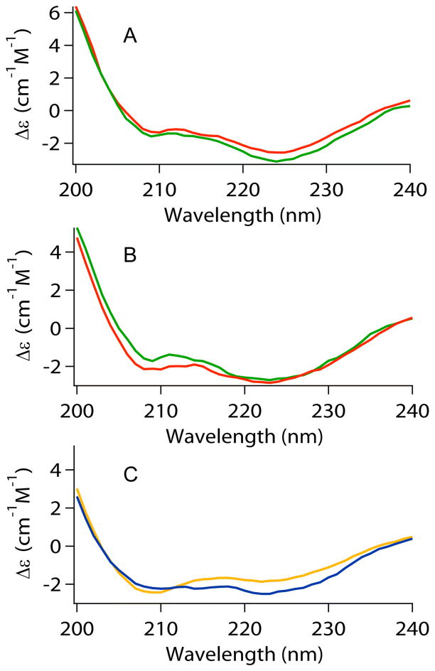
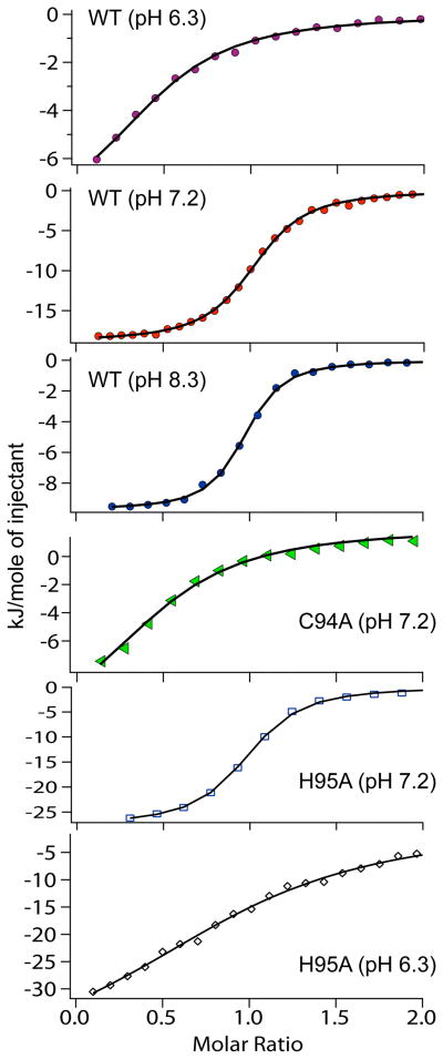
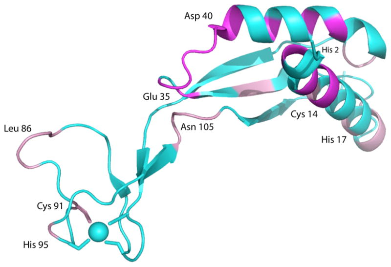
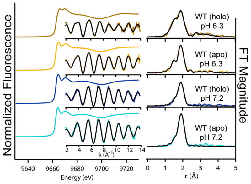
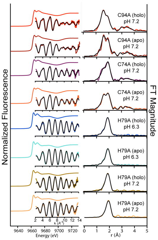
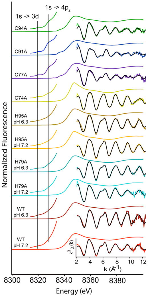
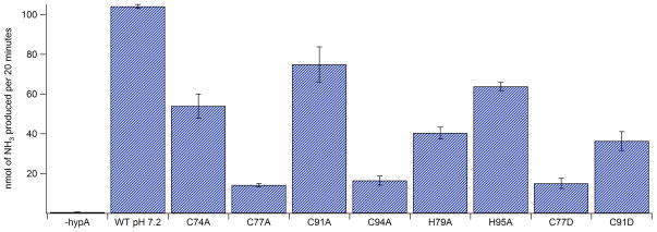
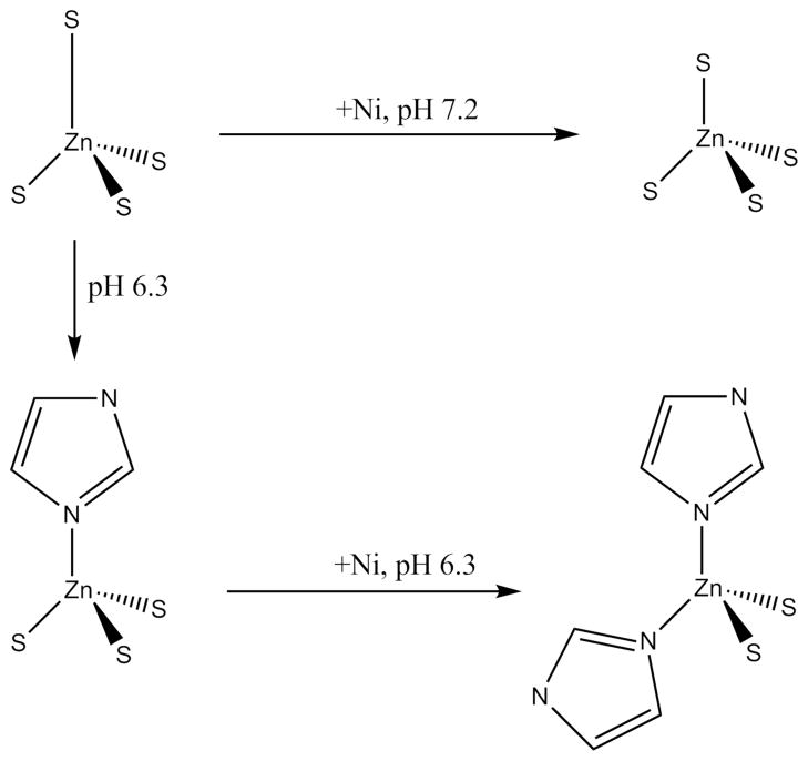
Helicobacter pylori , a pathogen that colonizes the human stomach, requires the nickel-containing metalloenzymes urease and NiFe-hydrogenase to survive this low pH environment. The maturation of both enzymes depends on the metallochaperone, HypA. HypA contains two metal sites, an intrinsic zinc site and a low-affinity nickel binding site. X-ray absorption spectroscopy (XAS) shows that the structure of the intrinsic zinc site of HypA is dynamic and able to sense both nickel loading and pH changes. At pH 6.3, an internal pH that occurs during acid shock, the zinc site undergoes unprecedented ligand substitutions to convert from a Zn(Cys)(4) site to a Zn(His)(2)(Cys)(2) site. NMR spectroscopy shows that binding of Ni(II) to HypA results in paramagnetic broadening of resonances near the N-terminus. NOEs between the beta-CH(2) protons of Zn cysteinyl ligands are consistent with a strand-swapped HypA dimer. Addition of nickel causes resonances from the zinc binding motif and other regions to double, indicating more than one conformation can exist in solution. Although the structure of the high-spin, 5-6 coordinate Ni(II) site is relatively unaffected by pH, the nickel binding stoichiometry is decreased from one per monomer to one per dimer at pH = 6.3. Mutation of any cysteine residue in the zinc binding motif results in a zinc site structure similar to that found for holo-WT-HypA at low pH and is unperturbed by the addition of nickel. Mutation of the histidines that flank the CXXC motifs results in a zinc site structure that is similar to holo-WT-HypA at neutral pH (Zn(Cys)(4)) and is no longer responsive to nickel binding or pH changes. Using an in vitro urease activity assay, it is shown that the recombinant protein is sufficient for recovery of urease activity in cell lysate from a HypA deletion mutant, and that mutations in the zinc-binding motif result in a decrease in recovered urease activity. The results are interpreted in terms of a model wherein HypA controls the flow of nickel traffic in the cell in response to nickel availability and pH.


Figures

Figure 1
Figure 1
Circular dichroism spectral comparisons of HypA proteins: (A) Apo- (red) vs. Holo- (green) WT-HypA at pH = 7.2. (B) Apo- (red) vs. Holo- (green) WT - HypA at pH = 6.3. (C) Apo-H95A-HypA (blue) vs. apo-C74A-HypA (orange) at pH = 7.2.
Figure 2
Figure 2
Isothermal titration calorimetry of HypA proteins. The integrated heat data for titrations of apo-WT-HypA and selected mutant HypA proteins with NiCl2 solutions is shown for titrations at different pH values. The symbols represent the integrated heat after each injection, while the solid line is the calculated fit of the data used for values in Table 1 (see materials and methods).
Figure 3
Figure 3
A plane from 13C-edited three-dimensional 1H,1H NOESY (800.13 MHz 1H) showing β-CH2 NOEs between Zn-ligating Cys residues in HypA as described in the text.
Figure 4
Figure 4
NMR results superimposed on the structure of monomeric HypA (PDB entry 2KDX). Resonances that are broadened to invisibility by addition of Ni(II) ions are shown in magenta. Resonances that are perturbed or doubled by binding Ni(II), indicating more than one conformation is present, are shown in pink. The Zn site is indicated by the blue sphere. See text for details.
Figure 5
Figure 5
Zinc K-edge XAS of apo- and holo-WT-HypA at pH 7.2 Left: Normalized XANES data with unfiltered k3-weighted EXAFS spectra (colored line) and best fits (black line) from Table 2. Right: Fourier-transformed EXAFS spectra and fits.
Figure 6
Figure 6
Zinc K-edge XAS data for selected mutant HypA proteins. Left: Normalized XANES spectra with unfiltered k3-weighted EXAFS spectra (colored line) and best fits (black line) from Table 2 inset. Right: Fourier-transformed EXAFS spectra and fits. Data for other Cys → Ala, Cys → Asp and H95A are shown in supporting information.
Figure 7
Figure 7
Nickel K-edge XAS data for holo-HypA proteins. XANES is shown as a single colored line. Left inset is an expansion of the pre-edge XANES region. Right insets are the unfiltered k3-weighted EXAFS data (colored line) and fits from Table 3 (black line).
Figure 8
Figure 8
In vitro urease activity assay for WT- and mutant HypA proteins. Activity is shown as the amount of ammonia produced per 20 minutes. The amount of ammonia produced was calibrated to a standard curve created using NH4Cl.
Figure 9
Figure 9
Sequence alignment of HypA and HypA homologue proteins from various bacteria. Black boxes represent strictly conserved residues, red boxes show the two conserved CxxC motifs and blue boxes depict the alignment with the flanking His residue from H. pylori. H. pylori HypA is 53%, 22%, 25%, 20% identical and 77%, 48%, 51%, 56% similar to HypA homologues from H. hepaticus, E. coli, and T. kodakaraensis, respectively.
Scheme 1
Scheme 1
Depiction of the coordination changes around the zinc atom depending on pH and Ni binding status.

References

    1. Marshall BJ, Warren JR. Lancet. 1984;1:1311. - PubMed
    1. Scott D, Weeks D, Melchers K, Sachs G. Gut. 1998;43(Suppl 1):S56. - PMC - PubMed
    1. Eaton KA, Brooks CL, Morgan DR, Krakowka S. Infect Immun. 1991;59:2470. - PMC - PubMed
    1. Eaton KA, Krakowka S. Infect Immun. 1994;62:3604. - PMC - PubMed
    1. Olson JW, Maier RJ. Science. 2002;298:1788. - PubMed

Publication types

LinkOut - more resources