. 2010 Sep 1;202(5):791-800.
doi: 10.1086/655225.

Increased pulmonary pressures and myocardial wall stress in children with severe malaria

Affiliations

Increased pulmonary pressures and myocardial wall stress in children with severe malaria

Jacqueline J Janka et al. J Infect Dis. .

Abstract

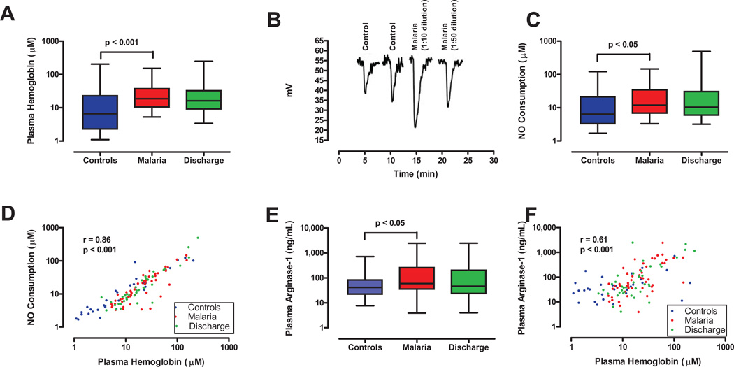
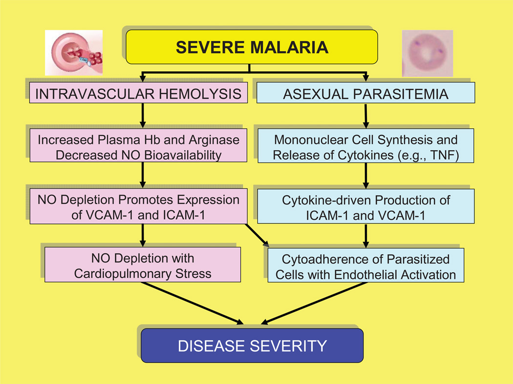
Background: Chronic intravascular hemolysis leads to nitric oxide (NO) depletion and pulmonary hypertension in sickle cell disease. To test whether this pathophysiology occurs in malaria, we examined in Mali 53 children who were admitted to the hospital with severe malaria (excluding cerebral malaria) and 31 age-matched controls.

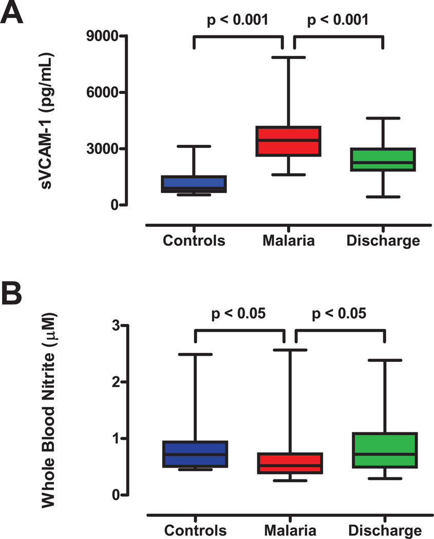
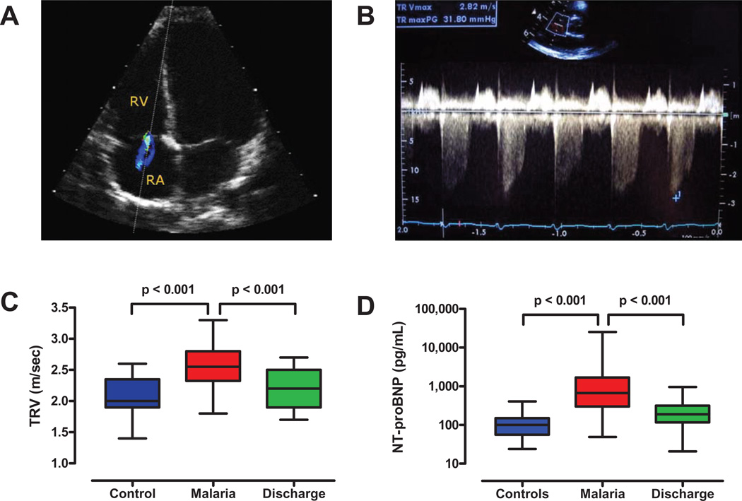
Methods: Severity of hemolysis was assessed from plasma levels of free hemoglobin and arginase-1. NO metabolism was assessed by whole-blood nitrite levels and plasma NO consumption. Effects on the cardiovascular system and endothelial function were assessed by using echocardiography to measure peak tricuspid regurgitant jet velocity and by evaluating plasma levels of N-terminal prohormone brain natriuretic peptide (NT-proBNP) and soluble vascular cell adhesion molecule-1.

Results: Children with severe malaria had higher plasma levels of hemoglobin and arginase-1, reduced whole-blood levels of nitrite, and increased NO consumption relative to controls. They also had increased pulmonary arterial pressures (P< .05) with elevated levels of NT-proBNP and soluble vascular cell adhesion molecule-1 (P< .001).

Conclusion: Children with severe malaria have increased pulmonary pressures and myocardial wall stress. These complications are consistent with NO depletion from intravascular hemolysis, and they indicate that the pathophysiologic cascade from intravascular hemolysis to NO depletion and its cardiopulmonary effects is activated in children with severe malaria.


Conflict of interest statement

Conflict of Interest

The authors declare that they do not have commercial or other associations that might pose a conflict of interest.

Figures

Figure 1
Figure 1. Intravascular hemolysis and nitric oxide resistance in malaria
A. Comparison of free plasma Hb levels for controls (n=31) at the time of admission, and for cases at the times of admission (n=52) and discharge (day 3–5, n=38). Plasma Hb levels were higher in cases than controls at the time of admission (p<0.001). B. NO consumption by plasma from a control subject and two dilutions of plasma from a patient with severe malaria. The downward displacement of the tracing from the NO analyzer represents the consumption of NO generated from the NO donor agent in the reaction vessel. These four tracings were obtained on the same day and combined in one figure for illustrative purposes. C. Comparison of NO consumption by plasma from controls (n=31) at the time of admission, and cases at the times of admission (n=52) and discharge (n=38). NO consumption was greater for cases than controls at the time of admission (p=0.04). D. Comparison of plasma free Hb and NO consumption results. Plasma Hb results from panel A are plotted on the abscissa with NO consumption results from panel C on the ordinate, demonstrating a positive correlation between the two (p<0.001). E. Comparison of plasma arginase-1 results for controls (n=31) and for cases at the times of admission (n=52) and discharge (n=37). Arginase levels were higher in cases than controls at admission (p<0.05), and showed a non-significant decline at the time of discharge. F. Comparison of plasma free Hb and arginase-1 results. Plasma Hb results from panel A are plotted on the abscissa with arginase-1 results from panel E on the ordinate, and demonstrate a positive correlation (p<0.001).
Figure 2
Figure 2. Markers of circulatory stress during severe malaria
A. Soluble vascular cell adhesion molecule (sVCAM-1), a marker of endothelial activation and adhesiveness. Plasma levels of sVCAM-1 were higher in cases (n=52) than controls (n=31, p<0.001) at admission, and decreased by the time of discharge (day 3–5, n=38, p<0.001). B. Comparison of whole blood nitrite levels in controls at admission, and in cases at times of admission and discharge. Whole blood nitrite levels, a marker of NO bioavailability, were lower in cases (n=50) than controls at admission (n=15; p<0.05), and rose by the time of discharge (p<0.05, n=26). Box and whisker plots indicate median, range and interquartile boundaries.
Figure 3
Figure 3. Echocardiographic evidence of elevated pulmonary arterial pressures in malaria
A. An ultrasound probe was used to visualize tricuspid regurgitation in Malian children with severe malaria. The dotted line indicates the path of sound waves from the probe across the tricuspid valve, where the regurgitant jet from the right ventricle (RV) to the right atrium (RA) is shown in color. B. Doppler echocardiogram demonstrating tricuspid regurgitation in a child with severe malaria. The lowest point of the downward deflection indicates the initial tricuspid regurgitant jet velocity (TRV) at the peak of right ventricular systole (scale is on the right side of the panel). This echocardiogram was interpreted as having a peak initial TRV of 2.8 meters per second. C. Tricuspid regurgitant jet velocities (TRVs) for controls (n=17) and cases (n=36) at the times of admission and discharge (day 3–5, n=19). TRVs for cases were greater than for controls (p<0.001) and fell between admission and discharge (p<0.001). D. N-terminal prohormone brain natriuretic peptide (NT-proBNP), a marker of cardiac muscle stretch and mechanical stress. Levels of NT-proBNP were higher in cases (n=50) than controls (n=31) at the time of admission (p<0.001), and fell in cases by the time of discharge (n=38, p<0.001).
Figure 4
Figure 4. Schematic diagram of the pathophysiology of severe malaria
Intravascular hemolysis produces increases in VCAM-1 and ICAM-1 from NO depletion that lead to increased pulmonary pressures and myocardial stress. In addition, both cytokine production in response to parasite factors and NO depletion from intravascular hemolysis lead to greater production of ICAM-1 and VCAM-1, which in turn increases the cytoadherence of parasitized red cells and produces endothelial activation.

References

    1. Laveran CL. Classics in infectious diseases: A newly discovered parasite in the blood of patients suffering from malaria. Parasitic etiology of attacks of malaria: Charles Louis Alphonse Laveran (1845–1922) Rev Infect Dis. 1982;4(4):908–911. - PubMed
    1. Geneva: World Health Organization; 2009. [Accessed 26 February 2010]. Roll Back Malaria: Global Malaria Action Plan: Key Facts, Figures and Strategies. Available at: http://www.rollbackmalaria.org/gmap/.
    1. Hay SI, Guerra CA, Gething PW, et al. A world malaria map: Plasmodium falciparum endemicity in 2007. PLoS Med. 2009;6(3):e1000048. - PMC - PubMed
    1. Korenromp EL, Williams BG, Gouws E, Dye C, Snow RW. Measurement of trends in childhood malaria mortality in Africa: an assessment of progress toward targets based on verbal autopsy. Lancet Infect Dis. 2003;3:349–358. - PubMed
    1. Pasvol G. Treatment of complicated and severe malaria. Br Med Bull. 2005;75–76:29–47. - PubMed

Publication types